GSK-3 is a Ser/Thr kinase first identified as an inactivator of Glycogen Synthase. GSK-3 acts as a multifunctional downstream switch that determines the output of numerous signaling pathways. There are two mammalian GSK-3 isoforms encoded by distinct genes, GSK-3 alpha and GSK-3 beta, which are structurally similar, but functionally non-identical. GSK-3a is inhibited by phosphorylation at S21 by Akt and other kinases. GSK-3 alpha and GSK-3 beta share 85% amino acid identity. Dysregulated GSK-3 has been implicated in several diseases including type II diabetes, Alzheimer's disease, bipolar disorder, and cancer.

Loading...
Key Product Details
Species Reactivity
Validated:
Human, Mouse, Rat
Cited:
Human, Mouse, Rat
Applications
Validated:
Western Blot, Intracellular Staining by Flow Cytometry, Immunocytochemistry, CyTOF-ready
Cited:
Immunohistochemistry, Western Blot, Flow Cytometry, Immunoprecipitation
Label
Unconjugated
Antibody Source
Polyclonal Rabbit IgG
Loading...
Product Specifications
Immunogen
E. coli-derived recombinant human GSK-3 beta
Accession # P49841
Accession # P49841
Specificity
Detects human, mouse, and rat GSK-3 beta and the closely related GSK-3 alpha.
Clonality
Polyclonal
Host
Rabbit
Isotype
IgG
Scientific Data Images for GSK-3 alpha/beta Antibody
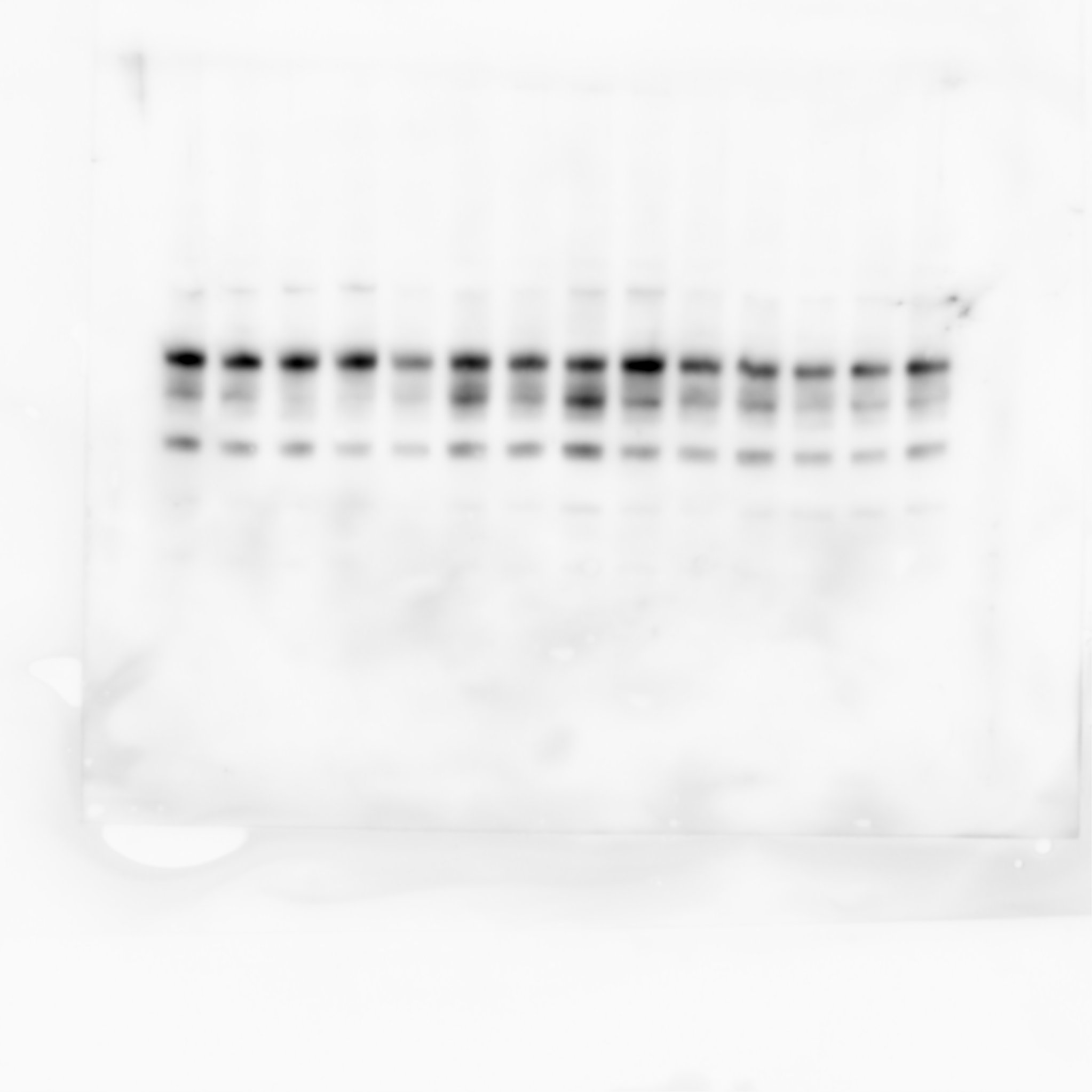
Detection of Human GSK‑3 alpha / beta by Western Blot.
Western blot shows lysates of HeLa human cervical epithelial carcinoma cell line, HT-29 human colon adenocarcinoma cell line, MDA-MB-468 and MCF-7 human breast cancer cell line. PVDF membrane was probed with 0.1 µg/mL of Rabbit Anti-Human/Mouse/ Rat GSK-3a/ beta Antigen Affinity-purified Polyclonal Antibody (Catalog # AF2157) followed by HRP-conjugated Anti-Rabbit IgG Secondary Antibody (Catalog # HAF008). Specific bands were detected for GSK-3a/ beta at approximately 46 and 51 kDa (as indicated). This experiment was conducted under reducing conditions and using Immunoblot Buffer Group 1.
GSK‑3 alpha / beta in HeLa Human Cell Line.
GSK-3a/ beta was detected in immersion fixed HeLa human cervical epithelial carcinoma cell line using Rabbit Anti-Human/Mouse/Rat GSK-3a/ beta Antigen Affinity-purified Polyclonal Antibody (Catalog # AF2157) at 10 µg/mL for 3 hours at room temperature. Cells were stained using the NorthernLights™ 557-conjugated Anti-Rabbit IgG Secondary Antibody (red; Catalog # NL004) and counterstained with DAPI (blue). Specific staining was localized to cytoplasm. View our protocol for Fluorescent ICC Staining of Cells on Coverslips.
Detection of GSK‑3 alpha / beta in HeLa Human Cell Line by Flow Cytometry.
HeLa human cervical epithelial carcinoma cell line was stained with Rabbit Anti-Human/Mouse/Rat GSK-3a/ beta Antigen Affinity-purified Polyclonal Antibody (Catalog # AF2157, filled histogram) or control antibody (Catalog # AB-105-C, open histogram), followed by Allophycocyanin-conjugated Anti-Rabbit IgG Secondary Antibody (Catalog # F0111). To facilitate intracellular staining, cells were fixed with paraformaldehyde and permeabilized with saponin.
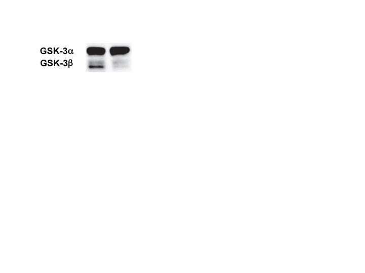
Detection of Mouse GSK-3 alpha/beta by Western Blot
GSK3 binds to scaffold formed by histidine-rich cytoplasmic loop domains within the ZIP6-ZIP10 heteromer.(a) Domain organization of ZIP10 and ZIP6 and deletion constructs used in this work to test the hypothesis that a cytoplasmic loop connecting transmembrane (TM) domains III and IV is essential for binding to GSK3 binding. (b) Control Western blot documenting ZIP6 expression products in eluates shown in panel ‘e’. (c) The co-expression of wild-type ZIP6 and ZIP10 proteins comprising intact cytoplasmic loop domains connecting TM domains III and IV is essential for binding of GSK3. GSK3-directed Western blot analysis of cellular lysates and co-immunoprecipitation eluates from in vivo formaldehyde crosslinked cells expressing different combinations of wild-type and/or mutant ZIP6/ZIP10. Note that in addition to protein bands reflecting the expected monomeric molecular weights of GSK3 and ZIP6, the Western blots in panels ‘d’ and ‘e’ revealed the appearance of high molecular weight crosslinked bands, whose appearance depended on the application of the in vivo formaldehyde crosslinking step. Image collected and cropped by CiteAb from the following publication (https://pubmed.ncbi.nlm.nih.gov/28098160), licensed under a CC-BY license. Not internally tested by R&D Systems.Applications for GSK-3 alpha/beta Antibody
Application
Recommended Usage
CyTOF-ready
Ready to be labeled using established conjugation methods. No BSA or other carrier proteins that could interfere with conjugation.
Immunocytochemistry
5-15 µg/mL
Sample: Immersion fixed HeLa human cervical epithelial carcinoma cell line
Sample: Immersion fixed HeLa human cervical epithelial carcinoma cell line
Intracellular Staining by Flow Cytometry
2.5 µg/106 cells
Sample: HeLa Human cell line fixed with paraformaldehyde and permeabilized with saponin
Sample: HeLa Human cell line fixed with paraformaldehyde and permeabilized with saponin
Western Blot
0.1 µg/mL
Sample: HeLa human cervical epithelial carcinoma cell line, HT-29 human colon adenocarcinoma cell line, MDA-MB-468 and MCF-7 human breast cancer cell line
Sample: HeLa human cervical epithelial carcinoma cell line, HT-29 human colon adenocarcinoma cell line, MDA-MB-468 and MCF-7 human breast cancer cell line
Reviewed Applications
Read 4 reviews rated 4.5 using AF2157 in the following applications:
Flow Cytometry Panel Builder
Bio-Techne Knows Flow Cytometry
Save time and reduce costly mistakes by quickly finding compatible reagents using the Panel Builder Tool.
Advanced Features
- Spectra Viewer - Custom analysis of spectra from multiple fluorochromes
- Spillover Popups - Visualize the spectra of individual fluorochromes
- Antigen Density Selector - Match fluorochrome brightness with antigen density

Formulation, Preparation, and Storage
Purification
Antigen Affinity-purified
Reconstitution
Reconstitute at 0.2 mg/mL in sterile PBS. For liquid material, refer to CoA for concentration.
Loading...
Formulation
Lyophilized from a 0.2 μm filtered solution in PBS with Trehalose. *Small pack size (SP) is supplied either lyophilized or as a 0.2 µm filtered solution in PBS.
Shipping
Lyophilized product is shipped at ambient temperature. Liquid small pack size (-SP) is shipped with polar packs. Upon receipt, store immediately at the temperature recommended below.
Stability & Storage
Use a manual defrost freezer and avoid repeated freeze-thaw cycles.
- 12 months from date of receipt, -20 to -70 °C as supplied.
- 1 month, 2 to 8 °C under sterile conditions after reconstitution.
- 6 months, -20 to -70 °C under sterile conditions after reconstitution.
Calculators
Background: GSK-3 alpha/beta
Long Name
Glycogen Synthase Kinase 3
Alternate Names
DKFZp686D0638, EC 2.7.11, EC 2.7.11.26, glycogen synthase kinase 3 alpha, glycogen synthase kinase-3 alpha, GSK-3 alpha
UniProt
Additional GSK-3 alpha/beta Products
Product Documents for GSK-3 alpha/beta Antibody
Certificate of Analysis
To download a Certificate of Analysis, please enter a lot or batch number in the search box below.
Note: Certificate of Analysis not available for kit components.
Product Specific Notices for GSK-3 alpha/beta Antibody
For research use only
Related Research Areas
Citations for GSK-3 alpha/beta Antibody
 Powered by Bioz
Customer Reviews for GSK-3 alpha/beta Antibody (4)
4.5 out of 5
4 Customer Ratings
Have you used GSK-3 alpha/beta Antibody?
Submit a review and receive an Amazon gift card!
$25/€18/£15/$25CAN/¥2500 Yen for a review with an image
$10/€7/£6/$10CAN/¥1110 Yen for a review without an image
Submit a review
Customer Images
Showing
1
-
4 of
4 reviews
Showing All
Filter By:
-
Application: Western BlotSample Tested: Lung single-cell suspensionSpecies: MouseVerified Customer | Posted 06/06/2019

-
Application: Western BlotSample Tested: Colon cancer cell lineSpecies: HumanVerified Customer | Posted 09/14/2018
-
Application: Western BlotSample Tested: IPS2 induced pluripotent stem cellsSpecies: MouseVerified Customer | Posted 01/25/2018
-
Application: Western BlotSample Tested: Pancreatic cancer cellsSpecies: HumanVerified Customer | Posted 12/27/2017
There are no reviews that match your criteria.
Protocols
Find general support by application which include: protocols, troubleshooting, illustrated assays, videos and webinars.
- 7-Amino Actinomycin D (7-AAD) Cell Viability Flow Cytometry Protocol
- Appropriate Fixation of IHC/ICC Samples
- Cellular Response to Hypoxia Protocols
- ClariTSA™ Fluorophore Kits
- Detection & Visualization of Antibody Binding
- Extracellular Membrane Flow Cytometry Protocol
- Flow Cytometry Protocol for Cell Surface Markers
- Flow Cytometry Protocol for Staining Membrane Associated Proteins
- Flow Cytometry Staining Protocols
- Flow Cytometry Troubleshooting Guide
- ICC Cell Smear Protocol for Suspension Cells
- ICC Immunocytochemistry Protocol Videos
- ICC for Adherent Cells
- Immunocytochemistry (ICC) Protocol
- Immunocytochemistry Troubleshooting
- Immunofluorescence of Organoids Embedded in Cultrex Basement Membrane Extract
- Immunohistochemistry (IHC) and Immunocytochemistry (ICC) Protocols
- Intracellular Flow Cytometry Protocol Using Alcohol (Methanol)
- Intracellular Flow Cytometry Protocol Using Detergents
- Intracellular Nuclear Staining Flow Cytometry Protocol Using Detergents
- Intracellular Staining Flow Cytometry Protocol Using Alcohol Permeabilization
- Intracellular Staining Flow Cytometry Protocol Using Detergents to Permeabilize Cells
- Preparing Samples for IHC/ICC Experiments
- Preventing Non-Specific Staining (Non-Specific Binding)
- Primary Antibody Selection & Optimization
- Propidium Iodide Cell Viability Flow Cytometry Protocol
- Protocol for Liperfluo
- Protocol for VisUCyte™ HRP Polymer Detection Reagent
- Protocol for the Characterization of Human Th22 Cells
- Protocol for the Characterization of Human Th9 Cells
- Protocol for the Fluorescent ICC Staining of Cell Smears - Graphic
- Protocol for the Fluorescent ICC Staining of Cultured Cells on Coverslips - Graphic
- Protocol for the Preparation and Fluorescent ICC Staining of Cells on Coverslips
- Protocol for the Preparation and Fluorescent ICC Staining of Non-adherent Cells
- Protocol for the Preparation and Fluorescent ICC Staining of Stem Cells on Coverslips
- Protocol for the Preparation of a Cell Smear for Non-adherent Cell ICC - Graphic
- Protocol: Annexin V and PI Staining by Flow Cytometry
- Protocol: Annexin V and PI Staining for Apoptosis by Flow Cytometry
- R&D Systems Quality Control Western Blot Protocol
- TUNEL and Active Caspase-3 Detection by IHC/ICC Protocol
- The Importance of IHC/ICC Controls
- Troubleshooting Guide: Fluorokine Flow Cytometry Kits
- Troubleshooting Guide: Western Blot Figures
- Western Blot Conditions
- Western Blot Protocol
- Western Blot Protocol for Cell Lysates
- Western Blot Troubleshooting
- Western Blot Troubleshooting Guide
- View all Protocols, Troubleshooting, Illustrated assays and Webinars
Loading...