HABP1 (hyaluronan binding protein1; also p32 and gC1qR) is a 33 kDa member of the MAM33 family of proteins. It is widely expressed and found on/in endothelial cells, platelets and dendritic cells. It has multiple binding partners, including complement C1q, hyaluronan, vitronectin and ARF. Full-length mouse HABP1 proprecursor is 279 amino acid (aa) in length. It contains a 70 aa mitochondrial targeting sequence (aa 1‑70) that is cleaved to generate a 209 aa mature segment. A hyaluronan binding site lies between Lys116‑Lys125 and a Tyr phosphorylation site exists at Tyr184. Although the prosegment is usually cleaved, the intact proprecursor is associated with sperm. HABP1 reportedly forms noncovalent homotrimers. Over aa 72‑279, mouse HABP1 shares 99% and 91% aa identity with rat and human HABP1, respectively.

Key Product Details
Species Reactivity
Validated:
Human, Mouse, Rat
Cited:
Human
Applications
Validated:
Western Blot, Simple Western
Cited:
Western Blot
Label
Unconjugated
Antibody Source
Polyclonal Goat IgG
Loading...
Product Specifications
Immunogen
E. coli-derived recombinant mouse HABP1/C1QBP
Leu72-Gln279
Accession # NP_031599
Leu72-Gln279
Accession # NP_031599
Specificity
Detects human, mouse, and rat HABP1/C1QBP in direct ELISAs and Western blots. In direct ELISAs, less than 1% cross-reactivity with recombinant human C1QR is observed.
Clonality
Polyclonal
Host
Goat
Isotype
IgG
Scientific Data Images for HABP1/C1QBP Antibody
Detection of Human/Mouse/Rat HABP1/C1QBP by Western Blot.
Western blot shows lysates of Jurkat human acute T cell leukemia cell line, Nalm-6 human Pre-B acute lymphocytic leukemia cell line, human liver tissue, mouse brain tissue and rat testis tissue. PVDF membrane was probed with 1 µg/mL of Goat Anti-Human/Mouse/Rat HABP1/C1QBP Antigen Affinity-purified Polyclonal Antibody (Catalog # AF5359) followed by HRP-conjugated Anti-Goat IgG Secondary Antibody (Catalog # HAF019). A specific band was detected for HABP1/C1QBP at approximately 35 kDa (as indicated). This experiment was conducted under reducing conditions and using Immunoblot Buffer Group 8.
Detection of Human and Mouse HABP1/C1QBP by Simple WesternTM.
Simple Western lane view shows lysates of Jurkat human acute T cell leukemia cell line and mouse brain (cortex) tissue, loaded at 0.2 mg/mL. A specific band was detected for HABP1/C1QBP at approximately 34 kDa (as indicated) using 10 µg/mL of Goat Anti-Human/Mouse/Rat HABP1/C1QBP Antigen Affinity-purified Polyclonal Antibody (Catalog # AF5359) followed by 1:50 dilution of HRP-conjugated Anti-Goat IgG Secondary Antibody (Catalog # HAF109). This experiment was conducted under reducing conditions and using the 12-230 kDa separation system.Applications for HABP1/C1QBP Antibody
Application
Recommended Usage
Simple Western
10 µg/mL
Sample: Jurkat human acute T cell leukemia cell line and mouse brain (cortex) tissue
Sample: Jurkat human acute T cell leukemia cell line and mouse brain (cortex) tissue
Western Blot
1 µg/mL
Sample: Jurkat human acute T cell leukemia cell line, Nalm‑6 human Pre‑B acute lymphocytic leukemia cell line, human liver tissue, mouse brain tissue and rat testis tissue
Sample: Jurkat human acute T cell leukemia cell line, Nalm‑6 human Pre‑B acute lymphocytic leukemia cell line, human liver tissue, mouse brain tissue and rat testis tissue
Reviewed Applications
Read 1 review rated 3 using AF5359 in the following applications:
Formulation, Preparation, and Storage
Purification
Antigen Affinity-purified
Reconstitution
Reconstitute at 0.2 mg/mL in sterile PBS. For liquid material, refer to CoA for concentration.
Loading...
Formulation
Lyophilized from a 0.2 μm filtered solution in PBS with Trehalose. *Small pack size (SP) is supplied either lyophilized or as a 0.2 µm filtered solution in PBS.
Shipping
Lyophilized product is shipped at ambient temperature. Liquid small pack size (-SP) is shipped with polar packs. Upon receipt, store immediately at the temperature recommended below.
Stability & Storage
Use a manual defrost freezer and avoid repeated freeze-thaw cycles.
- 12 months from date of receipt, -20 to -70 °C as supplied.
- 1 month, 2 to 8 °C under sterile conditions after reconstitution.
- 6 months, -20 to -70 °C under sterile conditions after reconstitution.
Calculators
Background: HABP1/C1QBP
Long Name
Hyaluronic Acid-binding Protein
Alternate Names
C1QBP, gC1qBP, gC1qR, SF2p32
Gene Symbol
C1QBP
UniProt
Additional HABP1/C1QBP Products
Product Documents for HABP1/C1QBP Antibody
Certificate of Analysis
To download a Certificate of Analysis, please enter a lot or batch number in the search box below.
Note: Certificate of Analysis not available for kit components.
Product Specific Notices for HABP1/C1QBP Antibody
For research use only
Related Research Areas
Citations for HABP1/C1QBP Antibody
 Powered by Bioz
Customer Reviews for HABP1/C1QBP Antibody (1)
3 out of 5
1 Customer Rating
Have you used HABP1/C1QBP Antibody?
Submit a review and receive an Amazon gift card!
$25/€18/£15/$25CAN/¥2500 Yen for a review with an image
$10/€7/£6/$10CAN/¥1110 Yen for a review without an image
Submit a review
Customer Images
Showing
1
-
1 of
1 review
Showing All
Filter By:
-
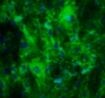
Application: ImmunohistochemistrySample Tested: HUVEC human umbilical vein endothelial cellsSpecies: HumanVerified Customer | Posted 12/08/2018

There are no reviews that match your criteria.
Protocols
Find general support by application which include: protocols, troubleshooting, illustrated assays, videos and webinars.
- Cellular Response to Hypoxia Protocols
- R&D Systems Quality Control Western Blot Protocol
- Troubleshooting Guide: Western Blot Figures
- Western Blot Conditions
- Western Blot Protocol
- Western Blot Protocol for Cell Lysates
- Western Blot Troubleshooting
- Western Blot Troubleshooting Guide
- View all Protocols, Troubleshooting, Illustrated assays and Webinars
Loading...