
Key Product Details
Species Reactivity
Validated:
Human, Mouse, Rat
Cited:
Human, Mouse, Coral - Stylophora pistillata (Hood Coral), Rabbit, Transgenic Mouse
Applications
Validated:
Immunohistochemistry, Western Blot, Simple Western
Cited:
Western Blot
Label
Unconjugated
Antibody Source
Polyclonal Rabbit IgG
Loading...
Product Specifications
Immunogen
E. coli-derived recombinant human JNK1
Met1-Gln384
Accession # P45983
Met1-Gln384
Accession # P45983
Specificity
Detects human, mouse, and rat p46 and p54 JNK in Western blots. Detects recombinant human (rh) JNK1, rhJNK2, and rhJNK3.
Clonality
Polyclonal
Host
Rabbit
Isotype
IgG
Scientific Data Images for JNK Pan Specific Antibody
Detection of Human/Mouse/Rat JNK by Western Blot.
Western blot shows lysates of HeLa human cervical epithelial carcinoma cell line, U937 human histiocytic lymphoma cell line, C6 rat glioma cell line, and C2C12 mouse myoblast cell line. PVDF membrane was probed with 0.2 µg/mL Rabbit Anti-Human/Mouse/Rat JNK Pan Specific Antigen Affinity-purified Polyclonal Antibody (Catalog # AF1387) followed by HRP-conjugated Anti-Rabbit IgG Secondary Antibody (Catalog # HAF008). Specific bands for JNK were detected at approximately 46 and 54 kDa (as indicated). This experiment was conducted under reducing conditions and using Immunoblot Buffer Group 1.
Detection of recombinant JNK family members by Western Blot.
Western blot shows recombinant human JNK1, JNK2, and JNK3 (1 ng/lane). PVDF membrane was probed with 0.2 µg/mL Rabbit Anti-Human/Mouse/Rat JNK Pan Specific Antigen Affinity-purified Polyclonal Antibody (Catalog # AF1387) followed by HRP-conjugated Anti-Rabbit IgG Secondary Antibody (Catalog # HAF008). This experiment was conducted under reducing conditions and using Immunoblot Buffer Group 1.
JNK in Human Prostate Cancer Tissue.
JNK was detected in immersion fixed paraffin-embedded sections of human prostate cancer tissue using Rabbit Anti-Human/Mouse/Rat JNK Antigen Affinity-purified Polyclonal Antibody (Catalog # AF1387) at 15 µg/mL overnight at 4 °C. Tissue was stained using the Anti-Rabbit HRP-DAB Cell & Tissue Staining Kit (brown; Catalog # CTS005) and counterstained with hematoxylin (blue). View our protocol for Chromogenic IHC Staining of immersion fixed paraffin-embedded Tissue Sections.
JNK in Rat Kidney.
JNK was detected in perfusion fixed frozen sections of rat kidney using Rabbit Anti-Human/Mouse/Rat JNK Antigen Affinity-purified Polyclonal Antibody (Catalog # AF1387) at 15 µg/mL overnight at 4 °C. Tissue was stained using the Anti-Rabbit HRP-DAB Cell & Tissue Staining Kit (brown; Catalog # CTS005) and counterstained with hematoxylin (blue). Lower panel shows a lack of labeling if primary antibodies are omitted and tissue is stained only with secondary antibody followed by incubation with detection reagents. View our protocol for Chromogenic IHC Staining of Frozen Tissue Sections.
Detection of Human JNK by Simple WesternTM.
Simple Western lane view shows lysates of HEK293T human embryonic kidney cell line, loaded at 0.2 mg/mL. A specific band was detected for JNK at approximately 47 kDa (as indicated) using 10 µg/mL of Rabbit Anti-Human/Mouse/Rat JNK Pan Specific Antigen Affinity-purified Polyclonal Antibody (Catalog # AF1387). This experiment was conducted under reducing conditions and using the 12-230 kDa separation system.
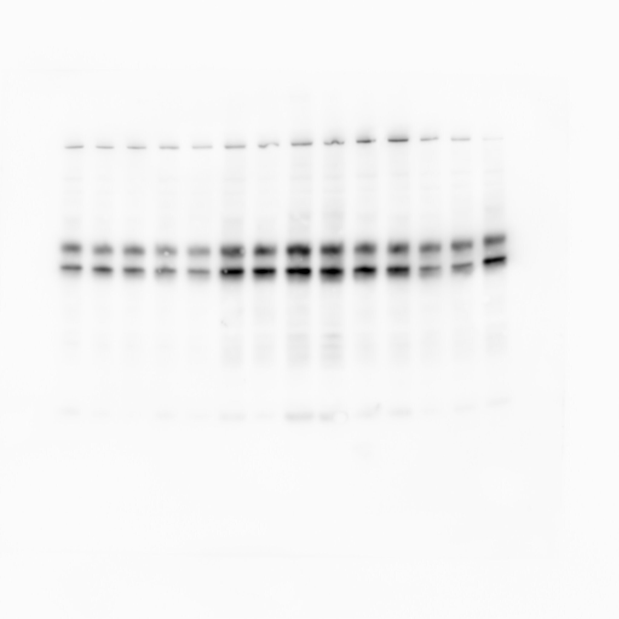
Detection of Human JNK1/2/3 by Western Blot
Time course of JNK activation by combined UVR and thermal stresses in human fibroblasts and coral.Immunoblot revealing activated (pJNK) and total JNK (JNK) in (a) human fibroblasts or (b) S. pistillata nubbins exposed to UVR and temperature rise for the indicated times. (a) Total JNK and actin served as loading controls for human fibroblasts. (b) Total JNK and amido black staining (a,b) served as loading controls for S. pistillata. Data are representative of three independent experiments. Image collected and cropped by CiteAb from the following open publication (https://pubmed.ncbi.nlm.nih.gov/28374828), licensed under a CC-BY license. Not internally tested by R&D Systems.
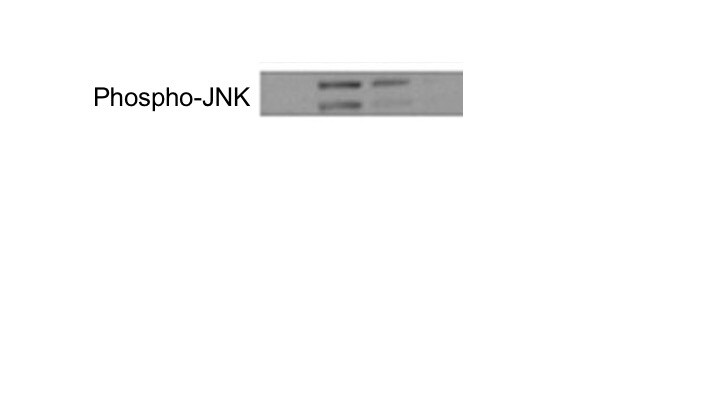
Detection of Human JNK1/2/3 by Western Blot
Detection of JNK activation in human fibroblasts and corals.Immunoblot revealing activated (pJNK) and total forms of JNK (JNK) present in (a) human fibroblasts and (b) S. pistillata nubbins incubated in the absence (control; ct) or presence of SP600125 (SP), anisomycin (Ani), or combination of both (Ani + SP). Molecular weight standards in kilodaltons (kDa) are indicated on the right side of the blots. (a) Total JNK and actin served as loading controls for human fibroblasts. (b) Total JNK and amido black staining (a,b) served as loading controls for S. pistillata. Data are representative of at least three independent experiments. Image collected and cropped by CiteAb from the following open publication (https://pubmed.ncbi.nlm.nih.gov/28374828), licensed under a CC-BY license. Not internally tested by R&D Systems.Applications for JNK Pan Specific Antibody
Application
Recommended Usage
Immunohistochemistry
5-15 µg/mL
Sample: Perfusion fixed frozen sections of rat kidney and thymus, and immersion fixed paraffin-embedded sections of human prostate cancer tissue
Sample: Perfusion fixed frozen sections of rat kidney and thymus, and immersion fixed paraffin-embedded sections of human prostate cancer tissue
Simple Western
10 µg/mL
Sample: HEK293T human embryonic kidney cell line
Sample: HEK293T human embryonic kidney cell line
Western Blot
0.2 µg/mL
Sample: HeLa human cervical epithelial carcinoma cell line, U937 human histiocytic lymphoma cell line, C6 rat glioma cell line, and C2C12 mouse myoblast cell line
Sample: HeLa human cervical epithelial carcinoma cell line, U937 human histiocytic lymphoma cell line, C6 rat glioma cell line, and C2C12 mouse myoblast cell line
Reviewed Applications
Read 2 reviews rated 5 using AF1387 in the following applications:
Formulation, Preparation, and Storage
Purification
Antigen Affinity-purified
Reconstitution
Reconstitute at 0.2 mg/mL in sterile PBS. For liquid material, refer to CoA for concentration.
Loading...
Formulation
Lyophilized from a 0.2 μm filtered solution in PBS with Trehalose. *Small pack size (SP) is supplied either lyophilized or as a 0.2 µm filtered solution in PBS.
Shipping
Lyophilized product is shipped at ambient temperature. Liquid small pack size (-SP) is shipped with polar packs. Upon receipt, store immediately at the temperature recommended below.
Stability & Storage
Use a manual defrost freezer and avoid repeated freeze-thaw cycles.
- 12 months from date of receipt, -20 to -70 °C as supplied.
- 1 month, 2 to 8 °C under sterile conditions after reconstitution.
- 6 months, -20 to -70 °C under sterile conditions after reconstitution.
Calculators
Background: JNK
Long Name
C-Jun N-terminal Kinase
Alternate Names
c-Jun N-terminal kinase 1, EC 2.7.11, EC 2.7.11.24, JNK1 alpha protein kinase, JNK1 beta protein kinase, JNK1JNK1A2, JNK-46, JUN N-terminal kinase, MAP kinase 8, MAPK 8, mitogen-activated protein kinase 8, mitogen-activated protein kinase 8 isoform JNK1 alpha1, mitogen-activated protein kinase 8 isoform JNK1 beta2, PRKM8JNK, protein kinase JNK1, SAPK1JNK21B1/2, Stress-activated protein kinase 1, Stress-activated protein kinase JNK1
UniProt
Additional JNK Products
Product Documents for JNK Pan Specific Antibody
Certificate of Analysis
To download a Certificate of Analysis, please enter a lot or batch number in the search box below.
Note: Certificate of Analysis not available for kit components.
Product Specific Notices for JNK Pan Specific Antibody
For research use only
Citations for JNK Pan Specific Antibody
 Powered by Bioz
Customer Reviews for JNK Pan Specific Antibody (2)
5 out of 5
2 Customer Ratings
Have you used JNK Pan Specific Antibody?
Submit a review and receive an Amazon gift card!
$25/€18/£15/$25CAN/¥2500 Yen for a review with an image
$10/€7/£6/$10CAN/¥1110 Yen for a review without an image
Submit a review
Customer Images
Showing
1
-
2 of
2 reviews
Showing All
Filter By:
-
Application: Western BlotSample Tested: Lung single-cell suspensionSpecies: MouseVerified Customer | Posted 06/07/2019

-
Application: Western BlotSample Tested: HEK293 human embryonic kidney cell lineSpecies: HumanVerified Customer | Posted 07/17/2018
There are no reviews that match your criteria.
Protocols
Find general support by application which include: protocols, troubleshooting, illustrated assays, videos and webinars.
- Antigen Retrieval Protocol (PIER)
- Antigen Retrieval for Frozen Sections Protocol
- Appropriate Fixation of IHC/ICC Samples
- Cellular Response to Hypoxia Protocols
- Chromogenic IHC Staining of Formalin-Fixed Paraffin-Embedded (FFPE) Tissue Protocol
- Chromogenic Immunohistochemistry Staining of Frozen Tissue
- ClariTSA™ Fluorophore Kits
- Detection & Visualization of Antibody Binding
- Fluorescent IHC Staining of Frozen Tissue Protocol
- Graphic Protocol for Heat-induced Epitope Retrieval
- Graphic Protocol for the Preparation and Fluorescent IHC Staining of Frozen Tissue Sections
- Graphic Protocol for the Preparation and Fluorescent IHC Staining of Paraffin-embedded Tissue Sections
- Graphic Protocol for the Preparation of Gelatin-coated Slides for Histological Tissue Sections
- IHC Sample Preparation (Frozen sections vs Paraffin)
- Immunofluorescent IHC Staining of Formalin-Fixed Paraffin-Embedded (FFPE) Tissue Protocol
- Immunohistochemistry (IHC) and Immunocytochemistry (ICC) Protocols
- Immunohistochemistry Frozen Troubleshooting
- Immunohistochemistry Paraffin Troubleshooting
- Preparing Samples for IHC/ICC Experiments
- Preventing Non-Specific Staining (Non-Specific Binding)
- Primary Antibody Selection & Optimization
- Protocol for Heat-Induced Epitope Retrieval (HIER)
- Protocol for Making a 4% Formaldehyde Solution in PBS
- Protocol for VisUCyte™ HRP Polymer Detection Reagent
- Protocol for the Preparation & Fixation of Cells on Coverslips
- Protocol for the Preparation and Chromogenic IHC Staining of Frozen Tissue Sections
- Protocol for the Preparation and Chromogenic IHC Staining of Frozen Tissue Sections - Graphic
- Protocol for the Preparation and Chromogenic IHC Staining of Paraffin-embedded Tissue Sections
- Protocol for the Preparation and Chromogenic IHC Staining of Paraffin-embedded Tissue Sections - Graphic
- Protocol for the Preparation and Fluorescent IHC Staining of Frozen Tissue Sections
- Protocol for the Preparation and Fluorescent IHC Staining of Paraffin-embedded Tissue Sections
- Protocol for the Preparation of Gelatin-coated Slides for Histological Tissue Sections
- R&D Systems Quality Control Western Blot Protocol
- TUNEL and Active Caspase-3 Detection by IHC/ICC Protocol
- The Importance of IHC/ICC Controls
- Troubleshooting Guide: Immunohistochemistry
- Troubleshooting Guide: Western Blot Figures
- Western Blot Conditions
- Western Blot Protocol
- Western Blot Protocol for Cell Lysates
- Western Blot Troubleshooting
- Western Blot Troubleshooting Guide
- View all Protocols, Troubleshooting, Illustrated assays and Webinars
Loading...