The Ataxia telangiectasia-mutated (ATM) protein kinase exists as a dimer in the cell nucleus. Changes in DNA structure induced by genotoxic stress lead to activation of ATM and phosphorylation of S1981 in trans. Once S1981 is phosphorylated, the dimer dissociates and active ATM monomers signal to downstream targets.
 by Western Blot.")
Key Product Details
Validated by
Biological Validation
Species Reactivity
Validated:
Human, Mouse, Rat
Cited:
Human, Mouse, Transgenic Mouse
Applications
Validated:
Western Blot, Simple Western
Cited:
Immunohistochemistry, Immunohistochemistry-Paraffin, Western Blot, Bioassay
Label
Unconjugated
Antibody Source
Polyclonal Rabbit IgG
Loading...
Product Specifications
Immunogen
Phosphopeptide containing human ATM S1981 site
Specificity
Detects human ATM when phosphorylated at S1981. Also detects the comparable phosphorylated sites in mouse ATM (S1987) and rat ATM (S1952).
Clonality
Polyclonal
Host
Rabbit
Isotype
IgG
Scientific Data Images for phospho-ATM (S1981) Antibody
Detection of Human/Mouse/Rat Phospho-ATM (S1981) by Western Blot.
Western blot shows lysates of HeLa human cervical epithelial carcinoma cell line untreated (-) or treated (+) with 1 µM camptothecin (CPT) for 1 hour. PVDF membrane was probed with 1 µg/mL Rabbit Anti-Human/Mouse/Rat Phospho-ATM (S1981) Antigen Affinity-purified Polyclonal Antibody (Catalog # AF1655) followed by HRP-conjugated Anti-Rabbit IgG Secondary Antibody (Catalog # HAF008). A specific band for Phospho-ATM (S1981) was detected at approximately 370 kDa (as indicated). The phospho-specificity of this antibody was supported by decreased labeling following treatment with 600 U lambda-phosphatase (lambda-PPase) for 1 hour. This experiment was conducted under reducing conditions and using Immunoblot Buffer Group 1. by Simple Western<SUP>TM</SUP>.")
Detection of Human Phospho-ATM (S1981) by Simple WesternTM.
Simple Western lane view shows lysates of HeLa human cervical epithelial carcinoma cell line untreated (-) or treated (+) with 1 µM Camptothecin (CPT) for 1 hour, loaded at 0.2 mg/mL. A specific band was detected for Phospho-ATM (S1981) at approximately 270 kDa (as indicated) using 10 µg/mL of Rabbit Anti-Human/Mouse/Rat Phospho-ATM (S1981) Antigen Affinity-purified Polyclonal Antibody (Catalog # AF1655). This experiment was conducted under reducing conditions and using the 66-440 kDa separation system.
Detection of Human ATM by Western Blot
Resveratrol activates ATM in transformed human cell lines.(a, b) Human HEK293T cells were treated with 0.01 mM ATM inhibitor (KU-55933) or vehicle (DMSO) for 1 hour, followed by incubation in media also containing 0.1 mM resveratrol (or DMSO), as indicated. After 30 minutes incubation with resveratrol, H2O2 (0.1 mM) or bleomycin (1 µg/ml) was added as indicated for 30 minutes before harvesting. Western blotting was performed with with antibodies directed against ATM, phospho-ATM Ser-1981, p53, and phospho-p53 Ser-15 as indicated. (c, d) Human HEK293T cells were treated with resveratrol, bleomycin, or both as in (a) and probed for gamma -H2AX foci by immunofluorescence. Cell images (291, 267, 216, and 260 cells, respectively) were analyzed for foci using Image J software, and the average number of foci per cell as well as the percentage of cells containing >5 foci were quantitated. (e, f) Human HCT116 cells were incubated with KU-55933, resveratrol, H2O2, and bleomycin as in (a, b) but additional phosphorylation targets were examined using antibodies directed against Smc1, phospho-Smc1 Ser-957, Kap1, phospho-Kap1 Ser-824, Nbs1, phospho-Nbs1 Ser-343, Chk2, and phospho-Chk2 Thr-68 as indicated. Image collected and cropped by CiteAb from the following publication (https://dx.plos.org/10.1371/journal.pone.0097969), licensed under a CC-BY license. Not internally tested by R&D Systems.
Detection of Human ATM by Western Blot
DEK is necessary for the activation of gamma H2AX after ionizing radiation.(a) MEFs were treated with 10 Gy of gamma ionizing radiation (IR) and stained for gamma H2AX. Representative cross sections from z-stack confocal images are shown. (b) Quantification of cells with >10 gamma H2AX foci in (A). At least 50 cells per time point were counted in each biological replicate, and all foci within the z-stack were quantified. DEK−/− MEFs were unable to activate gamma H2AX above baseline after irradiation. (paired t-test, n = 3, mean ± SEM). (c) Quantification of the average total number of gamma H2AX foci in each cell from (A). (paired t-test, n = 3, mean ± SEM) The number of foci per cell remained unchanged in DEK−/− MEFs after irradiation. (d) Western blot analysis of gamma H2AX in MEFs at 6 hs following irradiation with 10 Gy. (e) Western blot analysis of C33A human cancer cells infected with adenovirus 48 hr prior to receiving 10 Gy IR (see also Supplementary Fig. S1). Image collected and cropped by CiteAb from the following publication (https://pubmed.ncbi.nlm.nih.gov/28317934), licensed under a CC-BY license. Not internally tested by R&D Systems. Antibody by Western Blot")
Detection of Human Human/Mouse/Rat Phospho-ATM (S1981) Antibody by Western Blot
TCL cell lines show intrinsic DNA damage and basal activation of DDR. Healthy T lymphocytes isolated from healthy donors and malignant T cell lines (OCI-Ly12, KARPAS-299, SUP-T1, HH, HD-MAR-2, and JURKAT) were subjected to immunofluorescence for the evaluation of gamma H2AX or 53BP1 foci (A,B). The percentage (%) of cells displaying foci and the number of foci/cell in the subsets of cells with detectable foci are reported in (A,B), respectively. Data are expressed as the mean ± SD of independent experiments. Asterisks indicate statistically significant differences between each cell line and healthy T lymphocytes (* p < 0.05; ** p < 0.01; *** p < 0.001; ns: not significant). Representative immunofluorescence images (C,D) of experiments described in (A,B), original microscope magnification 100×. SUP-T1 cells were exposed to increasing concentrations of CHOEP (IC50, 4× and 8×, as in [5]) or to 20 μM etoposide for 3 h. After harvesting, untreated and treated cell lysates were analyzed by Western blot using the indicated antibodies (E). Image collected and cropped by CiteAb from the following publication (https://pubmed.ncbi.nlm.nih.gov/35409194), licensed under a CC-BY license. Not internally tested by R&D Systems.Applications for phospho-ATM (S1981) Antibody
Application
Recommended Usage
Simple Western
10 µg/mL
Sample: HeLa human cervical epithelial carcinoma cell line treated with Camptothecin (CPT)
Sample: HeLa human cervical epithelial carcinoma cell line treated with Camptothecin (CPT)
Western Blot
1 µg/mL
Sample: HeLa human cervical epithelial carcinoma cell line treated with camptothecin
Sample: HeLa human cervical epithelial carcinoma cell line treated with camptothecin
Reviewed Applications
Read 1 review rated 5 using AF1655 in the following applications:
Formulation, Preparation, and Storage
Purification
Antigen Affinity-purified
Reconstitution
Reconstitute at 0.2 mg/mL in sterile PBS. For liquid material, refer to CoA for concentration.
Loading...
Formulation
Lyophilized from a 0.2 μm filtered solution in PBS with Trehalose. *Small pack size (SP) is supplied either lyophilized or as a 0.2 µm filtered solution in PBS.
Shipping
Lyophilized product is shipped at ambient temperature. Liquid small pack size (-SP) is shipped with polar packs. Upon receipt, store immediately at the temperature recommended below.
Stability & Storage
Use a manual defrost freezer and avoid repeated freeze-thaw cycles.
- 12 months from date of receipt, -20 to -70 °C as supplied.
- 1 month, 2 to 8 °C under sterile conditions after reconstitution.
- 6 months, -20 to -70 °C under sterile conditions after reconstitution.
Calculators
Background: ATM
Long Name
Ataxia Telangiectasia-mutated
Alternate Names
TEL1, TELO1, TPLL
Gene Symbol
ATM
Additional ATM Products
Product Documents for phospho-ATM (S1981) Antibody
Certificate of Analysis
To download a Certificate of Analysis, please enter a lot or batch number in the search box below.
Note: Certificate of Analysis not available for kit components.
Product Specific Notices for phospho-ATM (S1981) Antibody
For research use only
Related Research Areas
Citations for phospho-ATM (S1981) Antibody
 Powered by Bioz
Customer Reviews for phospho-ATM (S1981) Antibody (1)
5 out of 5
1 Customer Rating
Have you used phospho-ATM (S1981) Antibody?
Submit a review and receive an Amazon gift card!
$25/€18/£15/$25CAN/¥2500 Yen for a review with an image
$10/€7/£6/$10CAN/¥1110 Yen for a review without an image
Submit a review
Customer Images
Showing
1
-
1 of
1 review
Showing All
Filter By:
-
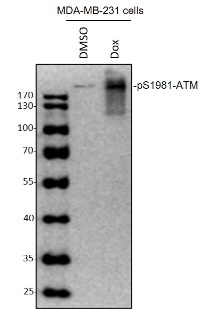
Application: Western BlotSample Tested: MDA MB 231 cellsSpecies: HumanVerified Customer | Posted 10/28/2020Western Blot: MDA-MB-231 cells were treated with DMSO or doxorubicin (1 μM) for 8 hours, whole cell lysates were loaded with 50 ug/lane. 10% SDS-PAGE. ATM [p Ser1981] Antibody (AF1655) primary antibody: 1:1000, 4℃, overnight.

There are no reviews that match your criteria.
Protocols
Find general support by application which include: protocols, troubleshooting, illustrated assays, videos and webinars.
- Cellular Response to Hypoxia Protocols
- R&D Systems Quality Control Western Blot Protocol
- Troubleshooting Guide: Western Blot Figures
- Western Blot Conditions
- Western Blot Protocol
- Western Blot Protocol for Cell Lysates
- Western Blot Troubleshooting
- Western Blot Troubleshooting Guide
- View all Protocols, Troubleshooting, Illustrated assays and Webinars
Loading...