MuSK (muscle-specific kinase) is a 100 kDa type I transmembrane (TM) protein belonging to the receptor tyrosine kinase family. It is found in the postsynaptic membrane of skeletal muscle motor endplates. MuSK does not bind agrin directly, but is part of an agrin receptor complex that promotes the clustering of acetylcholine receptor at the neuromuscular junction (NMJ). Over residues 22‑503, human MuSK shares 92% amino acid sequence identity with mouse MuSK. Human MuSK apparently has multiple isoforms. One contains deletions at residues 307‑394 and 454‑461, while a second is a short soluble form that contains residues 120‑209 plus a unique 24 aa C‑terminal tail.

Key Product Details
Species Reactivity
Validated:
Human
Cited:
Mouse
Applications
Validated:
Western Blot
Cited:
Western Blot
Label
Unconjugated
Antibody Source
Polyclonal Goat IgG
Loading...
Product Specifications
Immunogen
Mouse myeloma cell line NS0-derived recombinant human MuSK
Glu22-Ser503
Accession # O15146
Glu22-Ser503
Accession # O15146
Specificity
Detects human MuSK in direct ELISAs and Western blots. In direct ELISAs, less than 40% cross-reactivity with recombinant rat MuSK is observed.
Clonality
Polyclonal
Host
Goat
Isotype
IgG
Scientific Data Images for Human MuSK Antibody
Detection of Mouse MuSK by Western Blot
Sorbs1 is enriched at AChR aggregates, and Sorbs1 RNAi blocks AChR clustering in vitro. (A) Treatment of myotubes with siRNA directed against Sorbs1 blocks AChR clustering in C2C12 cells. Montages containing sixteen fields at a magnification of 10× were analyzed with ImageJ software (NIH). (B) Sorbs1 siRNA significantly reduces Sorbs1 protein expression in myotubes. (C) Sorbs1 protein is highly enriched at sites where AChRs aggregate. (D) Agrin stimulates tyrosine phosphorylation of MuSK and Dok-7 at similar levels in myotubes treated with Sorbs1 siRNA. Image collected and cropped by CiteAb from the following publication (https://pubmed.ncbi.nlm.nih.gov/26527617), licensed under a CC-BY license. Not internally tested by R&D Systems.Applications for Human MuSK Antibody
Application
Recommended Usage
Western Blot
0.1 µg/mL
Sample: Recombinant Human MuSK
Sample: Recombinant Human MuSK
Reviewed Applications
Read 2 reviews rated 3 using AF3904 in the following applications:
Formulation, Preparation, and Storage
Purification
Antigen Affinity-purified
Reconstitution
Reconstitute at 0.2 mg/mL in sterile PBS. For liquid material, refer to CoA for concentration.
Loading...
Formulation
Lyophilized from a 0.2 μm filtered solution in PBS with Trehalose. *Small pack size (SP) is supplied either lyophilized or as a 0.2 µm filtered solution in PBS.
Shipping
Lyophilized product is shipped at ambient temperature. Liquid small pack size (-SP) is shipped with polar packs. Upon receipt, store immediately at the temperature recommended below.
Stability & Storage
Use a manual defrost freezer and avoid repeated freeze-thaw cycles.
- 12 months from date of receipt, -20 to -70 °C as supplied.
- 1 month, 2 to 8 °C under sterile conditions after reconstitution.
- 6 months, -20 to -70 °C under sterile conditions after reconstitution.
Calculators
Background: MuSK
Long Name
Muscle-specific Receptor Tyrosine Kinase
Alternate Names
EC 2.7.10, EC 2.7.10.1, MGC126323, MGC126324, muscle, skeletal, receptor tyrosine kinase, MuSK, skeletal receptor tyrosine-protein kinase
Gene Symbol
MUSK
UniProt
Additional MuSK Products
Product Documents for Human MuSK Antibody
Certificate of Analysis
To download a Certificate of Analysis, please enter a lot or batch number in the search box below.
Note: Certificate of Analysis not available for kit components.
Product Specific Notices for Human MuSK Antibody
For research use only
Related Research Areas
Citations for Human MuSK Antibody
 Powered by Bioz
Customer Reviews for Human MuSK Antibody (2)
3 out of 5
2 Customer Ratings
Have you used Human MuSK Antibody?
Submit a review and receive an Amazon gift card!
$25/€18/£15/$25CAN/¥2500 Yen for a review with an image
$10/€7/£6/$10CAN/¥1110 Yen for a review without an image
Submit a review
Customer Images

Showing
1
-
2 of
2 reviews
Showing All
Filter By:
-
Application: Simple WesternSample Tested: Differentiated C2C12 myoblast cellsSpecies: MouseVerified Customer | Posted 05/19/2017C2C12 differentiated myotubes, following IP with an anti-MuSK antibody (different from this Ab), SDS-PAGE, and transferred into PVDF membrane. After blocking with 5% BSA/TBST, blot was probed with 1:400 of AF562 (4C, o/n). Subsequent probing with a HRP-anti-goat secondary Ab yielded excellent publishable results, with 100 kDa MuSK band visible.

-
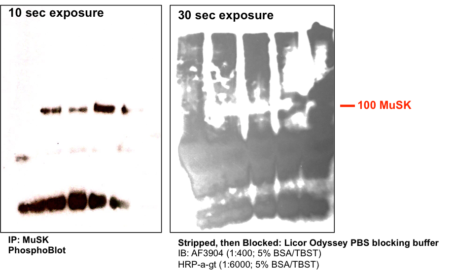
Application: Western BlotSample Tested: Differentiated C2C12 myoblast cellsSpecies: MouseVerified Customer | Posted 05/16/2017Differentiated C2C12 mouse skeletal muscle lysates IP'ed for MuSK using a donated Ab. IP works well and I can detect phosphorylation of MuSK using a general phosphotyrosine Ab, but upon stripping, blocking, and probing with this Ab, I am unable to detect MuSK (perhaps due to high background signal). Probing with a different MuSK Ab is much cleaner
Bio-Techne ResponseTechnical Service is following up
There are no reviews that match your criteria.
Protocols
Find general support by application which include: protocols, troubleshooting, illustrated assays, videos and webinars.
- Cellular Response to Hypoxia Protocols
- R&D Systems Quality Control Western Blot Protocol
- Troubleshooting Guide: Western Blot Figures
- Western Blot Conditions
- Western Blot Protocol
- Western Blot Protocol for Cell Lysates
- Western Blot Troubleshooting
- Western Blot Troubleshooting Guide
- View all Protocols, Troubleshooting, Illustrated assays and Webinars
Loading...