Nicastrin (NCT) is a 150‑160 kDa member of the Nicastrin family of proteins. It is a component of the aspartyl protease gamma -secretase complex and serves to stabilize and direct gamma -secretase components to proper positions in the plasma membrane. The gamma -secretase complex mediates the cleavage of intramembrane proteins such as Notch-1 and APP. Mature human Nicastrin is a 676 amino acid type I transmembrane glycoprotein. It contains a 636 aa extracellular domain (aa 34‑669) that shows a 58 aa sequence (aa 312‑369) which interacts with gamma -secretase substrates. There are multiple splice variants of NCT. One shows a deletion of aa 195‑322 and 394‑709, a second shows a 29 aa substitution for the C-terminal 604 aa and a third shows a deletion of aa 200‑709 accompanied by an insertion of 33 aa after Leu30. Over aa 34‑669, human NCT shares 90% aa identity with mouse NCT.

Key Product Details
Species Reactivity
Human
Applications
Immunocytochemistry
Label
Unconjugated
Antibody Source
Monoclonal Mouse IgG1 Clone # 716918
Loading...
Product Specifications
Immunogen
Chinese hamster ovary cell line CHO-derived recombinant human Nicastrin
Asn34-Glu669
Accession # Q92542
Asn34-Glu669
Accession # Q92542
Specificity
Detects human Nicastrin in direct ELISAs.
Clonality
Monoclonal
Host
Mouse
Isotype
IgG1
Scientific Data Images for Human Nicastrin Antibody (716918)
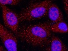
Nicastrin in HepG2 Human Cell Line.
Nicastrin was detected in immersion fixed HepG2 human hepatocellular carcinoma cell line using Mouse Anti-Human Nicastrin Monoclonal Antibody (Catalog # MAB5378) at 10 µg/mL for 3 hours at room temperature. Cells were stained using the NorthernLights™ 557-conjugated Anti-Mouse IgG Secondary Antibody (red; Catalog # NL007) and counterstained with DAPI (blue). Specific staining was localized to cytoplasm. View our protocol for Fluorescent ICC Staining of Cells on Coverslips.Applications for Human Nicastrin Antibody (716918)
Application
Recommended Usage
Immunocytochemistry
8-25 µg/mL
Sample: Immersion fixed HepG2 human hepatocellular carcinoma cell line
Sample: Immersion fixed HepG2 human hepatocellular carcinoma cell line
Reviewed Applications
Read 1 review rated 5 using MAB5378 in the following applications:
Formulation, Preparation, and Storage
Purification
Protein A or G purified from hybridoma culture supernatant
Reconstitution
Sterile PBS to a final concentration of 0.5 mg/mL. For liquid material, refer to CoA for concentration.
Loading...
Formulation
Lyophilized from a 0.2 μm filtered solution in PBS with Trehalose. *Small pack size (SP) is supplied either lyophilized or as a 0.2 µm filtered solution in PBS.
Shipping
Lyophilized product is shipped at ambient temperature. Liquid small pack size (-SP) is shipped with polar packs. Upon receipt, store immediately at the temperature recommended below.
Stability & Storage
Use a manual defrost freezer and avoid repeated freeze-thaw cycles.
- 12 months from date of receipt, -20 to -70 °C as supplied.
- 1 month, 2 to 8 °C under sterile conditions after reconstitution.
- 6 months, -20 to -70 °C under sterile conditions after reconstitution.
Calculators
Background: Nicastrin
Alternate Names
APH2, NCSTN
Gene Symbol
NCSTN
UniProt
Additional Nicastrin Products
Product Documents for Human Nicastrin Antibody (716918)
Certificate of Analysis
To download a Certificate of Analysis, please enter a lot or batch number in the search box below.
Note: Certificate of Analysis not available for kit components.
Product Specific Notices for Human Nicastrin Antibody (716918)
For research use only
Related Research Areas
Customer Reviews for Human Nicastrin Antibody (716918) (1)
5 out of 5
1 Customer Rating
Have you used Human Nicastrin Antibody (716918)?
Submit a review and receive an Amazon gift card!
$25/€18/£15/$25CAN/¥2500 Yen for a review with an image
$10/€7/£6/$10CAN/¥1110 Yen for a review without an image
Submit a review
Customer Images
Showing
1
-
1 of
1 review
Showing All
Filter By:
-
Application: ImmunocytochemistrySample Tested: HepG2 human hepatocellular carcinoma cell lineSpecies: HumanVerified Customer | Posted 03/22/2022HepG2 human hepatocellular carcinoma cell lineIncubated at 10 ug/mL for 3 hours at room temperature

There are no reviews that match your criteria.
Protocols
Find general support by application which include: protocols, troubleshooting, illustrated assays, videos and webinars.
- Appropriate Fixation of IHC/ICC Samples
- Cellular Response to Hypoxia Protocols
- ClariTSA™ Fluorophore Kits
- Detection & Visualization of Antibody Binding
- ICC Cell Smear Protocol for Suspension Cells
- ICC Immunocytochemistry Protocol Videos
- ICC for Adherent Cells
- Immunocytochemistry (ICC) Protocol
- Immunocytochemistry Troubleshooting
- Immunofluorescence of Organoids Embedded in Cultrex Basement Membrane Extract
- Immunohistochemistry (IHC) and Immunocytochemistry (ICC) Protocols
- Preparing Samples for IHC/ICC Experiments
- Preventing Non-Specific Staining (Non-Specific Binding)
- Primary Antibody Selection & Optimization
- Protocol for VisUCyte™ HRP Polymer Detection Reagent
- Protocol for the Fluorescent ICC Staining of Cell Smears - Graphic
- Protocol for the Fluorescent ICC Staining of Cultured Cells on Coverslips - Graphic
- Protocol for the Preparation and Fluorescent ICC Staining of Cells on Coverslips
- Protocol for the Preparation and Fluorescent ICC Staining of Non-adherent Cells
- Protocol for the Preparation and Fluorescent ICC Staining of Stem Cells on Coverslips
- Protocol for the Preparation of a Cell Smear for Non-adherent Cell ICC - Graphic
- TUNEL and Active Caspase-3 Detection by IHC/ICC Protocol
- The Importance of IHC/ICC Controls
- View all Protocols, Troubleshooting, Illustrated assays and Webinars
Loading...
Associated Pathways

