Human Occludin Antibody (690213)
R&D Systems | Catalog # MAB7074

Key Product Details
Species Reactivity
Validated:
Human
Cited:
Human
Applications
Validated:
Immunocytochemistry
Cited:
Immunocytochemistry
Label
Unconjugated
Antibody Source
Monoclonal Mouse IgG1 Clone # 690213
Loading...
Product Specifications
Immunogen
NS0 mouse myeloma cell line transfected with human Occludin
Accession # Q16625
Accession # Q16625
Specificity
Detects human Occludin in direct ELISAs.
Clonality
Monoclonal
Host
Mouse
Isotype
IgG1
Scientific Data Images for Human Occludin Antibody (690213)
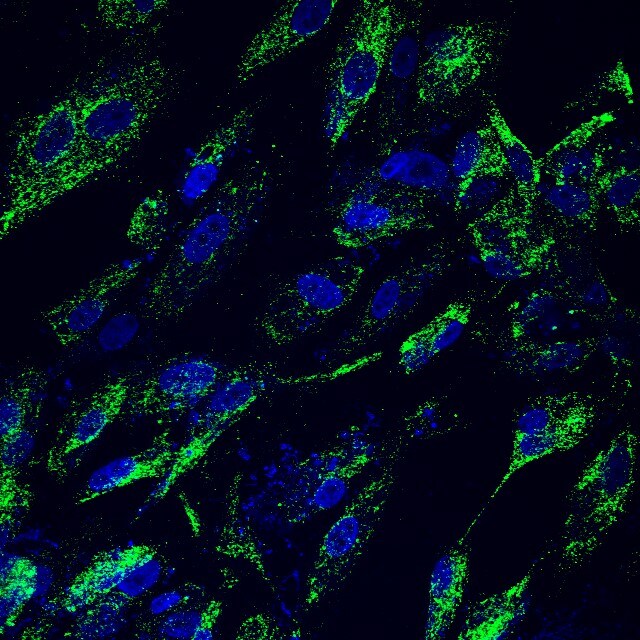
Occludin in HUVEC Human Cells.
Occludin was detected in immersion fixed HUVEC human umbilical vein endothelial cells using Mouse Anti-Human Occludin Monoclonal Antibody (Catalog # MAB7074) at 10 µg/mL for 3 hours at room temperature. Cells were stained using the Northern-Lights™ 557-conjugated Anti-Mouse IgG Secondary Antibody (red; Catalog # NL007) and counterstained with DAPI (blue). Specific staining was localized to cytoplasm. View our protocol for Fluorescent ICC Staining of Cells on Coverslips.
Detection of Human Occludin by Western Blot
Analysis of barrier formation in endothelial monolayer.(A) Expression of endothelial junctional molecules CD31, CD144 and ZO-1 (from left to right) and actin (far right) in hCMVEC monolayer 48 h after seeding. (B) The permeability of hCMVEC monolayer to FITC-dextran 48 h after seeding (mean ± SEM, n = 4). (C) Time course analysis of endothelial tight junction molecule occludin expression at 24, 48 and 72 h post seeding of hCMVECs using Western blot using a mouse monoclonal anti-occludin (left) and rabbit polyclonal anti-occludin (right). (D) Real time measurement of electrical resistance across the endothelial monolayer as an indicator of endothelial barrier integrity. The barrier resistance (Rb) and the basolateral adhesion (Alpha) due to focal adhesions can be modelled using the ECIS Z theta software when the experiment is run using multi-frequency mode (250 to 64000 Hz). The modelling reveals that the basolateral adhesion occurs very fast and junction formation begins around 10 hours after seeding. Overall barrier resistance is the summation of both Rb and alpha, but this software function can determine whether barrier changes are predominantly due to the Rb. Data are mean ± SEM (n = 4). Image collected and cropped by CiteAb from the following publication (https://dx.plos.org/10.1371/journal.pone.0180267), licensed under a CC-BY license. Not internally tested by R&D Systems.Applications for Human Occludin Antibody (690213)
Application
Recommended Usage
Immunocytochemistry
8-25 µg/mL
Sample: Immersion fixed HUVEC human umbilical vein endothelial cells
Sample: Immersion fixed HUVEC human umbilical vein endothelial cells
Reviewed Applications
Read 2 reviews rated 5 using MAB7074 in the following applications:
Formulation, Preparation, and Storage
Purification
Protein A or G purified from hybridoma culture supernatant
Reconstitution
Sterile PBS to a final concentration of 0.5 mg/mL. For liquid material, refer to CoA for concentration.
Loading...
Formulation
Lyophilized from a 0.2 μm filtered solution in PBS with Trehalose. *Small pack size (SP) is supplied either lyophilized or as a 0.2 µm filtered solution in PBS.
Shipping
Lyophilized product is shipped at ambient temperature. Liquid small pack size (-SP) is shipped with polar packs. Upon receipt, store immediately at the temperature recommended below.
Stability & Storage
Use a manual defrost freezer and avoid repeated freeze-thaw cycles.
- 12 months from date of receipt, -20 to -70 °C as supplied.
- 1 month, 2 to 8 °C under sterile conditions after reconstitution.
- 6 months, -20 to -70 °C under sterile conditions after reconstitution.
Calculators
Background: Occludin
Alternate Names
BLCPMG, OCLN
Gene Symbol
OCLN
UniProt
Additional Occludin Products
Product Documents for Human Occludin Antibody (690213)
Certificate of Analysis
To download a Certificate of Analysis, please enter a lot or batch number in the search box below.
Note: Certificate of Analysis not available for kit components.
Product Specific Notices for Human Occludin Antibody (690213)
For research use only
Citations for Human Occludin Antibody (690213)
 Powered by Bioz
Customer Reviews for Human Occludin Antibody (690213) (2)
5 out of 5
2 Customer Ratings
Have you used Human Occludin Antibody (690213)?
Submit a review and receive an Amazon gift card!
$25/€18/£15/$25CAN/¥2500 Yen for a review with an image
$10/€7/£6/$10CAN/¥1110 Yen for a review without an image
Submit a review
Customer Images
Showing
1
-
2 of
2 reviews
Showing All
Filter By:
-
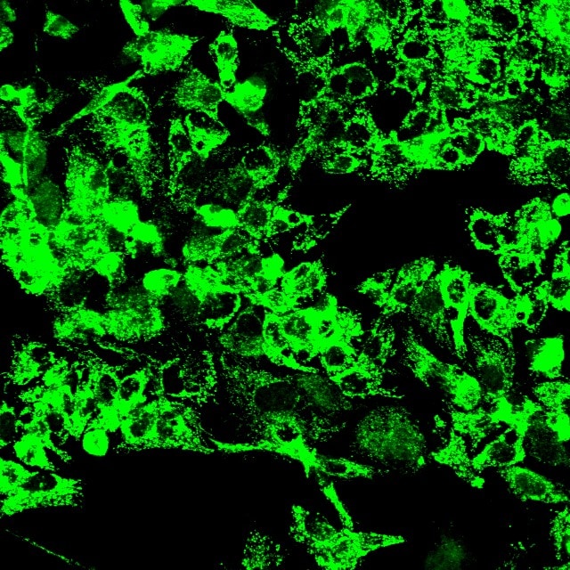
Application: ImmunocytochemistrySample Tested: pig SC cellsSpecies: PigVerified Customer | Posted 06/07/2017MAB7074 Occludin antibody label pig SC cells with alexa488

-
Application: ImmunocytochemistrySample Tested: Hep2G cellsSpecies: PigVerified Customer | Posted 06/07/2017MAB7074 Occludin antibody labeling Hep2G cells with Alexa 488
There are no reviews that match your criteria.
Protocols
Find general support by application which include: protocols, troubleshooting, illustrated assays, videos and webinars.
- Appropriate Fixation of IHC/ICC Samples
- Cellular Response to Hypoxia Protocols
- ClariTSA™ Fluorophore Kits
- Detection & Visualization of Antibody Binding
- ICC Cell Smear Protocol for Suspension Cells
- ICC Immunocytochemistry Protocol Videos
- ICC for Adherent Cells
- Immunocytochemistry (ICC) Protocol
- Immunocytochemistry Troubleshooting
- Immunofluorescence of Organoids Embedded in Cultrex Basement Membrane Extract
- Immunohistochemistry (IHC) and Immunocytochemistry (ICC) Protocols
- Preparing Samples for IHC/ICC Experiments
- Preventing Non-Specific Staining (Non-Specific Binding)
- Primary Antibody Selection & Optimization
- Protocol for VisUCyte™ HRP Polymer Detection Reagent
- Protocol for the Fluorescent ICC Staining of Cell Smears - Graphic
- Protocol for the Fluorescent ICC Staining of Cultured Cells on Coverslips - Graphic
- Protocol for the Preparation and Fluorescent ICC Staining of Cells on Coverslips
- Protocol for the Preparation and Fluorescent ICC Staining of Non-adherent Cells
- Protocol for the Preparation and Fluorescent ICC Staining of Stem Cells on Coverslips
- Protocol for the Preparation of a Cell Smear for Non-adherent Cell ICC - Graphic
- TUNEL and Active Caspase-3 Detection by IHC/ICC Protocol
- The Importance of IHC/ICC Controls
- View all Protocols, Troubleshooting, Illustrated assays and Webinars
Loading...
Associated Pathways


