Human phospho-p53 (S15) Antibody
R&D Systems | Catalog # AF1043
 by Western Blot.")
Key Product Details
Validated by
Biological Validation
Species Reactivity
Validated:
Human
Cited:
Human, Mouse
Applications
Validated:
Immunohistochemistry, Western Blot, Immunocytochemistry, Immunoprecipitation
Cited:
Immunohistochemistry, Western Blot
Label
Unconjugated
Antibody Source
Polyclonal Rabbit IgG
Loading...
Product Specifications
Immunogen
Phosphopeptide containing human p53 S15 site
Specificity
Detects human, monkey, and rat p53 when phosphorylated at S15 and mouse p53 when phosphorylated at S18.
Clonality
Polyclonal
Host
Rabbit
Isotype
IgG
Scientific Data Images for Human phospho-p53 (S15) Antibody
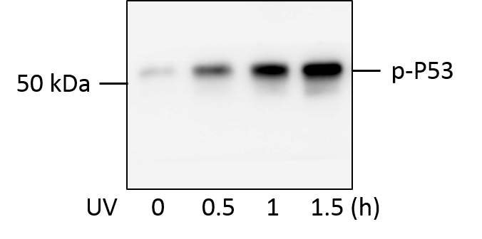
Detection of Human Phospho-p53 (S15) by Western Blot.
Western blot shows lysates of CEM human T-lymphoblastoid cell line untreated or exposed to 100 J/m2UV-C for the indicated time. PVDF membrane was probed with 0.2 µg/mL Human Phospho-p53 (S15) Antigen Affinity-purified Polyclonal Antibody (Catalog # AF1043) followed by HRP-conjugated Anti-Rabbit IgG Secondary Antibody (Catalog # HAF008). A specific band for Phospho-p53 (S15) was detected at approximately 53 kDa (as indicated). The phospho-specificity of this antibody was supported by decreased labeling following treatment with 600 U lambda-phosphatase (lambda-PPase) for 1 hour. For additional reference, duplicate lysates were probed with 1:5000 dilution Human/Mouse/Rat p53 HRP-conjugated Antigen Affinity-purified Polyclonal Antibody (lower panel, Catalog # HAF1355). This experiment was conducted under reducing conditions and using Immunoblot Buffer Group 1. in HeLa Human Cell Line.")
Phospho-p53 (S15) in HeLa Human Cell Line.
p53 phosphorylated at S15 was detected in immersion fixed HeLa human cervical epithelial carcinoma cell line treated with UV light (left panel) or untreated (right panel) using Rabbit Anti-Human Phospho-p53 (S15) Antigen Affinity-purified Polyclonal Antibody (Catalog # AF1043) at 10 µg/mL for 3 hours at room temperature. Cells were stained using the NorthernLights™ 557-conjugated Anti-Rabbit IgG Secondary Antibody (red; Catalog # NL004) and counterstained with DAPI (blue). Specific staining was localized to nuclei. Cells were co-stained using Rat Anti-Tubulin (Catalog # NB600-506, Novus Biologicals) and NorthernLights™ 493-conjugated Anti-Rat IgG Secondary Antibody (green; Catalog # NL015). View our protocol for Fluorescent ICC Staining of Cells on Coverslips. in Human Breast Cancer Tissue.")
Phospho-p53 (S15) in Human Breast Cancer Tissue.
p53 phosphorylated at S15 was detected in immersion fixed paraffin-embedded sections of human breast cancer tissue using Human Phospho-p53 (S15) Antigen Affinity-purified Polyclonal Antibody (Catalog # AF1043) at 15 µg/mL overnight at 4 °C. Tissue was stained using the Anti-Rabbit HRP-DAB Cell & Tissue Staining Kit (brown; Catalog # CTS005) and counterstained with hematoxylin (blue). View our protocol for Chromogenic IHC Staining of immersion fixed paraffin-embedded Tissue Sections. in Human Breast Cancer Tissue.")
Phospho-p53 (S15) in Human Breast Cancer Tissue.
p53 phosphorylated at S15 was detected in immersion fixed paraffin-embedded sections of human breast cancer tissue using Human Phospho-p53 (S15) Antigen Affinity-purified Polyclonal Antibody (Catalog # AF1043) at 15 µg/mL overnight at 4 °C. Tissue was stained using the Anti-Rabbit HRP-DAB Cell & Tissue Staining Kit (brown; Catalog # CTS005) and counterstained with hematoxylin (blue). View our protocol for Chromogenic IHC Staining of immersion fixed paraffin-embedded Tissue Sections. by Western Blot")
Detection of Phospho-p53 (S15) by Western Blot
The increase of HDAC9 was associated with bone and fat imbalance in bone aging. a Micro-CT analyses of bone mass of trabecular and cortical bone thickness in the femora of 2-month-old (young) and 16-month-old (aged) mice. Bone mineral density (BMD), trabecular bone volume (BV/TV), and cortical bone thickness (Ct.Th) were performed. Scale bar = 1 mm. b–d Immunofluorescent staining of OCN (b), PPAR-gamma (c), and TRAP (d) was performed in the bone marrow from young and aged mice, and the positive signals were quantitatively analyzed. Scale bar = 50 μm. e Expressions of the senescence-related proteins, p53 and p-p53, in the bone marrow from young and aged mice were examined by western blotting. f Expressions of the senescence-related proteins, p53 and p-p53, in BMMSCs from young and aged mice were examined by western blotting. g Alizarin Red staining was performed, and quantification of mineralized nodules was analyzed in young and aged BMMSCs. h Oil Red O staining was performed, and quantification of lipid droplet positive ratio areas was analyzed in young and aged BMMSCs. Scale bars = 100 μm. i, j Expression of HDAC9 (i) and acetylation sites of H3K, including H3K9, H3K18, and H4K16 (j), were examined by western blotting. The data are presented as the means ± SD of each independent experiment performed in triplicate. *P < 0.05, **P < 0.01, ***P < 0.001, unpaired two-tailed Student’s t test Image collected and cropped by CiteAb from the following open publication (https://pubmed.ncbi.nlm.nih.gov/32620134), licensed under a CC-BY license. Not internally tested by R&D Systems. by Western Blot")
Detection of Phospho-p53 (S15) by Western Blot
Downregulation of HDAC9 rescued lineage differentiation imbalance and ameliorated senescence in aged BMMSCs. a Alizarin Red staining was performed, and osteogenesis-related proteins were detected by western blotting in aged BMMSCs transfected with HDAC9 siRNA. b Oil Red O staining was performed, and adipogenesis-related proteins were detected by western blotting in aged BMMSCs transfected with HDAC9 siRNA. c Alizarin Red staining was performed, and osteogenic-related proteins were detected by western blotting in young BMMSCs and young BMMSCs transfected with HDAC9 siRNA. d Oil Red O staining was performed, and adipogenic-related proteins were detected by western blotting in young BMMSCs and young BMMSCs transfected with HDAC9 siRNA. e, f Expressions of the senescence-related proteins p53 and p-p53 in BMMSCs cultured in vitro from aged mice (e) and young mice (f) were examined by western blotting. The data are presented as the means ± SD of each independent experiment performed in triplicate. *P < 0.05, **P < 0.01, ***P < 0.001, one-way analysis of variance (ANOVA) Image collected and cropped by CiteAb from the following open publication (https://pubmed.ncbi.nlm.nih.gov/32620134), licensed under a CC-BY license. Not internally tested by R&D Systems. by Western Blot")
Detection of Phospho-p53 (S15) by Western Blot
The increase of HDAC9 was associated with bone and fat imbalance in bone aging. a Micro-CT analyses of bone mass of trabecular and cortical bone thickness in the femora of 2-month-old (young) and 16-month-old (aged) mice. Bone mineral density (BMD), trabecular bone volume (BV/TV), and cortical bone thickness (Ct.Th) were performed. Scale bar = 1 mm. b–d Immunofluorescent staining of OCN (b), PPAR-gamma (c), and TRAP (d) was performed in the bone marrow from young and aged mice, and the positive signals were quantitatively analyzed. Scale bar = 50 μm. e Expressions of the senescence-related proteins, p53 and p-p53, in the bone marrow from young and aged mice were examined by western blotting. f Expressions of the senescence-related proteins, p53 and p-p53, in BMMSCs from young and aged mice were examined by western blotting. g Alizarin Red staining was performed, and quantification of mineralized nodules was analyzed in young and aged BMMSCs. h Oil Red O staining was performed, and quantification of lipid droplet positive ratio areas was analyzed in young and aged BMMSCs. Scale bars = 100 μm. i, j Expression of HDAC9 (i) and acetylation sites of H3K, including H3K9, H3K18, and H4K16 (j), were examined by western blotting. The data are presented as the means ± SD of each independent experiment performed in triplicate. *P < 0.05, **P < 0.01, ***P < 0.001, unpaired two-tailed Student’s t test Image collected and cropped by CiteAb from the following open publication (https://pubmed.ncbi.nlm.nih.gov/32620134), licensed under a CC-BY license. Not internally tested by R&D Systems.Applications for Human phospho-p53 (S15) Antibody
Application
Recommended Usage
Immunocytochemistry
5-15 µg/mL
Sample: Immersion fixed HeLa human cervical epithelial carcinoma cell line treated with UV light
Sample: Immersion fixed HeLa human cervical epithelial carcinoma cell line treated with UV light
Immunohistochemistry
5-15 µg/mL
Sample: Immersion fixed paraffin-embedded sections of human breast cancer tissue subjected to Antigen Retrieval Reagent-Basic (Catalog # CTS013)
Sample: Immersion fixed paraffin-embedded sections of human breast cancer tissue subjected to Antigen Retrieval Reagent-Basic (Catalog # CTS013)
Immunoprecipitation
1 µg/500 µg cell lysate
Sample: CEM human T-lymphoblastoid cell line exposed to UV-C and MCF‑7 human breast cancer cell line treated with camptothecin (CPT), see our available Western blot detection antibodies
Sample: CEM human T-lymphoblastoid cell line exposed to UV-C and MCF‑7 human breast cancer cell line treated with camptothecin (CPT), see our available Western blot detection antibodies
Western Blot
0.2 µg/mL
Sample: CEM human T-lymphoblastoid cell line exposed to UV-C
Sample: CEM human T-lymphoblastoid cell line exposed to UV-C
Reviewed Applications
Read 3 reviews rated 4.3 using AF1043 in the following applications:
Formulation, Preparation, and Storage
Purification
Antigen Affinity-purified
Reconstitution
Reconstitute at 0.2 mg/mL in sterile PBS. For liquid material, refer to CoA for concentration.
Loading...
Formulation
Lyophilized from a 0.2 μm filtered solution in PBS with Trehalose. *Small pack size (SP) is supplied either lyophilized or as a 0.2 µm filtered solution in PBS.
Shipping
Lyophilized product is shipped at ambient temperature. Liquid small pack size (-SP) is shipped with polar packs. Upon receipt, store immediately at the temperature recommended below.
Stability & Storage
Use a manual defrost freezer and avoid repeated freeze-thaw cycles.
- 12 months from date of receipt, -20 to -70 °C as supplied.
- 1 month, 2 to 8 °C under sterile conditions after reconstitution.
- 6 months, -20 to -70 °C under sterile conditions after reconstitution.
Calculators
Background: p53
Alternate Names
BCC7, LFS1, TP53, TRP53
Gene Symbol
TP53
Additional p53 Products
Product Documents for Human phospho-p53 (S15) Antibody
Certificate of Analysis
To download a Certificate of Analysis, please enter a lot or batch number in the search box below.
Note: Certificate of Analysis not available for kit components.
Product Specific Notices for Human phospho-p53 (S15) Antibody
For research use only
Citations for Human phospho-p53 (S15) Antibody
 Powered by Bioz
Customer Reviews for Human phospho-p53 (S15) Antibody (3)
4.3 out of 5
3 Customer Ratings
Have you used Human phospho-p53 (S15) Antibody?
Submit a review and receive an Amazon gift card!
$25/€18/£15/$25CAN/¥2500 Yen for a review with an image
$10/€7/£6/$10CAN/¥1110 Yen for a review without an image
Submit a review
Customer Images
Showing
1
-
3 of
3 reviews
Showing All
Filter By:
-
Application: Western BlotSample Tested: HepG2 human hepatocellular carcinoma cell lineSpecies: HumanVerified Customer | Posted 03/02/2023Suggested concentration was to low, increased to get better results

-
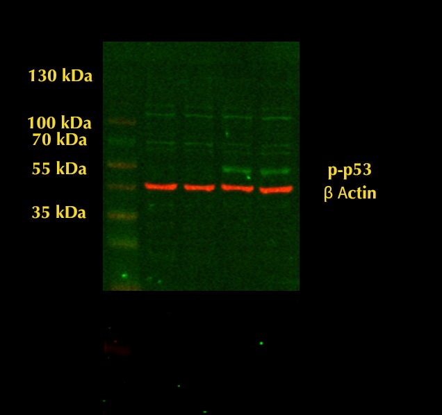
Application: Western BlotSample Tested: Cancer CellsSpecies: HumanVerified Customer | Posted 07/11/2018PC3 cells were treated with UV light for the indicated times. Total cell lysates were subjected to western blot. PVDF membrane were probed with 1mm/ml Human Phospho-p53 (S15) Antibody (AF1043). A specific band was detected for p-P53 at approximately 53kDa. This experiment was conducted under reducing conditions
-
Application: Western BlotSample Tested: MDA-MB-231 human breast cancer cell line and T47D human breast cancer cell lineSpecies: HumanVerified Customer | Posted 08/16/2017
There are no reviews that match your criteria.
Protocols
Find general support by application which include: protocols, troubleshooting, illustrated assays, videos and webinars.
- Antigen Retrieval Protocol (PIER)
- Antigen Retrieval for Frozen Sections Protocol
- Appropriate Fixation of IHC/ICC Samples
- Cellular Response to Hypoxia Protocols
- Chromogenic IHC Staining of Formalin-Fixed Paraffin-Embedded (FFPE) Tissue Protocol
- Chromogenic Immunohistochemistry Staining of Frozen Tissue
- ClariTSA™ Fluorophore Kits
- Detection & Visualization of Antibody Binding
- Fluorescent IHC Staining of Frozen Tissue Protocol
- Graphic Protocol for Heat-induced Epitope Retrieval
- Graphic Protocol for the Preparation and Fluorescent IHC Staining of Frozen Tissue Sections
- Graphic Protocol for the Preparation and Fluorescent IHC Staining of Paraffin-embedded Tissue Sections
- Graphic Protocol for the Preparation of Gelatin-coated Slides for Histological Tissue Sections
- ICC Cell Smear Protocol for Suspension Cells
- ICC Immunocytochemistry Protocol Videos
- ICC for Adherent Cells
- IHC Sample Preparation (Frozen sections vs Paraffin)
- Immunocytochemistry (ICC) Protocol
- Immunocytochemistry Troubleshooting
- Immunofluorescence of Organoids Embedded in Cultrex Basement Membrane Extract
- Immunofluorescent IHC Staining of Formalin-Fixed Paraffin-Embedded (FFPE) Tissue Protocol
- Immunohistochemistry (IHC) and Immunocytochemistry (ICC) Protocols
- Immunohistochemistry Frozen Troubleshooting
- Immunohistochemistry Paraffin Troubleshooting
- Immunoprecipitation Protocol
- Preparing Samples for IHC/ICC Experiments
- Preventing Non-Specific Staining (Non-Specific Binding)
- Primary Antibody Selection & Optimization
- Protocol for Heat-Induced Epitope Retrieval (HIER)
- Protocol for Making a 4% Formaldehyde Solution in PBS
- Protocol for VisUCyte™ HRP Polymer Detection Reagent
- Protocol for the Fluorescent ICC Staining of Cell Smears - Graphic
- Protocol for the Fluorescent ICC Staining of Cultured Cells on Coverslips - Graphic
- Protocol for the Preparation & Fixation of Cells on Coverslips
- Protocol for the Preparation and Chromogenic IHC Staining of Frozen Tissue Sections
- Protocol for the Preparation and Chromogenic IHC Staining of Frozen Tissue Sections - Graphic
- Protocol for the Preparation and Chromogenic IHC Staining of Paraffin-embedded Tissue Sections
- Protocol for the Preparation and Chromogenic IHC Staining of Paraffin-embedded Tissue Sections - Graphic
- Protocol for the Preparation and Fluorescent ICC Staining of Cells on Coverslips
- Protocol for the Preparation and Fluorescent ICC Staining of Non-adherent Cells
- Protocol for the Preparation and Fluorescent ICC Staining of Stem Cells on Coverslips
- Protocol for the Preparation and Fluorescent IHC Staining of Frozen Tissue Sections
- Protocol for the Preparation and Fluorescent IHC Staining of Paraffin-embedded Tissue Sections
- Protocol for the Preparation of Gelatin-coated Slides for Histological Tissue Sections
- Protocol for the Preparation of a Cell Smear for Non-adherent Cell ICC - Graphic
- R&D Systems Quality Control Western Blot Protocol
- TUNEL and Active Caspase-3 Detection by IHC/ICC Protocol
- The Importance of IHC/ICC Controls
- Troubleshooting Guide: Immunohistochemistry
- Troubleshooting Guide: Western Blot Figures
- Western Blot Conditions
- Western Blot Protocol
- Western Blot Protocol for Cell Lysates
- Western Blot Troubleshooting
- Western Blot Troubleshooting Guide
- View all Protocols, Troubleshooting, Illustrated assays and Webinars
Loading...
Associated Pathways


