Plexin A4 is a 220‑230 kDa member of the plexin A subfamily, plexin family of proteins (1). It is found on sensory, autonomic and motor neurons and oligodendrocytes, plus T cells and dendritic cells (1‑8). Mature human Plexin A4 is an 1871 amino acid (aa) type I transmembrane glycoprotein with a 23 aa signal sequence, a 1214 aa extracellular domain (ECD), and a 636 aa cytoplasmic region. The ECD contains one Sema-domain (aa 51‑482), three PSI domains (aa 509‑856) and four IPT regions (aa 858‑1230) that contain a phosphoserine at aa 946 (1). Of three isoform variants, one shows a 65 aa substitution for aa 458‑1894, a second shows an 80 aa substitution for aa 1292‑1894, and a third shows the just mentioned 80 aa substitution coupled to a 14 aa substitution for aa 1‑535 (9). The human Plexin A4 ECD shares 97% aa identity with mouse, equine, canine, and bovine Plexin A4. Full‑length Plexin A4 also shares 67% aa identity with the most related family member, Plexin A2. Plexin A4 regulates cell migration, activation and axon guidance via repulsion (1‑5). It serves as a receptor for transmembrane semaphorins, Sema6A and 6B, and as a coreceptor with neuropilin-1 for the secreted semaphorin, Sema3A (1‑8). During development, it plays a role in nerve migration and midline crossing and down‑regulates dendrite formation (2‑8). It is often co‑expressed with Plexin A3, which can also engage class 6 semaphorins but prefers Sema3F/neuropilin‑2 to Sema3A/neuropilin-1 (3, 8). Thus, Plexins A3 and A4 are redundant in some functions, but unique in others. In T cells, Plexin A4 engages Sema3A and negatively regulates TCR signals (6).
Human Plexin A4 Antibody (707206)
R&D Systems | Catalog # MAB58561

Key Product Details
Species Reactivity
Validated:
Human
Cited:
Mouse
Applications
Validated:
Flow Cytometry, CyTOF-ready
Cited:
Flow Cytometry
Label
Unconjugated
Antibody Source
Monoclonal Mouse IgG1 Clone # 707206
Loading...
Product Specifications
Immunogen
Chinese hamster ovary cell line CHO-derived recombinant human Plexin A4
Thr24-Pro1237
Accession # Q9HCM2
Thr24-Pro1237
Accession # Q9HCM2
Specificity
Detects human Plexin A4 in direct ELISAs. In direct ELISAs, no cross-reactivity with recombinant human Plexin A1, recombinant mouse (rm) Plexin A1 or rmPlexin A2 is observed.
Clonality
Monoclonal
Host
Mouse
Isotype
IgG1
Scientific Data Images for Human Plexin A4 Antibody (707206)
Detection of Plexin A4 in SH-SY5Y cells by Flow Cytometry
SH-SY5Y (filled histogram) and Jurkat (open histogram) cells were stained with Mouse Anti-Human Plexin A4 Monoclonal Antibody (Catalog # MAB58561) followed by Allophycocyanin-conjugated Anti-Mouse IgG Secondary Antibody (Catalog # F0101B). View our protocol for Staining Membrane-associated Proteins.
Detection of Plexin A4 in Human Blood Lymphocytes by Flow Cytometry.
Human peripheral blood lymphocytes were stained with Mouse Anti-Human Plexin A4 Monoclonal Antibody (Catalog # MAB58561) followed by Phycoerythrin-conjugated Anti-Mouse IgG Secondary Antibody (Catalog # F0102B) and Mouse Anti-Human CD3e APC-conjugated Monoclonal Antibody (Catalog # FAB100A). Quadrant markers were set based on control antibody staining (Catalog # MAB002).Applications for Human Plexin A4 Antibody (707206)
Application
Recommended Usage
CyTOF-ready
Ready to be labeled using established conjugation methods. No BSA or other carrier proteins that could interfere with conjugation.
Flow Cytometry
0.25 µg/106 cells
Sample: SH-SH5Y human neuroblastoma cell line or Human peripheral blood lymphocytes
Sample: SH-SH5Y human neuroblastoma cell line or Human peripheral blood lymphocytes
Reviewed Applications
Read 1 review rated 4 using MAB58561 in the following applications:
Flow Cytometry Panel Builder
Bio-Techne Knows Flow Cytometry
Save time and reduce costly mistakes by quickly finding compatible reagents using the Panel Builder Tool.
Advanced Features
- Spectra Viewer - Custom analysis of spectra from multiple fluorochromes
- Spillover Popups - Visualize the spectra of individual fluorochromes
- Antigen Density Selector - Match fluorochrome brightness with antigen density

Formulation, Preparation, and Storage
Purification
Protein A or G purified from hybridoma culture supernatant
Reconstitution
Sterile PBS to a final concentration of 0.5 mg/mL. For liquid material, refer to CoA for concentration.
Loading...
Formulation
Lyophilized from a 0.2 μm filtered solution in PBS with Trehalose. See Certificate of Analysis for details.
*Small pack size (-SP) is supplied either lyophilized or as a 0.2 µm filtered solution in PBS.
*Small pack size (-SP) is supplied either lyophilized or as a 0.2 µm filtered solution in PBS.
Shipping
Lyophilized product is shipped at ambient temperature. Liquid small pack size (-SP) is shipped with polar packs. Upon receipt, store immediately at the temperature recommended below.
Stability & Storage
Use a manual defrost freezer and avoid repeated freeze-thaw cycles.
- 12 months from date of receipt, -20 to -70 °C as supplied.
- 1 month, 2 to 8 °C under sterile conditions after reconstitution.
- 6 months, -20 to -70 °C under sterile conditions after reconstitution.
Calculators
Background: Plexin A4
References
- Suto, F. et al. (2003) Mech. Dev. 120:385.
- Suto, F. et al. (2005) J. Neurosci. 25:3628.
- Faulkner, R.L. et al. (2008) Neural Dev. 3:21.
- Waimey, K.E. et al. (2008) Dev. Biol. 315:448.
- Runker, A.E. et al. (2008) Neural Dev. 3:34.
- Yamamoto, M. et al. (2008) Int. Immunol. 20:413.
- Okada, A. et al. (2007) Biochem. Biophys. Res. Commun. 352:158.
- Yaron, A. et al. (2005) Neuron 45:513.
- Protein Accession # NP_861440, EAW83796, EAL24077.
Alternate Names
PLEXA4, PLXNA4
Gene Symbol
PLXNA4
UniProt
Additional Plexin A4 Products
Product Documents for Human Plexin A4 Antibody (707206)
Certificate of Analysis
To download a Certificate of Analysis, please enter a lot or batch number in the search box below.
Note: Certificate of Analysis not available for kit components.
Product Specific Notices for Human Plexin A4 Antibody (707206)
For research use only
Citations for Human Plexin A4 Antibody (707206)
 Powered by Bioz
Customer Reviews for Human Plexin A4 Antibody (707206) (1)
4 out of 5
1 Customer Rating
Have you used Human Plexin A4 Antibody (707206)?
Submit a review and receive an Amazon gift card!
$25/€18/£15/$25CAN/¥2500 Yen for a review with an image
$10/€7/£6/$10CAN/¥1110 Yen for a review without an image
Submit a review
Customer Images
Showing
1
-
1 of
1 review
Showing All
Filter By:
-
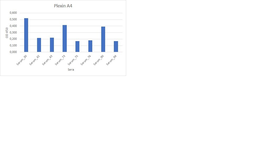
Application: ELISASample Tested: Serum and PlasmaSpecies: HumanVerified Customer | Posted 02/13/2023

There are no reviews that match your criteria.
Protocols
Find general support by application which include: protocols, troubleshooting, illustrated assays, videos and webinars.
- 7-Amino Actinomycin D (7-AAD) Cell Viability Flow Cytometry Protocol
- Extracellular Membrane Flow Cytometry Protocol
- Flow Cytometry Protocol for Cell Surface Markers
- Flow Cytometry Protocol for Staining Membrane Associated Proteins
- Flow Cytometry Staining Protocols
- Flow Cytometry Troubleshooting Guide
- Intracellular Flow Cytometry Protocol Using Alcohol (Methanol)
- Intracellular Flow Cytometry Protocol Using Detergents
- Intracellular Nuclear Staining Flow Cytometry Protocol Using Detergents
- Intracellular Staining Flow Cytometry Protocol Using Alcohol Permeabilization
- Intracellular Staining Flow Cytometry Protocol Using Detergents to Permeabilize Cells
- Propidium Iodide Cell Viability Flow Cytometry Protocol
- Protocol for Liperfluo
- Protocol for the Characterization of Human Th22 Cells
- Protocol for the Characterization of Human Th9 Cells
- Protocol: Annexin V and PI Staining by Flow Cytometry
- Protocol: Annexin V and PI Staining for Apoptosis by Flow Cytometry
- Troubleshooting Guide: Fluorokine Flow Cytometry Kits
- View all Protocols, Troubleshooting, Illustrated assays and Webinars
Loading...