Trypsin is a general term for any of three 24 kDa gene products that belong to the peptidase S1 family of enzymes. Trypsin in Greek means “rubbing or friction”, and it was chosen here because the first trypsins were extracted from pancreas via a glycerin-based rubbing maceration. Trypsin-1 (cationic), -2 (anionic), and -3 (mesotrypsin) are synthesized as 26 kDa trypsinogens (plus a 35 kDa trypsinogen-3 isoform) that are 247 amino acids (aa) in length. The first 15 aa constitute a signal sequence, followed by an enterokinase-cleavable eight aa propeptide, and a 224 aa mature molecule. Asp194 is linked to enzyme activity, and Tyr154 is sulfated. Over their mature regions, the three trypsins share 84% aa identity. Mouse trypsin-1 shares 74% aa identity with the human trypsin consensus sequence. Trypsin-1 and -2 cleave peptide bonds carboxylterminal to a Lys or
Human Trypsin Pan Specific (PRSS1/2/3) Antibody
R&D Systems | Catalog # AF3586

Key Product Details
Species Reactivity
Validated:
Human
Cited:
Human
Applications
Validated:
Immunohistochemistry, Western Blot, Neutralization, Immunoprecipitation
Cited:
Immunohistochemistry, Immunohistochemistry-Paraffin, Immunocytochemistry
Label
Unconjugated
Antibody Source
Polyclonal Sheep IgG
Loading...
Product Specifications
Immunogen
Mouse myeloma cell line NS0-derived recombinant human Trypsin 2
Ala16-Ser247
Accession # P07478
Ala16-Ser247
Accession # P07478
Specificity
Detects human Trypsin Pan Specific (PRSS1/2/3) in direct ELISAs and Western blots.
Clonality
Polyclonal
Host
Sheep
Isotype
IgG
Scientific Data Images for Human Trypsin Pan Specific (PRSS1/2/3) Antibody
Detection of Human Trypsin by Western Blot.
Western blot shows lysates of human pancreas. PVDF membrane was probed with 1 µg/mL of Sheep Anti-Human Trypsin Pan Specific (PRSS1/2/3) Antigen Affinity-purified Polyclonal Antibody (Catalog # AF3586) followed by HRP-conjugated Anti-Sheep IgG Secondary Antibody (HAF016). A specific band was detected for Trypsin at approximately 24 kDa (as indicated). This experiment was conducted under reducing conditions and using Western Blot Buffer Group 1.
Detection of Human Trypsin by Western Blot.
Western blot shows recombinant human Trypsin 1, 2, and 3. PVDF membrane was probed with 1 µg/mL of Sheep Anti-Human Trypsin Pan Specific (PRSS1/2/3) Antigen Affinity-purified Polyclonal Antibody (Catalog # AF3586) followed by HRP-conjugated Anti-Sheep IgG Secondary Antibody (HAF016). A specific band was detected for Trypsin at approximately 24 kDa (as indicated). This experiment was conducted under reducing conditions and using Western Blot Buffer Group 1.
Trypsin in Human Pancreas.
Trypsin was detected in immersion fixed paraffin-embedded sections of human pancreas using Sheep Anti-Human Trypsin Pan Specific (PRSS1/2/3) Antigen Affinity-purified Polyclonal Antibody (Catalog # AF3586) at 15 µg/mL overnight at 4 °C. Tissue was stained using the Anti-Sheep HRP-DAB Cell & Tissue Staining Kit (brown; Catalog # CTS019) and counterstained with hematoxylin (blue). Specific staining was localized to exocrine cells. View our protocol for Chromogenic IHC Staining of Paraffin-embedded Tissue Sections.
Trypsin in Human Pancreatic Cancer Tissue.
Trypsin was detected in immersion fixed paraffin-embedded sections of human pancreatic cancer tissue using Sheep Anti-Human Trypsin Pan Specific (PRSS1/2/3) Antigen Affinity-purified Polyclonal Antibody (Catalog # AF3586) at 5 µg/mL overnight at 4 °C. Tissue was stained using the Anti-Sheep HRP-DAB Cell & Tissue Staining Kit (brown; Catalog # CTS019) and counterstained with hematoxylin (blue). Specific staining was localized to cytoplasm of cancer cells. View our protocol for Chromogenic IHC Staining of Paraffin-embedded Tissue Sections.
Detection of Trypsin by Immunoprecipitation
Human Trypsin 2/PRSS2 was immunoprecipitated from 500 μg of human pancreas lysates with 12.5 ug Mouse Anti-Human Trypsin 2/PRSS2 Monoclonal Antibody (Catalog # MAB3586). The Trypsin 2/PRSS2-antibody complexes were absorbed using Protein G Sepharose. Immunoprecipitated human Trypsin 2/PRSS2 was detected by Western blot using 2 µg/mL of Sheep Anti-Human Trypsin Pan Specific (PRSS1/2/3) Antigen Affinity-purified Polyclonal Antibody (AF3586) under non-reducing conditions and using Western Blot Buffer Group 1.Applications for Human Trypsin Pan Specific (PRSS1/2/3) Antibody
Application
Recommended Usage
Immunohistochemistry
5-15 µg/mL
Sample: Immersion fixed paraffin-embedded sections of human pancreas and immersion fixed paraffin-embedded sections of human pancreatic cancer tissue
Sample: Immersion fixed paraffin-embedded sections of human pancreas and immersion fixed paraffin-embedded sections of human pancreatic cancer tissue
Immunoprecipitation
12.5 µg/500 µg cell lysate
Sample: Human pancreas tissue
Sample: Human pancreas tissue
Western Blot
1 µg/mL
Sample: Human pancreas and recombinant human Trypsin 1, 2, and 3
Sample: Human pancreas and recombinant human Trypsin 1, 2, and 3
Neutralization
Measured by its ability to neutralize Recombinant Human Trypsin 2/PRSS2 (0.02 µg/mL, Catalog # 3586-SE) cleavage of the fluorogenic peptide substrate Mca-RPKPVE-Nval-WRK(Dnp)-NH2 (10 µM, Catalog # ES002 ). The Neutralization Dose (ND50) is typically 0.3 µg/mL.
Reviewed Applications
Read 3 reviews rated 4.7 using AF3586 in the following applications:
Formulation, Preparation, and Storage
Purification
Antigen Affinity-purified
Reconstitution
Reconstitute at 0.2 mg/mL in sterile PBS. For liquid material, refer to CoA for concentration.
Loading...
Formulation
Lyophilized from a 0.2 μm filtered solution in PBS with Trehalose. *Small pack size (SP) is supplied either lyophilized or as a 0.2 µm filtered solution in PBS.
Shipping
Lyophilized product is shipped at ambient temperature. Liquid small pack size (-SP) is shipped with polar packs. Upon receipt, store immediately at the temperature recommended below.
Stability & Storage
Use a manual defrost freezer and avoid repeated freeze-thaw cycles.
- 12 months from date of receipt, -20 to -70 °C as supplied.
- 1 month, 2 to 8 °C under sterile conditions after reconstitution.
- 6 months, -20 to -70 °C under sterile conditions after reconstitution.
Calculators
Background: Trypsin
Alternate Names
TRY2, TRY8, TRYP2
UniProt
Additional Trypsin Products
Product Documents for Human Trypsin Pan Specific (PRSS1/2/3) Antibody
Certificate of Analysis
To download a Certificate of Analysis, please enter a lot or batch number in the search box below.
Note: Certificate of Analysis not available for kit components.
Product Specific Notices for Human Trypsin Pan Specific (PRSS1/2/3) Antibody
For research use only
Related Research Areas
Citations for Human Trypsin Pan Specific (PRSS1/2/3) Antibody
 Powered by Bioz
Customer Reviews for Human Trypsin Pan Specific (PRSS1/2/3) Antibody (3)
4.7 out of 5
3 Customer Ratings
Have you used Human Trypsin Pan Specific (PRSS1/2/3) Antibody?
Submit a review and receive an Amazon gift card!
$25/€18/£15/$25CAN/¥2500 Yen for a review with an image
$10/€7/£6/$10CAN/¥1110 Yen for a review without an image
Submit a review
Customer Images
Showing
1
-
3 of
3 reviews
Showing All
Filter By:
-
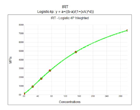
Application: Luminex XMAP technologySample Tested: Adult pancreasSpecies: HumanVerified Customer | Posted 04/17/2018product was used as a primary antibody attached to a solid phase. Works only when combined with Trypsin-1 from another vendor as 50/50 rate

-
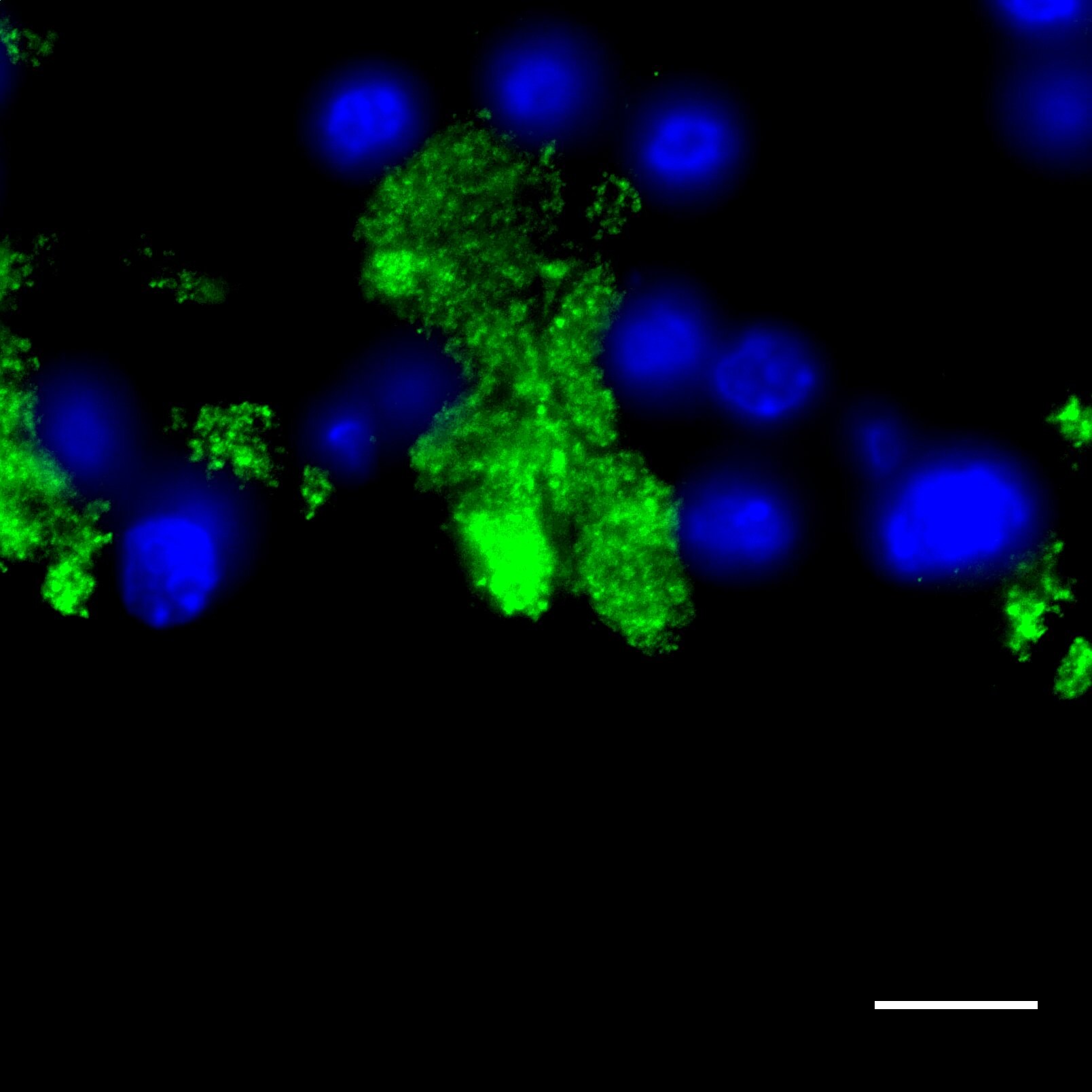
Application: ImmunocytochemistrySample Tested: Mouse pancreas in OCTSpecies: MouseVerified Customer | Posted 06/09/2017cryosection of mouse pancreas, primary 1/100, secondary 1/200 488. Epi-fluorescence 60 x. signal weak
-
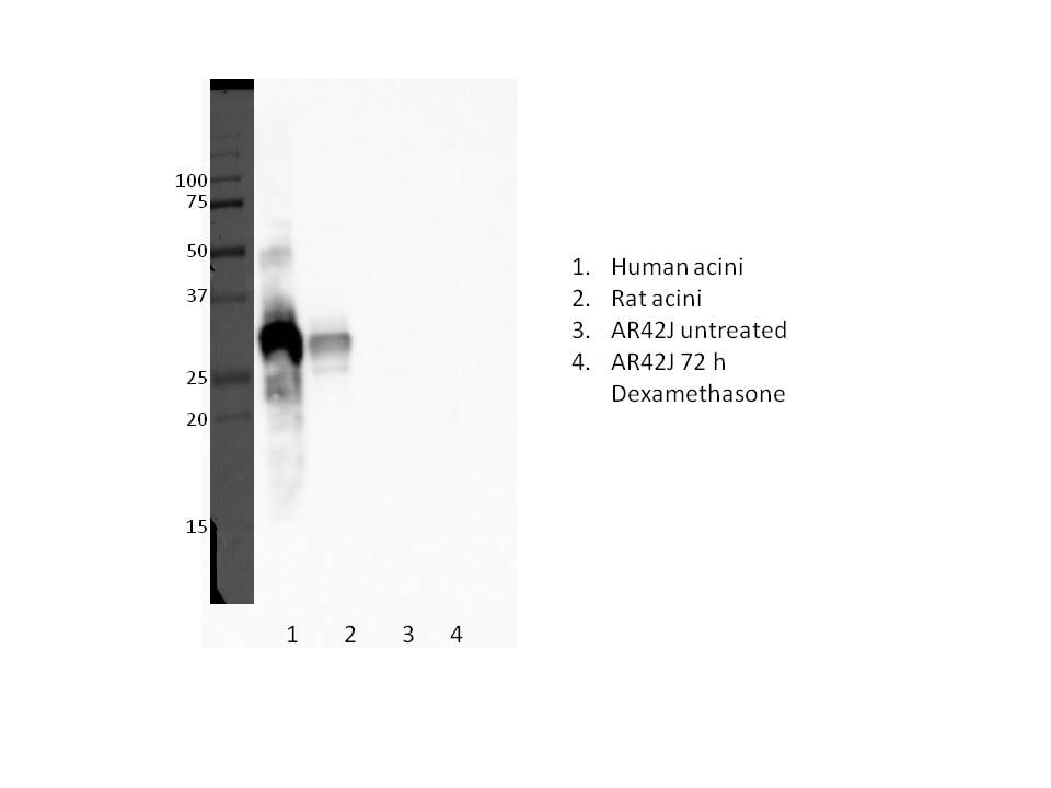
Application: Western BlotSample Tested: human acini, rat acini and AR42J rat acinar-like cellsSpecies: Human and RatVerified Customer | Posted 05/24/20171/1000, 12% gel, 50 ug protein
There are no reviews that match your criteria.
Protocols
Find general support by application which include: protocols, troubleshooting, illustrated assays, videos and webinars.
- Antigen Retrieval Protocol (PIER)
- Antigen Retrieval for Frozen Sections Protocol
- Appropriate Fixation of IHC/ICC Samples
- Cellular Response to Hypoxia Protocols
- Chromogenic IHC Staining of Formalin-Fixed Paraffin-Embedded (FFPE) Tissue Protocol
- Chromogenic Immunohistochemistry Staining of Frozen Tissue
- ClariTSA™ Fluorophore Kits
- Detection & Visualization of Antibody Binding
- Fluorescent IHC Staining of Frozen Tissue Protocol
- Graphic Protocol for Heat-induced Epitope Retrieval
- Graphic Protocol for the Preparation and Fluorescent IHC Staining of Frozen Tissue Sections
- Graphic Protocol for the Preparation and Fluorescent IHC Staining of Paraffin-embedded Tissue Sections
- Graphic Protocol for the Preparation of Gelatin-coated Slides for Histological Tissue Sections
- IHC Sample Preparation (Frozen sections vs Paraffin)
- Immunofluorescent IHC Staining of Formalin-Fixed Paraffin-Embedded (FFPE) Tissue Protocol
- Immunohistochemistry (IHC) and Immunocytochemistry (ICC) Protocols
- Immunohistochemistry Frozen Troubleshooting
- Immunohistochemistry Paraffin Troubleshooting
- Immunoprecipitation Protocol
- Preparing Samples for IHC/ICC Experiments
- Preventing Non-Specific Staining (Non-Specific Binding)
- Primary Antibody Selection & Optimization
- Protocol for Heat-Induced Epitope Retrieval (HIER)
- Protocol for Making a 4% Formaldehyde Solution in PBS
- Protocol for VisUCyte™ HRP Polymer Detection Reagent
- Protocol for the Preparation & Fixation of Cells on Coverslips
- Protocol for the Preparation and Chromogenic IHC Staining of Frozen Tissue Sections
- Protocol for the Preparation and Chromogenic IHC Staining of Frozen Tissue Sections - Graphic
- Protocol for the Preparation and Chromogenic IHC Staining of Paraffin-embedded Tissue Sections
- Protocol for the Preparation and Chromogenic IHC Staining of Paraffin-embedded Tissue Sections - Graphic
- Protocol for the Preparation and Fluorescent IHC Staining of Frozen Tissue Sections
- Protocol for the Preparation and Fluorescent IHC Staining of Paraffin-embedded Tissue Sections
- Protocol for the Preparation of Gelatin-coated Slides for Histological Tissue Sections
- R&D Systems Quality Control Western Blot Protocol
- TUNEL and Active Caspase-3 Detection by IHC/ICC Protocol
- The Importance of IHC/ICC Controls
- Troubleshooting Guide: Immunohistochemistry
- Troubleshooting Guide: Western Blot Figures
- Western Blot Conditions
- Western Blot Protocol
- Western Blot Protocol for Cell Lysates
- Western Blot Troubleshooting
- Western Blot Troubleshooting Guide
- View all Protocols, Troubleshooting, Illustrated assays and Webinars
Loading...