VIAAT (Vesicular inhibitory amino acid transporter; also VGAT and SLC32A1) is a 56-58 kDa member of the amino acid/polyamine transporter 2 family of proteins. It is expressed in inhibitory neurons throughout the CNS, in both resting and activated lymphocytes, and in pancreatic alpha -cells (in rodent). VIAAT is believed to transport both GABA and glycine across synaptic vesicle membranes in an electrical and pH gradient-dependent manner. Once transported, these neurotransmitters are released at the presynaptic membrane, acting as inhibitory factors in the mature nervous system, and excitatory factors in the immature nervous system. Human VIAAT is a 525 amino acid (aa) 10-transmembrane nonglycosylated protein. The N-terminus (aa 1-133) and C-terminus (aa 511-525) are cytoplasmic. There is a 52 kDa short form that is not well characterized. Over aa 2-133, human VIAAT shares 95% aa identity with mouse VIAAT.

Key Product Details
Species Reactivity
Human
Applications
Immunocytochemistry
Label
Unconjugated
Antibody Source
Monoclonal Mouse IgG1 Clone # 731307
Loading...
Product Specifications
Immunogen
E. coli-derived recombinant human VIAAT/SLC32A1
Ala2-Phe133
Accession # Q9H598
Ala2-Phe133
Accession # Q9H598
Specificity
Detects human VIAAT/SLC32A1 in direct ELISAs.
Clonality
Monoclonal
Host
Mouse
Isotype
IgG1
Scientific Data Images for Human VIAAT/SLC32A1 Antibody
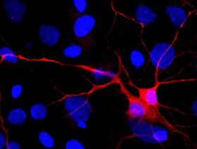
VIAAT/SLC32A1 in Rat Cortical Stem Cells.
VIAAT/SLC32A1 was detected in immersion fixed 7-day differentiated rat cortical stem cells using Mouse Anti-Human VIAAT/SLC32A1 Monoclonal Antibody (Catalog # MAB6847) at 10 µg/mL for 3 hours at room temperature. Cells were stained using the NorthernLights™ 557-conjugated Anti-Mouse IgG Secondary Antibody (red; Catalog # NL007) and counterstained with DAPI (blue). Specific staining was localized to neurons. View our protocol for Fluorescent ICC Staining of Cells on Coverslips.Applications for Human VIAAT/SLC32A1 Antibody
Application
Recommended Usage
Immunocytochemistry
8-25 µg/mL
Sample: Immersion fixed 7-day differentiated rat cortical stem cells
Sample: Immersion fixed 7-day differentiated rat cortical stem cells
Reviewed Applications
Read 1 review rated 5 using MAB6847 in the following applications:
Formulation, Preparation, and Storage
Purification
Protein A or G purified from hybridoma culture supernatant
Reconstitution
Sterile PBS to a final concentration of 0.5 mg/mL. For liquid material, refer to CoA for concentration.
Loading...
Formulation
Lyophilized from a 0.2 μm filtered solution in PBS with Trehalose. *Small pack size (SP) is supplied either lyophilized or as a 0.2 µm filtered solution in PBS.
Shipping
Lyophilized product is shipped at ambient temperature. Liquid small pack size (-SP) is shipped with polar packs. Upon receipt, store immediately at the temperature recommended below.
Stability & Storage
Use a manual defrost freezer and avoid repeated freeze-thaw cycles.
- 12 months from date of receipt, -20 to -70 °C as supplied.
- 1 month, 2 to 8 °C under sterile conditions after reconstitution.
- 6 months, -20 to -70 °C under sterile conditions after reconstitution.
Calculators
Background: VIAAT/SLC32A1
Long Name
Vesicular Inhibitory Amino Acid Transporter
Alternate Names
SLC32A1, VGAT
Gene Symbol
SLC32A1
UniProt
Additional VIAAT/SLC32A1 Products
Product Documents for Human VIAAT/SLC32A1 Antibody
Certificate of Analysis
To download a Certificate of Analysis, please enter a lot or batch number in the search box below.
Note: Certificate of Analysis not available for kit components.
Product Specific Notices for Human VIAAT/SLC32A1 Antibody
For research use only
Related Research Areas
Citations for Human VIAAT/SLC32A1 Antibody
 Powered by Bioz
Customer Reviews for Human VIAAT/SLC32A1 Antibody (1)
5 out of 5
1 Customer Rating
Have you used Human VIAAT/SLC32A1 Antibody?
Submit a review and receive an Amazon gift card!
$25/€18/£15/$25CAN/¥2500 Yen for a review with an image
$10/€7/£6/$10CAN/¥1110 Yen for a review without an image
Submit a review
Customer Images
Showing
1
-
1 of
1 review
Showing All
Filter By:
-
Application: Immunocytochemistry/ImmunofluorescenceSample Tested: Cortical stem cellsSpecies: RatVerified Customer | Posted 11/01/2022

There are no reviews that match your criteria.
Protocols
Find general support by application which include: protocols, troubleshooting, illustrated assays, videos and webinars.
- Appropriate Fixation of IHC/ICC Samples
- Cellular Response to Hypoxia Protocols
- ClariTSA™ Fluorophore Kits
- Detection & Visualization of Antibody Binding
- ICC Cell Smear Protocol for Suspension Cells
- ICC Immunocytochemistry Protocol Videos
- ICC for Adherent Cells
- Immunocytochemistry (ICC) Protocol
- Immunocytochemistry Troubleshooting
- Immunofluorescence of Organoids Embedded in Cultrex Basement Membrane Extract
- Immunohistochemistry (IHC) and Immunocytochemistry (ICC) Protocols
- Preparing Samples for IHC/ICC Experiments
- Preventing Non-Specific Staining (Non-Specific Binding)
- Primary Antibody Selection & Optimization
- Protocol for VisUCyte™ HRP Polymer Detection Reagent
- Protocol for the Fluorescent ICC Staining of Cell Smears - Graphic
- Protocol for the Fluorescent ICC Staining of Cultured Cells on Coverslips - Graphic
- Protocol for the Preparation and Fluorescent ICC Staining of Cells on Coverslips
- Protocol for the Preparation and Fluorescent ICC Staining of Non-adherent Cells
- Protocol for the Preparation and Fluorescent ICC Staining of Stem Cells on Coverslips
- Protocol for the Preparation of a Cell Smear for Non-adherent Cell ICC - Graphic
- TUNEL and Active Caspase-3 Detection by IHC/ICC Protocol
- The Importance of IHC/ICC Controls
- View all Protocols, Troubleshooting, Illustrated assays and Webinars
Loading...