
Key Product Details
Species Reactivity
Validated:
Human
Cited:
Human
Applications
Validated:
Flow Cytometry, Immunocytochemistry, CyTOF-ready
Cited:
Western Blot, Flow Cytometry, Immunocytochemistry
Label
Unconjugated
Antibody Source
Monoclonal Mouse IgG1 Clone # 639914
Loading...
Product Specifications
Immunogen
E. coli-derived recombinant human ZEB1
Glu430-Ser575
Accession # P37275
Glu430-Ser575
Accession # P37275
Specificity
Detects human ZEB1 in direct ELISAs.
Clonality
Monoclonal
Host
Mouse
Isotype
IgG1
Scientific Data Images for Human ZEB1 Antibody (639914)
ZEB1 in MDA‑MB‑231 Human Cell Line.
ZEB1 was detected in immersion fixed MDA-MB-231 human breast cancer cell line using Mouse Anti-Human ZEB1 Monoclonal Antibody (Catalog # MAB6708) at 10 µg/mL for 3 hours at room temperature. Cells were stained using the NorthernLights™ 557-conjugated Anti-Mouse IgG Secondary Antibody (red, upper panel; Catalog # NL007) and counterstained with DAPI (blue, lower panel). Specific staining was localized to nuclei and cytoplasm. View our protocol for Fluorescent ICC Staining of Cells on Coverslips.
Detection of ZEB-1 in MDA‑MB‑231 Human Cell Line by Flow Cytometry
MDA-MB-231 human breast cancer cell line was stained with Mouse Anti-Human ZEB1 Monoclonal Antibody (Catalog # MAB6708, filled histogram) or isotype control antibody (Catalog # MAB002, open histogram), followed by Allophycocyanin-conjugated Anti-Mouse IgG Secondary Antibody (Catalog # F0101B). To facilitate intracellular staining, cells were fixed with paraformaldehyde and permeabilized with saponin.
Detection of Human Human ZEB1 Antibody by Western Blot
RAB25 is a ZEB1 target gene. (A) RAB25 expression positively correlated with E-cadherin but negatively with ZEB1 in a series of lung cancer cell lines. RNA expression was measured by quantitative real-time PCR in 22 NSCLC cell lines, two NHBE cultures, and two immortalized human airway primary cell lines (BAES2B and FC6625-2 3KT). Cells are ranked from left to right with increasing RAB25 mRNA level. Values are expressed as percent of the geometric mean between GAPDH and actin mRNA. The experiment was done twice with qRT-PCR in duplicate. (B) RAB25 mRNA level is decreased by ZEB1 overexpression in H358 FlipIn ZEB1 cells (left) and by TGF beta treatment in H358 EV control cells (right). Values are expressed as percent of GAPDH for three independent experiments with qRT-PCR in duplicate. Bars = SD. (C) Western blot: RAB25 protein level is decreased by ZEB1 overexpression during 1 to 5 days of DOX treatment in H358 FlipIn ZEB1cells. E-cadherin is decreased as well. Actin is the loading control. Protein molecular weights are indicated in kDa on the left. Image collected and cropped by CiteAb from the following publication (https://pubmed.ncbi.nlm.nih.gov/24216980), licensed under a CC-BY license. Not internally tested by R&D Systems.Applications for Human ZEB1 Antibody (639914)
Application
Recommended Usage
CyTOF-ready
Ready to be labeled using established conjugation methods. No BSA or other carrier proteins that could interfere with conjugation.
Flow Cytometry
2.5 µg/106 cells
Sample: MDA‑MB‑231 human breast cancer cell line
Sample: MDA‑MB‑231 human breast cancer cell line
Immunocytochemistry
8-25 µg/mL
Sample: Immersion fixed MDA‑MB‑231 human breast cancer cell line
Sample: Immersion fixed MDA‑MB‑231 human breast cancer cell line
Reviewed Applications
Read 2 reviews rated 4.5 using MAB6708 in the following applications:
Flow Cytometry Panel Builder
Bio-Techne Knows Flow Cytometry
Save time and reduce costly mistakes by quickly finding compatible reagents using the Panel Builder Tool.
Advanced Features
- Spectra Viewer - Custom analysis of spectra from multiple fluorochromes
- Spillover Popups - Visualize the spectra of individual fluorochromes
- Antigen Density Selector - Match fluorochrome brightness with antigen density

Formulation, Preparation, and Storage
Purification
Protein A or G purified from hybridoma culture supernatant
Reconstitution
Sterile PBS to a final concentration of 0.5 mg/mL. For liquid material, refer to CoA for concentration.
Loading...
Formulation
Lyophilized from a 0.2 μm filtered solution in PBS with Trehalose. *Small pack size (SP) is supplied either lyophilized or as a 0.2 µm filtered solution in PBS.
Shipping
Lyophilized product is shipped at ambient temperature. Liquid small pack size (-SP) is shipped with polar packs. Upon receipt, store immediately at the temperature recommended below.
Stability & Storage
Use a manual defrost freezer and avoid repeated freeze-thaw cycles.
- 12 months from date of receipt, -20 to -70 °C as supplied.
- 1 month, 2 to 8 °C under sterile conditions after reconstitution.
- 6 months, -20 to -70 °C under sterile conditions after reconstitution.
Calculators
Background: ZEB1
Long Name
Zinc Finger E-box Binding Homeobox 1
Alternate Names
AREB6, BZP, DELTAEF1, FECD6, NIL2A, PPCD3, TCF8, ZFHEP
Gene Symbol
ZEB1
UniProt
Additional ZEB1 Products
Product Documents for Human ZEB1 Antibody (639914)
Certificate of Analysis
To download a Certificate of Analysis, please enter a lot or batch number in the search box below.
Note: Certificate of Analysis not available for kit components.
Product Specific Notices for Human ZEB1 Antibody (639914)
For research use only
Citations for Human ZEB1 Antibody (639914)
 Powered by Bioz
Customer Reviews for Human ZEB1 Antibody (639914) (2)
4.5 out of 5
2 Customer Ratings
Have you used Human ZEB1 Antibody (639914)?
Submit a review and receive an Amazon gift card!
$25/€18/£15/$25CAN/¥2500 Yen for a review with an image
$10/€7/£6/$10CAN/¥1110 Yen for a review without an image
Submit a review
Customer Images
Showing
1
-
2 of
2 reviews
Showing All
Filter By:
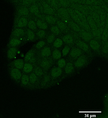
-
Application: Flow CytometrySample Tested: Pancreas tissueSpecies: HumanVerified Customer | Posted 06/27/2018
-
Application: Immunocytochemistry/ImmunofluorescenceSample Tested: Urinary bladder transitional cell carcinoma cell lineSpecies: HumanVerified Customer | Posted 11/17/2017

There are no reviews that match your criteria.
Protocols
Find general support by application which include: protocols, troubleshooting, illustrated assays, videos and webinars.
- 7-Amino Actinomycin D (7-AAD) Cell Viability Flow Cytometry Protocol
- Appropriate Fixation of IHC/ICC Samples
- Cellular Response to Hypoxia Protocols
- ClariTSA™ Fluorophore Kits
- Detection & Visualization of Antibody Binding
- Extracellular Membrane Flow Cytometry Protocol
- Flow Cytometry Protocol for Cell Surface Markers
- Flow Cytometry Protocol for Staining Membrane Associated Proteins
- Flow Cytometry Staining Protocols
- Flow Cytometry Troubleshooting Guide
- ICC Cell Smear Protocol for Suspension Cells
- ICC Immunocytochemistry Protocol Videos
- ICC for Adherent Cells
- Immunocytochemistry (ICC) Protocol
- Immunocytochemistry Troubleshooting
- Immunofluorescence of Organoids Embedded in Cultrex Basement Membrane Extract
- Immunohistochemistry (IHC) and Immunocytochemistry (ICC) Protocols
- Intracellular Flow Cytometry Protocol Using Alcohol (Methanol)
- Intracellular Flow Cytometry Protocol Using Detergents
- Intracellular Nuclear Staining Flow Cytometry Protocol Using Detergents
- Intracellular Staining Flow Cytometry Protocol Using Alcohol Permeabilization
- Intracellular Staining Flow Cytometry Protocol Using Detergents to Permeabilize Cells
- Preparing Samples for IHC/ICC Experiments
- Preventing Non-Specific Staining (Non-Specific Binding)
- Primary Antibody Selection & Optimization
- Propidium Iodide Cell Viability Flow Cytometry Protocol
- Protocol for Liperfluo
- Protocol for VisUCyte™ HRP Polymer Detection Reagent
- Protocol for the Characterization of Human Th22 Cells
- Protocol for the Characterization of Human Th9 Cells
- Protocol for the Fluorescent ICC Staining of Cell Smears - Graphic
- Protocol for the Fluorescent ICC Staining of Cultured Cells on Coverslips - Graphic
- Protocol for the Preparation and Fluorescent ICC Staining of Cells on Coverslips
- Protocol for the Preparation and Fluorescent ICC Staining of Non-adherent Cells
- Protocol for the Preparation and Fluorescent ICC Staining of Stem Cells on Coverslips
- Protocol for the Preparation of a Cell Smear for Non-adherent Cell ICC - Graphic
- Protocol: Annexin V and PI Staining by Flow Cytometry
- Protocol: Annexin V and PI Staining for Apoptosis by Flow Cytometry
- TUNEL and Active Caspase-3 Detection by IHC/ICC Protocol
- The Importance of IHC/ICC Controls
- Troubleshooting Guide: Fluorokine Flow Cytometry Kits
- View all Protocols, Troubleshooting, Illustrated assays and Webinars
Loading...