IKK beta Antibody (10AG2) - BSA Free
Novus Biologicals | Catalog # NB100-56509
Key Product Details
Validated by
Biological Validation
Species Reactivity
Validated:
Human, Mouse, Rat
Cited:
Human, Mouse
Applications
Validated:
Immunohistochemistry, Immunohistochemistry-Paraffin, Western Blot, Immunoblotting, Flow Cytometry, Immunocytochemistry/ Immunofluorescence, Simple Western, Immunoprecipitation, CyTOF-ready
Cited:
Immunohistochemistry-Paraffin, Western Blot, Immunocytochemistry/ Immunofluorescence, Immunoprecipitation, IF/IHC
Label
Unconjugated
Antibody Source
Monoclonal Mouse IgG1 Clone # 10AG2
Format
BSA Free
Loading...
Product Specifications
Immunogen
Full-length recombinant human IKK beta protein (NP_001547).
Reactivity Notes
Use in Rat reported in scientific literature (PMID:34333329)
Clonality
Monoclonal
Host
Mouse
Isotype
IgG1
Theoretical MW
87 kDa.
Disclaimer note: The observed molecular weight of the protein may vary from the listed predicted molecular weight due to post translational modifications, post translation cleavages, relative charges, and other experimental factors.
Disclaimer note: The observed molecular weight of the protein may vary from the listed predicted molecular weight due to post translational modifications, post translation cleavages, relative charges, and other experimental factors.
Scientific Data Images for IKK beta Antibody (10AG2) - BSA Free
![Western Blot: IKK beta Antibody (10AG2)BSA Free [NB100-56509]](https://resources.rndsystems.com/images/products/IKK-beta-Antibody-10AG2-Western-Blot-NB100-56509-img0015.jpg "Western Blot: IKK beta Antibody (10AG2)BSA Free [NB100-56509]")
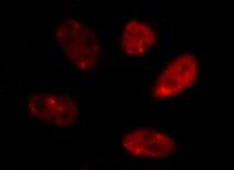
![Immunocytochemistry/ Immunofluorescence: IKK beta Antibody (10AG2) - BSA Free [NB100-56509]](https://resources.rndsystems.com/images/products/IKK-beta-Antibody-10AG2-Immunocytochemistry-Immunofluorescence-NB100-56509-img0016.jpg "Immunocytochemistry/ Immunofluorescence: IKK beta Antibody (10AG2) - BSA Free [NB100-56509]")
Immunocytochemistry/ Immunofluorescence: IKK beta Antibody (10AG2) - BSA Free [NB100-56509]
Immunocytochemistry/Immunofluorescence: IKK beta Antibody (10AG2) [NB100-56509] - Analysis of IKK beta Antibody in HeLa cells. Antibody dilution 5 ug/ml. Image from verified customer review.![Immunohistochemistry-Paraffin: IKK beta Antibody (10AG2) - BSA Free [NB100-56509]](https://resources.rndsystems.com/images/products/IKK-beta-Antibody-10AG2-Immunohistochemistry-Paraffin-NB100-56509-img0014.jpg "Immunohistochemistry-Paraffin: IKK beta Antibody (10AG2) - BSA Free [NB100-56509]")
Immunohistochemistry-Paraffin: IKK beta Antibody (10AG2) - BSA Free [NB100-56509]
Immunohistochemistry-Paraffin: IKK beta Antibody (10AG2) [NB100-56509] - Analysis of a FFPE tissue section of human placenta using 1:200 dilution of IKK beta clone 10AG2 antibody. The staining was developed using HRP labeled anti-rabbit secondary antibody and DAB reagent, and nuclei of cells were counter-stained with hematoxylin.![Flow Cytometry: IKK beta Antibody (10AG2) - BSA Free [NB100-56509]](https://resources.rndsystems.com/images/products/IKK-beta-Antibody-10AG2-Flow-Cytometry-NB100-56509-img0012.jpg "Flow Cytometry: IKK beta Antibody (10AG2) - BSA Free [NB100-56509]")
Flow Cytometry: IKK beta Antibody (10AG2) - BSA Free [NB100-56509]
Flow Cytometry: IKK beta Antibody (10AG2) [NB100-56509] - Analysis of PE conjugate of NB100-56509. An intracellular stain was performed on Jurkat cells with IKK Beta antibody (10AG2) NB100-56509PE (blue) and a matched isotype control NBP2-27287AF488 (orange). Cells were fixed with 4% PFA and then permeablized wi![Western Blot: IKK beta Antibody (10AG2)BSA Free [NB100-56509]](https://resources.rndsystems.com/images/products/IKK-beta-Antibody-10AG2-Western-Blot-NB100-56509-img0007.jpg "Western Blot: IKK beta Antibody (10AG2)BSA Free [NB100-56509]")
Western Blot: IKK beta Antibody (10AG2)BSA Free [NB100-56509]
Western Blot: IKK beta Antibody (10AG2) [NB100-56509] - Analysis of A) human Daudi, B) HeLa, and C) mouse NIH3T3 lysate probed with IKKbeta antibody at 2 ug/ml.![Immunocytochemistry/ Immunofluorescence: IKK beta Antibody (10AG2) - BSA Free [NB100-56509]](https://resources.rndsystems.com/images/products/IKK-beta-Antibody-10AG2-Immunocytochemistry-Immunofluorescence-NB100-56509-img0013.jpg "Immunocytochemistry/ Immunofluorescence: IKK beta Antibody (10AG2) - BSA Free [NB100-56509]")
Immunocytochemistry/ Immunofluorescence: IKK beta Antibody (10AG2) - BSA Free [NB100-56509]
Immunocytochemistry/Immunofluorescence: IKK beta Antibody (10AG2) [NB100-56509] - U-87 cells were fixed for 10 minutes using 10% formalin and then permeabilized for 5 minutes using 1X PBS + 0.5% Triton-X100. The cells were incubated with anti-IKK beta Antibody (10AG2) at 2 ug/ml overnight at 4C and detected with an anti-mouse Dylight 488 (Green) at a 1:500 dilution. Actin was detected with Phalloidin 568 (Red) at a 1:500 dilution. Nuclei were counterstained with DAPI (Blue). Cells were imaged using a 40X objective.![Flow Cytometry: IKK beta Antibody (10AG2) - BSA Free [NB100-56509]](https://resources.rndsystems.com/images/products/IKK-beta-Antibody-10AG2-Flow-Cytometry-NB100-56509-img0010.jpg "Flow Cytometry: IKK beta Antibody (10AG2) - BSA Free [NB100-56509]")
Flow Cytometry: IKK beta Antibody (10AG2) - BSA Free [NB100-56509]
Flow Cytometry: IKK beta Antibody (10AG2) [NB100-56509] - Analysis using the Alexa Fluor (R) 488 conjugate of NB100-56509. Staining of IKKbeta in HEK 293 cells using 0.1 ug of Alexa Fluor 488-conjugated antibody. Green histogram represents the isotype control, red represents the IKKbeta antibody.![Flow Cytometry: IKK beta Antibody (10AG2) - BSA Free [NB100-56509]](https://resources.rndsystems.com/images/products/IKK-beta-Antibody-10AG2-Flow-Cytometry-NB100-56509-img0011.jpg "Flow Cytometry: IKK beta Antibody (10AG2) - BSA Free [NB100-56509]")
Flow Cytometry: IKK beta Antibody (10AG2) - BSA Free [NB100-56509]
Flow Cytometry: IKK beta Antibody (10AG2) [NB100-56509] - Analysis using Alexa Fluor (R) 647 conjugate of NB100-56509. An intracellular stain was performed on HeLa cells with IKK beta antibody (10AG2) NB100-56509 (blue) and a matched isotype control NBP2-27287 (orange). Cells were fixed with 4% PFA and then permeablized with 0.1% saponin. 1 ug of antibody was added to 100 uL of staining buffer and cells were incubated for 30 minutes at room temperature. Both antibodies were conjugated to Alexa Fluor 647.![Simple Western: IKK beta Antibody (10AG2)BSA Free [NB100-56509]](https://resources.rndsystems.com/images/products/IKK-beta-Antibody-10AG2-Simple-Western-NB100-56509-img0008.jpg "Simple Western: IKK beta Antibody (10AG2)BSA Free [NB100-56509]")
Simple Western: IKK beta Antibody (10AG2)BSA Free [NB100-56509]
Simple Western: IKK beta Antibody (10AG2) [NB100-56509] - Lane view shows a specific band for IKK beta in 1.0 mg/ml of HeLa lysate. This experiment was performed under reducing conditions using the 12-230 kDa separation system. - BSA Free [NB100-56509] -")
Western Blot: IKK beta Antibody (10AG2) - BSA Free [NB100-56509] -
Western Blot: IKK beta Antibody (10AG2) - BSA Free [NB100-56509] - TRAM is required for the pro-inflammatory polarization of macrophages by SLD-LPS. (A,B) Flow cytometry analysis of Ly6C (A) or CD200R (B) expression in WT or TRAM−/− live-cell-gated macrophages with geometric MFI quantification on right. Pink = PBS, Blue = 100 pg/ml LPS. (C–E) Representative Western blots of IKK-beta (C), p-p65 (D), & total NF-kappa B p65 (E) expression in WT & TRAM−/− monocytes treated with either PBS or 100 pg/mL LPS for 5 days. Quantification is depicted on the right. (F) Ratio of p-p65/total p65 (p-p65 levels from (D) divided by corresponding total p65 levels). All data are representative of at least 3 separate experiments (n = 3) for (A,B) & n = 6 for (C–F). ANOVA with Tukey's multiple comparisons test (A,B); unpaired student t-test (C–F) *p < 0.05, **p < 0.01, ***p < 0.001, ****p < 0.0001, n.s, non-significant; MFI, Mean fluorescence intensity. Image collected & cropped by CiteAb from the following publication (https://pubmed.ncbi.nlm.nih.gov/32765513), licensed under a CC-BY license. Not internally tested by Novus Biologicals.Applications for IKK beta Antibody (10AG2) - BSA Free
Application
Recommended Usage
Flow Cytometry
0.25 - 0.5 ug/ 10^6 cells
Immunoblotting
reported in scientific literature (PMID 20103608)
Immunocytochemistry/ Immunofluorescence
2-5 ug/ml. Use reported in scientific literature (PMID 24825920)
Immunohistochemistry
1:10 - 1:500
Immunohistochemistry-Paraffin
1:10 - 1:500. Use reported in scientific literature (Page et al)
Immunoprecipitation
1:10 - 1:500
Simple Western
1:12.5
Western Blot
2 - 4 ug/ml
Application Notes
In Simple Western only 10 - 15 ul of the recommended dilution is used per data point.
See Simple Western Antibody Database for Simple Western validation: Tested in HeLa lysate 1.0 mg/mL, separated by Size, antibody dilution of 1:12.5, apparent MW was 90 kDa. Separated by Size-Wes, Sally Sue/Peggy Sue.
The observed molecular weight of the protein may vary from the listed predicted molecular weight due to post translational modifications, post translation cleavages, relative charges, and other experimental factors. This antibody is CyTOF ready.
Reviewed Applications
Read 2 reviews rated 4.5 using NB100-56509 in the following applications:
Flow Cytometry Panel Builder
Bio-Techne Knows Flow Cytometry
Save time and reduce costly mistakes by quickly finding compatible reagents using the Panel Builder Tool.
Advanced Features
- Spectra Viewer - Custom analysis of spectra from multiple fluorochromes
- Spillover Popups - Visualize the spectra of individual fluorochromes
- Antigen Density Selector - Match fluorochrome brightness with antigen density

Formulation, Preparation, and Storage
Purification
Protein G purified
Formulation
PBS
Format
BSA Free
Preservative
0.02% Sodium Azide
Concentration
1.0 mg/ml
Shipping
The product is shipped with polar packs. Upon receipt, store it immediately at the temperature recommended below.
Stability & Storage
Store at -20C. Avoid freeze-thaw cycles.
Background: IKK beta
Long Name
IkB Kinase beta
Alternate Names
IkBKB, IKK2, NFKBIKB
Entrez Gene IDs
3551 (Human)
Gene Symbol
IKBKB
UniProt
Additional IKK beta Products
Product Documents for IKK beta Antibody (10AG2) - BSA Free
Certificate of Analysis
To download a Certificate of Analysis, please enter a lot or batch number in the search box below.
Product Specific Notices for IKK beta Antibody (10AG2) - BSA Free
This product is for research use only and is not approved for use in humans or in clinical diagnosis. Primary Antibodies are guaranteed for 1 year from date of receipt.
Citations for IKK beta Antibody (10AG2) - BSA Free
 Powered by Bioz
Customer Reviews for IKK beta Antibody (10AG2) - BSA Free (2)
4.5 out of 5
2 Customer Ratings
Have you used IKK beta Antibody (10AG2) - BSA Free?
Submit a review and receive an Amazon gift card!
$25/€18/£15/$25CAN/¥2500 Yen for a review with an image
$10/€7/£6/$10CAN/¥1110 Yen for a review without an image
Submit a review
Customer Images
-(0025-mg)_NB100-56509_7366.jpg)
Showing
1
-
2 of
2 reviews
Showing All
Filter By:
-
Application: ImmunocytochemistrySample Tested: HeLa cellsSpecies: HumanVerified Customer | Posted 01/26/2022IKK beta Antibody in HeLa cellsDilution 5 ug/ml

-
Application: Western BlotSample Tested: Cell lysateSpecies: MouseVerified Customer | Posted 05/06/2014mAb anti-IKK beta (10AG2)  
There are no reviews that match your criteria.
Protocols
View specific protocols for IKK beta Antibody (10AG2) - BSA Free (NB100-56509):
Protocol for Flow Cytometry Intracellular Staining
Sample Preparation.
1. Grow cells to 60-85% confluency. Flow cytometry requires between 2 x 10⁵ and 1 x 10⁶ cells for optimal performance.
2. If cells are adherent, harvest gently by washing once with staining buffer and then scraping. Avoid using trypsin as this can disrupt certain epitopes of interest. If enzymatic harvest is required, use Accutase, Collagenase, or TrypLE Express for a less damaging option.
3. Reserve 100 uL for counting, then transfer cell volume into a 50 mL conical tube and centrifuge for 8 minutes at 400 RCF.
a. Count cells using a hemocytometer and a 1:1 trypan blue exclusion stain to determine cell viability before starting the flow protocol. If cells appear blue, do not proceed.
4. Re-suspend cells to a concentration of 1 x 10⁶ cells/mL in staining buffer (NBP2-26247).
5. Aliquot out 100 uL samples in accordance with your experimental samples.
Tip: When cell surface and intracellular staining are required in the same sample, it is advisable that the cell surface staining be performed first since the fixation and permeablization steps might reduce the availability of surface antigens.
Intracellular Staining.
Tip: When performing intracellular staining, it is important to use appropriate fixation and permeabilization reagents based upon the target and its subcellular location. Generally, our Intracellular Flow Assay Kit (NBP2-29450) is a good place to start as it contains an optimized combination of reagents for intracellular staining as well as an inhibitor of intracellular protein transport (necessary if staining secreted proteins). Certain targets may require more gentle or transient permeabilization protocols such as the commonly employed methanol or saponin-based methods.
Protocol for Cytoplasmic Targets:
1. Fix the cells by adding 100 uL fixation solution (such as 4% PFA) to each sample for 10-15 minutes.
2. Permeabilize cells by adding 100 uL of a permeabization buffer to every 1 x 10⁶ cells present in the sample. Mix well and incubate at room temperature for 15 minutes.
a. For cytoplasmic targets, use a gentle permeabilization solution such as 1X PBS + 0.5% Saponin or 1X PBS + 0.5% Tween-20.
b. To maintain the permeabilized state throughout your experiment, use staining buffer + 0.1% of the permeabilization reagent (i.e. 0.1% Tween-20 or 0.1% Saponin).
3. Following the 15 minute incubation, add 2 mL of the staining buffer + 0.1% permeabilizer to each sample.
4. Centrifuge for 1 minute at 400 RCF.
5. Discard supernatant and re-suspend in 100 uL of staining buffer + 0.1% permeabilizer.
6. Add appropriate amount of each antibody (eg. 1 test or 1 ug per sample, as experimentally determined).
7. Mix well and incubate at room temperature for 30 minutes- 1 hour. Gently mix samples every 10-15 minutes.
8. Following the primary/conjugate incubation, add 1-2 mL/sample of staining buffer +0.1% permeabilizer and centrifuge for 1 minute at 400 RCF.
9. Wash twice by re-suspending cells in staining buffer (2 mL for tubes or 200 uL for wells) and centrifuging at 400 RCF for 5 minutes. Discard supernatant.
10. Add appropriate amount of secondary antibody (as experimentally determined) to each sample.
11. Incubate at room temperature in dark for 20 minutes.
12. Add 1-2 mL of staining buffer and centrifuge at 400 RCF for 1 minute and discard supernatant.
13. Wash twice by re-suspending cells in staining buffer (2 mL for tubes or 200 uL for wells) and centrifuging at 400 RCF for 5 minutes. Discard supernatant.
14. Resuspend in an appropriate volume of staining buffer (usually 500 uL per sample) and proceed with analysis on your flow cytometer.
Immunocytochemistry Protocol
Culture cells to appropriate density in 35 mm culture dishes or 6-well plates.
1. Remove culture medium and wash the cells briefly in PBS. Add 10% formalin to the dish and fix at room temperature for 10 minutes.
2. Remove the formalin and wash the cells in PBS.
3. Permeablize the cells with 0.1% Triton X100 or other suitable detergent for 10 min.
4. Remove the permeablization buffer and wash three times for 10 minutes each in PBS. Be sure to not let the specimen dry out.
5. To block nonspecific antibody binding, incubate in 10% normal goat serum from 1 hour to overnight at room temperature.
6. Add primary antibody at appropriate dilution and incubate overnight at 4C.
7. Remove primary antibody and replace with PBS. Wash three times for 10 minutes each.
8. Add secondary antibody at appropriate dilution. Incubate for 1 hour at room temperature.
9. Remove secondary antibody and replace with PBS. Wash three times for 10 minutes each.
10. Counter stain DNA with DAPi if required.
Immunohistochemistry-Paraffin Embedded Sections
Antigen Unmasking:
Bring slides to a boil in 10 mM sodium citrate buffer (pH 6.0) then maintain at a sub-boiling temperature for 10 minutes. Cool slides on bench-top for 30 minutes (keep slides in the sodium citrate buffer all the time).
Staining:
1. Wash sections in deionized water three times for 5 minutes each.
2. Wash sections in PBS for 5 minutes.
3. Block each section with 100-400 ul blocking solution (1% BSA in PBS) for 1 hour at room temperature.
4. Remove blocking solution and add 100-400 ul diluted primary antibody. Incubate overnight at 4 C.
5. Remove antibody solution and wash sections in wash buffer three times for 5 minutes each.
6. Add 100-400 ul HRP polymer conjugated secondary antibody. Incubate 30 minutes at room temperature.
7. Wash sections three times in wash buffer for 5 minutes each.
8. Add 100-400 ul DAB substrate to each section and monitor staining closely.
9. As soon as the sections develop, immerse slides in deionized water.
10. Counterstain sections in hematoxylin.
11. Wash sections in deionized water two times for 5 minutes each.
12. Dehydrate sections.
13. Mount coverslips.
Western Blot Protocol
1. Perform SDS-PAGE on samples to be analyzed, loading 10-25 ug of total protein per lane.
2. Transfer proteins to PVDF membrane according to the instructions provided by the manufacturer of the membrane and transfer apparatus.
3. Stain the membrane with Ponceau S (or similar product) to assess transfer success, and mark molecular weight standards where appropriate.
4. Rinse the blot TBS -0.05% Tween 20 (TBST).
5. Block the membrane in 5% Non-fat milk in TBST (blocking buffer) for at least 1 hour.
6. Wash the membrane in TBST three times for 10 minutes each.
7. Dilute primary antibody in blocking buffer and incubate overnight at 4C with gentle rocking.
8. Wash the membrane in TBST three times for 10 minutes each.
9. Incubate the membrane in diluted HRP conjugated secondary antibody in blocking buffer (as per manufacturer's instructions) for 1 hour at room temperature.
10. Wash the blot in TBST three times for 10 minutes each (this step can be repeated as required to reduce background).
11. Apply the detection reagent of choice in accordance with the manufacturers instructi
Find general support by application which include: protocols, troubleshooting, illustrated assays, videos and webinars.
- 7-Amino Actinomycin D (7-AAD) Cell Viability Flow Cytometry Protocol
- Antigen Retrieval Protocol (PIER)
- Antigen Retrieval for Frozen Sections Protocol
- Appropriate Fixation of IHC/ICC Samples
- Cellular Response to Hypoxia Protocols
- Chromogenic IHC Staining of Formalin-Fixed Paraffin-Embedded (FFPE) Tissue Protocol
- Chromogenic Immunohistochemistry Staining of Frozen Tissue
- ClariTSA™ Fluorophore Kits
- Detection & Visualization of Antibody Binding
- Extracellular Membrane Flow Cytometry Protocol
- Flow Cytometry Protocol for Cell Surface Markers
- Flow Cytometry Protocol for Staining Membrane Associated Proteins
- Flow Cytometry Staining Protocols
- Flow Cytometry Troubleshooting Guide
- Fluorescent IHC Staining of Frozen Tissue Protocol
- Graphic Protocol for Heat-induced Epitope Retrieval
- Graphic Protocol for the Preparation and Fluorescent IHC Staining of Frozen Tissue Sections
- Graphic Protocol for the Preparation and Fluorescent IHC Staining of Paraffin-embedded Tissue Sections
- Graphic Protocol for the Preparation of Gelatin-coated Slides for Histological Tissue Sections
- ICC Cell Smear Protocol for Suspension Cells
- ICC Immunocytochemistry Protocol Videos
- ICC for Adherent Cells
- IHC Sample Preparation (Frozen sections vs Paraffin)
- Immunocytochemistry (ICC) Protocol
- Immunocytochemistry Troubleshooting
- Immunofluorescence of Organoids Embedded in Cultrex Basement Membrane Extract
- Immunofluorescent IHC Staining of Formalin-Fixed Paraffin-Embedded (FFPE) Tissue Protocol
- Immunohistochemistry (IHC) and Immunocytochemistry (ICC) Protocols
- Immunohistochemistry Frozen Troubleshooting
- Immunohistochemistry Paraffin Troubleshooting
- Immunoprecipitation Protocol
- Intracellular Flow Cytometry Protocol Using Alcohol (Methanol)
- Intracellular Flow Cytometry Protocol Using Detergents
- Intracellular Nuclear Staining Flow Cytometry Protocol Using Detergents
- Intracellular Staining Flow Cytometry Protocol Using Alcohol Permeabilization
- Intracellular Staining Flow Cytometry Protocol Using Detergents to Permeabilize Cells
- Preparing Samples for IHC/ICC Experiments
- Preventing Non-Specific Staining (Non-Specific Binding)
- Primary Antibody Selection & Optimization
- Propidium Iodide Cell Viability Flow Cytometry Protocol
- Protocol for Heat-Induced Epitope Retrieval (HIER)
- Protocol for Liperfluo
- Protocol for Making a 4% Formaldehyde Solution in PBS
- Protocol for VisUCyte™ HRP Polymer Detection Reagent
- Protocol for the Characterization of Human Th22 Cells
- Protocol for the Characterization of Human Th9 Cells
- Protocol for the Fluorescent ICC Staining of Cell Smears - Graphic
- Protocol for the Fluorescent ICC Staining of Cultured Cells on Coverslips - Graphic
- Protocol for the Preparation & Fixation of Cells on Coverslips
- Protocol for the Preparation and Chromogenic IHC Staining of Frozen Tissue Sections
- Protocol for the Preparation and Chromogenic IHC Staining of Frozen Tissue Sections - Graphic
- Protocol for the Preparation and Chromogenic IHC Staining of Paraffin-embedded Tissue Sections
- Protocol for the Preparation and Chromogenic IHC Staining of Paraffin-embedded Tissue Sections - Graphic
- Protocol for the Preparation and Fluorescent ICC Staining of Cells on Coverslips
- Protocol for the Preparation and Fluorescent ICC Staining of Non-adherent Cells
- Protocol for the Preparation and Fluorescent ICC Staining of Stem Cells on Coverslips
- Protocol for the Preparation and Fluorescent IHC Staining of Frozen Tissue Sections
- Protocol for the Preparation and Fluorescent IHC Staining of Paraffin-embedded Tissue Sections
- Protocol for the Preparation of Gelatin-coated Slides for Histological Tissue Sections
- Protocol for the Preparation of a Cell Smear for Non-adherent Cell ICC - Graphic
- Protocol: Annexin V and PI Staining by Flow Cytometry
- Protocol: Annexin V and PI Staining for Apoptosis by Flow Cytometry
- R&D Systems Quality Control Western Blot Protocol
- TUNEL and Active Caspase-3 Detection by IHC/ICC Protocol
- The Importance of IHC/ICC Controls
- Troubleshooting Guide: Fluorokine Flow Cytometry Kits
- Troubleshooting Guide: Immunohistochemistry
- Troubleshooting Guide: Western Blot Figures
- Western Blot Conditions
- Western Blot Protocol
- Western Blot Protocol for Cell Lysates
- Western Blot Troubleshooting
- Western Blot Troubleshooting Guide
- View all Protocols, Troubleshooting, Illustrated assays and Webinars
Loading...
Associated Pathways


Notch Signaling Pathways
 Pathogen or Damage-activated C-Type Lectin Receptor Signaling Pathways
 Toll-Like Receptor Signaling Pathways
