IL-1 beta/IL-1F2 Antibody [Biotin]
Novus Biologicals | Catalog # NBP1-42768
Key Product Details
Species Reactivity
Validated:
Mouse, Rat
Cited:
Mouse
Applications
Validated:
Immunohistochemistry, Immunohistochemistry-Paraffin, Western Blot, ELISA, Flow Cytometry
Cited:
Flow Cytometry
Label
Biotin
Antibody Source
Polyclonal Rabbit IgG
Loading...
Product Specifications
Immunogen
This IL-1 beta/IL-1F2 Antibody was prepared by repeated immunizations with recombinant mouse IL-1 beta/IL-1F2 produced in E.coli. The MW of recombinant mouse IL-1 beta/IL-1F2 was 17 kDa. (Uniprot: P10749)
Specificity
This antibody is primarily directed against mature, 17,000 MW mouse IL-1beta and is useful in determining its presence in various assays. The antibody does not recognize human IL-1beta or mouse IL-1alpha based on a neutralization assay. In ELISA formats and other immunoreactive assays, reactivity occurs with rat IL-1beta. This antibody will recognize 10% of the non-denatured (native) precursor 31,000 MW mouse IL-1beta containing samples but will primarily detect all of the 17,000 MW mature molecule. However, in immunoblot analysis, the usual procedure of heating the sample in SDS with or without reducing agents will facilitate denaturing of the 31,000 MW IL- 1beta precursor molecule. Denatured 31,000 precursor IL-1beta will be recognized by this antibody.
Clonality
Polyclonal
Host
Rabbit
Isotype
IgG
Description
Store vial at 4C prior to restoration. For extended storage aliquot contents and freeze at -20C or below. Avoid cycles of freezing and thawing. Centrifuge product if not completely clear after standing at room temperature. This product is stable for several weeks at 4C as an undiluted liquid. Dilute only prior to immediate use.
This is an IgG preparation of whole rabbit serum purified by DEAE fractionation
This is an IgG preparation of whole rabbit serum purified by DEAE fractionation
Scientific Data Images for IL-1 beta/IL-1F2 Antibody [Biotin]
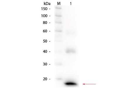
Western Blot: IL-1 beta/IL-1F2 Antibody [Biotin] [NBP1-42768] - Lane 1: Mouse IL-1Beta Recombinant Protein. Load: 50 ng per lane. Primary antibody: Rabbit anti-Mouse IL-1Beta Antibody Biotin Conjugated at 1:1,000 overnight at 4C. Secondary antibody: HRP streptavidin secondary antibody at 1:40,000 for 30 min at RT. Block: incubated with blocking buffer for 30 minutes at RT. Predicted/Observed size: 17.5 kDa, 17.5 kDa for proteolytically processed Mouse Il-1Beta.

Western Blot: IL-1 beta/IL-1F2 Antibody [Biotin] [NBP1-42768] - Lane 1: Mouse IL-1 beta Recombinant Protein. Load: 50 ng per lane. Used at a dilution of 1:1,000 overnight at 4C.
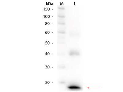
Western Blot: IL-1 beta/IL-1F2 Antibody [Biotin] [NBP1-42768] - Lane 1: Mouse IL-1Beta Recombinant Protein. Load: 50 ng per lane. Primary antibody: Rabbit anti-Mouse IL-1Beta Antibody Biotin Conjugated at 1:1,000 overnight at 4C. Secondary antibody: HRP streptavidin secondary antibody at 1:40,000 for 30 min at RT. Block: incubated with blocking buffer for 30 minutes at RT. Predicted/Observed size: 17.5 kDa, 17.5 kDa for proteolytically processed Mouse Il-1Beta.
Applications for IL-1 beta/IL-1F2 Antibody [Biotin]
Application
Recommended Usage
ELISA
1:20000-1:100000
Flow Cytometry
1:10-1:1000
Immunohistochemistry
1:1000-1:5000
Immunohistochemistry-Paraffin
1:10-1:500
Western Blot
1:2000 - 1:10000
Application Notes
This product has been tested in dot blot and western blot and is suitable for use in neutralizations, ELISA, radioimmunoassays, flow cytometry, immunohistochemistry, and immunoprecipitation. It recognizes the 17,000 MW mature IL-1beta. For immunoblots, typically, IL-1beta is detected from supernatants or lysates of 2 x 10E6 endotoxin-stimulated peripheral blood mononuclear cells (PBMC). PBMC are stimulated for 24 hours with 1% (v/v) serum plus 10 ng/mL E.coli LPS. For immunoprecipitation pre-clearing the preparation with a non-specific Rabbit IgG to reduce background is suggested. For immunohistochemistry either paraffin fixation or cryofixation can be used for sample preparation to stain intracellular IL-1beta. For ELISA use HRP Conjugated Anti-Rabbit IgG [H&L] (Goat) (611-1302) for detection. In ELISA formats this antibody is best used as the second antibody in combination with a monoclonal antibody as a capture antibody. This antibody is also useful for neutralization of mouse and rat IL-1beta activity in bioassays. It does not neutralize the biological activity IL-1alpha. It does not neutralize the biological activity of human or primate IL-1beta. For neutralization, it is recommended to incubate the sample with a dilution of the antibody for at least 4 hours before being tested. A control of similarly diluted normal rabbit IgG is recommended. This antibody can be used for FACS analysis. Caution should be exhibited as the F( c) domain of the rabbit IgG molecule may interact with cells non-specifically.
Reviewed Applications
Read 1 review rated 5 using NBP1-42768 in the following applications:
Flow Cytometry Panel Builder
Bio-Techne Knows Flow Cytometry
Save time and reduce costly mistakes by quickly finding compatible reagents using the Panel Builder Tool.
Advanced Features
- Spectra Viewer - Custom analysis of spectra from multiple fluorochromes
- Spillover Popups - Visualize the spectra of individual fluorochromes
- Antigen Density Selector - Match fluorochrome brightness with antigen density

Formulation, Preparation, and Storage
Purification
Column Chromatography
Reconstitution
Reconstitute with 100 ul deionized water (or equivalent)
Formulation
Lyophilized from 0.02 M Potassium Phosphate, 0.15 M Sodium Chloride, pH 7.2, 10 mg/mL Bovine Serum Albumin (BSA) - Immunoglobulin and Protease free
Preservative
0.01% Sodium Azide
Concentration
LYOPH mg/ml
Shipping
The product is shipped with polar packs. Upon receipt, store it immediately at the temperature recommended below.
Stability & Storage
Store lyophilized antibody at 4C in the dark. Aliquot reconstituted liquid and store at -20C. Avoid freeze-thaw cycles.
Calculators
Background: IL-1 beta/IL-1F2
IL-1 beta binding to its receptor IL-1RI and the downstream signaling contributes to a dual pathophysiological role (3). On one hand, IL-1 beta signaling activates immune cells and drives CD4+ T cell polarization to T helper type 1 (Th1) and Th17 cells, resulting in anti-tumor responses and mediation of acute inflammation (2,3). However, IL-1 beta also supports tumor growth and metastasis driven by multiple mechanisms including chronic inflammation, an immunosuppressive tumor microenvironment (TME), and angiogenesis (3). Additionally, IL-1 beta signaling been implicated in the pathogenesis of neuroinflammatory diseases of the central nervous system (CNS) such as multiple sclerosis (MS), Alzheimer's disease, and diabetic retinopathy (DR) (2). Mouse studies have shown regression of tumors treated with IL-1 as well as protective effects of IL-1 beta in instances of induced colitis and colon carcinoma (3). Conversely, blocking IL-1 beta has also shown promising effect in cancer treatment, especially when combined with chemotherapeutics (2,3). Approved IL-1 beta monoclonal antibody canakinumab has shown significant therapeutic promise in the treatment of DR (2). Given its multifaceted role in disease, IL-1 beta is a promising therapeutic target.
References
1. Lopez-Castejon G, Brough D. Understanding the mechanism of IL-1beta secretion. Cytokine Growth Factor Rev. 2011;22(4):189-195. https://doi.org/10.1016/j.cytogfr.2011.10.001
2. Mendiola AS, Cardona AE. The IL-1beta phenomena in neuroinflammatory diseases. J Neural Transm (Vienna). 2018;125(5):781-795. https://doi.org/10.1007/s00702-017-1732-9
3. Bent R, Moll L, Grabbe S, Bros M. Interleukin-1 Beta-A Friend or Foe in Malignancies?. Int J Mol Sci. 2018;19(8):2155. https://doi.org/doi:10.3390/ijms19082155
4. Krumm B, Xiang Y, Deng J. Structural biology of the IL-1 superfamily: key cytokines in the regulation of immune and inflammatory responses. Protein Sci. 2014;23(5):526-538. https://doi.org/10.1002/pro.2441
5. He Y, Hara H, Nunez G. Mechanism and Regulation of NLRP3 Inflammasome Activation. Trends Biochem Sci. 2016;41(12):1012-1021. https://doi.org/10.1016/j.tibs.2016.09.002
6. Uniprot (P01584)
Long Name
Interleukin 1 beta
Alternate Names
IL-1b, IL-1F2, IL1 beta, IL1B
Gene Symbol
IL1B
UniProt
Additional IL-1 beta/IL-1F2 Products
Product Documents for IL-1 beta/IL-1F2 Antibody [Biotin]
Certificate of Analysis
To download a Certificate of Analysis, please enter a lot or batch number in the search box below.
Product Specific Notices for IL-1 beta/IL-1F2 Antibody [Biotin]
This product is for research use only and is not approved for use in humans or in clinical diagnosis. Primary Antibodies are guaranteed for 1 year from date of receipt.
Related Research Areas
Citations for IL-1 beta/IL-1F2 Antibody [Biotin]
 Powered by Bioz
Customer Reviews for IL-1 beta/IL-1F2 Antibody [Biotin] (1)
5 out of 5
1 Customer Rating
Have you used IL-1 beta/IL-1F2 Antibody [Biotin]?
Submit a review and receive an Amazon gift card!
$25/€18/£15/$25CAN/¥2500 Yen for a review with an image
$10/€7/£6/$10CAN/¥1110 Yen for a review without an image
Submit a review
Customer Images
![IL-1 beta/IL-1F2 Antibody [Biotin] NBP1-42768](https://resources.rndsystems.com/images/reviews/review_nbp1-42768_29871.png)
Showing
1
-
1 of
1 review
Showing All
Filter By:
-
Application: Flow CytometrySample Tested: Bone marrod derived cellsSpecies: MouseVerified Customer | Posted 02/27/2017IL1b expression in stimulated BMDM-derived monocytes.Single cell suspension was blocked with Fc block for 30 minutes, prior to the permeabilization with BD cytoperm/fix kit for an additional of 30 minutes. Cells were then washed and incubated with the biotinylated-conjugated IL1b antibody (1:100 in PERM buffer) for 30 minutes on ice. Cells were then briefly washed and analyzed by flow cytometry.
![IL-1 beta/IL-1F2 Antibody [Biotin] NBP1-42768](data:image/png;base64,R0lGODlhAQABAAD/ACwAAAAAAQABAAACADs=)
There are no reviews that match your criteria.
Protocols
Find general support by application which include: protocols, troubleshooting, illustrated assays, videos and webinars.
- 7-Amino Actinomycin D (7-AAD) Cell Viability Flow Cytometry Protocol
- Antigen Retrieval Protocol (PIER)
- Antigen Retrieval for Frozen Sections Protocol
- Appropriate Fixation of IHC/ICC Samples
- Cellular Response to Hypoxia Protocols
- Chromogenic IHC Staining of Formalin-Fixed Paraffin-Embedded (FFPE) Tissue Protocol
- Chromogenic Immunohistochemistry Staining of Frozen Tissue
- ClariTSA™ Fluorophore Kits
- Detection & Visualization of Antibody Binding
- ELISA Sample Preparation & Collection Guide
- ELISA Troubleshooting Guide
- Extracellular Membrane Flow Cytometry Protocol
- Flow Cytometry Protocol for Cell Surface Markers
- Flow Cytometry Protocol for Staining Membrane Associated Proteins
- Flow Cytometry Staining Protocols
- Flow Cytometry Troubleshooting Guide
- Fluorescent IHC Staining of Frozen Tissue Protocol
- Graphic Protocol for Heat-induced Epitope Retrieval
- Graphic Protocol for the Preparation and Fluorescent IHC Staining of Frozen Tissue Sections
- Graphic Protocol for the Preparation and Fluorescent IHC Staining of Paraffin-embedded Tissue Sections
- Graphic Protocol for the Preparation of Gelatin-coated Slides for Histological Tissue Sections
- How to Run an R&D Systems DuoSet ELISA
- How to Run an R&D Systems Quantikine ELISA
- How to Run an R&D Systems Quantikine™ QuicKit™ ELISA
- IHC Sample Preparation (Frozen sections vs Paraffin)
- Immunofluorescent IHC Staining of Formalin-Fixed Paraffin-Embedded (FFPE) Tissue Protocol
- Immunohistochemistry (IHC) and Immunocytochemistry (ICC) Protocols
- Immunohistochemistry Frozen Troubleshooting
- Immunohistochemistry Paraffin Troubleshooting
- Intracellular Flow Cytometry Protocol Using Alcohol (Methanol)
- Intracellular Flow Cytometry Protocol Using Detergents
- Intracellular Nuclear Staining Flow Cytometry Protocol Using Detergents
- Intracellular Staining Flow Cytometry Protocol Using Alcohol Permeabilization
- Intracellular Staining Flow Cytometry Protocol Using Detergents to Permeabilize Cells
- Preparing Samples for IHC/ICC Experiments
- Preventing Non-Specific Staining (Non-Specific Binding)
- Primary Antibody Selection & Optimization
- Propidium Iodide Cell Viability Flow Cytometry Protocol
- Protocol for Heat-Induced Epitope Retrieval (HIER)
- Protocol for Liperfluo
- Protocol for Making a 4% Formaldehyde Solution in PBS
- Protocol for VisUCyte™ HRP Polymer Detection Reagent
- Protocol for the Characterization of Human Th22 Cells
- Protocol for the Characterization of Human Th9 Cells
- Protocol for the Preparation & Fixation of Cells on Coverslips
- Protocol for the Preparation and Chromogenic IHC Staining of Frozen Tissue Sections
- Protocol for the Preparation and Chromogenic IHC Staining of Frozen Tissue Sections - Graphic
- Protocol for the Preparation and Chromogenic IHC Staining of Paraffin-embedded Tissue Sections
- Protocol for the Preparation and Chromogenic IHC Staining of Paraffin-embedded Tissue Sections - Graphic
- Protocol for the Preparation and Fluorescent IHC Staining of Frozen Tissue Sections
- Protocol for the Preparation and Fluorescent IHC Staining of Paraffin-embedded Tissue Sections
- Protocol for the Preparation of Gelatin-coated Slides for Histological Tissue Sections
- Protocol: Annexin V and PI Staining by Flow Cytometry
- Protocol: Annexin V and PI Staining for Apoptosis by Flow Cytometry
- Quantikine HS ELISA Kit Assay Principle, Alkaline Phosphatase
- Quantikine HS ELISA Kit Principle, Streptavidin-HRP Polymer
- R&D Systems Quality Control Western Blot Protocol
- Sandwich ELISA (Colorimetric) – Biotin/Streptavidin Detection Protocol
- Sandwich ELISA (Colorimetric) – Direct Detection Protocol
- TUNEL and Active Caspase-3 Detection by IHC/ICC Protocol
- The Importance of IHC/ICC Controls
- Troubleshooting Guide: ELISA
- Troubleshooting Guide: Fluorokine Flow Cytometry Kits
- Troubleshooting Guide: Immunohistochemistry
- Troubleshooting Guide: Western Blot Figures
- Western Blot Conditions
- Western Blot Protocol
- Western Blot Protocol for Cell Lysates
- Western Blot Troubleshooting
- Western Blot Troubleshooting Guide
- View all Protocols, Troubleshooting, Illustrated assays and Webinars
FAQs for IL-1 beta/IL-1F2 Antibody [Biotin]
Showing
1
-
1 of
1 FAQ
Showing All
-
Q: I bought the product NBP1-42768 and I'm not sure about the reconstitution and the concentration. I have 100ug, so if I add 50ul, I will have a concentration of 2mg/ml, right?
A: Centrifuge vial prior to adding liquid. Add 100ul of deionized water and resuspend. The final concentration will be 1mg/ml.
Loading...
Associated Pathways


Innate Lymphoid Cell Differentiation Pathways
 NOD-like Receptor Signaling Pathways
 Th17 Differentiation Pathway
 Toll-Like Receptor Signaling Pathways
