Jak1 [p Tyr1022, p Tyr1023] Antibody - BSA Free
Novus Biologicals | Catalog # NBP3-05436
Loading...
Key Product Details
Validated by
Biological Validation
Species Reactivity
Human, Mouse
Applications
Western Blot, Immunocytochemistry/ Immunofluorescence
Label
Unconjugated
Antibody Source
Polyclonal Rabbit IgG
Format
BSA Free
Loading...
Product Specifications
Immunogen
A synthetic phosphorylated peptide around Y1022 & Y1023 of human JAK1 (NP_002218.2). KEYYTV
Modification
p Tyr1022, p Tyr1023
Clonality
Polyclonal
Host
Rabbit
Isotype
IgG
Theoretical MW
133 kDa.
Disclaimer note: The observed molecular weight of the protein may vary from the listed predicted molecular weight due to post translational modifications, post translation cleavages, relative charges, and other experimental factors.
Disclaimer note: The observed molecular weight of the protein may vary from the listed predicted molecular weight due to post translational modifications, post translation cleavages, relative charges, and other experimental factors.
Scientific Data Images for Jak1 [p Tyr1022, p Tyr1023] Antibody - BSA Free
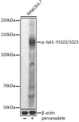
Western Blot: Jak1 [p Tyr1022, p Tyr1023] Antibody [NBP3-05436] - Western blot analysis of extracts of RAW264.7 cells, using Jak1 antibody (NBP3-05436) at 1:1000 dilution.RAW264.7 cells were treated by Pervanadate (1 mM) at 37C for 30 minutes. Secondary antibody: HRP Goat Anti-Rabbit IgG (H+L) at 1:10000 dilution. Lysates/proteins: 25ug per lane. Blocking buffer: 3% nonfat dry milk in TBST. Detection: ECL Basic Kit. Exposure time: 10s.
![Immunocytochemistry/ Immunofluorescence: Jak1 [p Tyr1022, p Tyr1023] Antibody - BSA Free [NBP3-05436]](https://resources.rndsystems.com/images/products/Jak1-[p-Tyr1022--p-Tyr1023]-Antibody-Immunocytochemistry-Immunofluorescence-NBP3-05436-img0001.jpg "Immunocytochemistry/ Immunofluorescence: Jak1 [p Tyr1022, p Tyr1023] Antibody - BSA Free [NBP3-05436]")
Immunocytochemistry/ Immunofluorescence: Jak1 [p Tyr1022, p Tyr1023] Antibody - BSA Free [NBP3-05436]
Immunocytochemistry/Immunofluorescence: Jak1 [p Tyr1022, p Tyr1023] Antibody [NBP3-05436] - Immunofluorescence analysis of NIH/3T3 cells using Jak1 antibody (NBP3-05436) at dilution of 1:100. Blue: DAPI for nuclear staining.Applications for Jak1 [p Tyr1022, p Tyr1023] Antibody - BSA Free
Application
Recommended Usage
Immunocytochemistry/ Immunofluorescence
1:50 - 1:200
Western Blot
1:500 - 1:1000
Formulation, Preparation, and Storage
Purification
Affinity purified
Formulation
PBS (pH 7.3), 50% glycerol
Format
BSA Free
Preservative
0.02% Sodium Azide
Concentration
Please see the vial label for concentration. If unlisted please contact technical services.
Shipping
The product is shipped with polar packs. Upon receipt, store it immediately at the temperature recommended below.
Stability & Storage
Store at -20C. Avoid freeze-thaw cycles.
Background: Jak1
Long Name
Janus Kinase 1
Alternate Names
EC 2.7.10, JAK-1, JAK1Atyrosine-protein kinase JAK1, JAK1B, Janus kinase 1EC 2.7.10.2, JTK3
Gene Symbol
JAK1
Additional Jak1 Products
Product Documents for Jak1 [p Tyr1022, p Tyr1023] Antibody - BSA Free
Certificate of Analysis
To download a Certificate of Analysis, please enter a lot or batch number in the search box below.
Product Specific Notices for Jak1 [p Tyr1022, p Tyr1023] Antibody - BSA Free
This product is for research use only and is not approved for use in humans or in clinical diagnosis. Primary Antibodies are guaranteed for 1 year from date of receipt.
Customer Reviews for Jak1 [p Tyr1022, p Tyr1023] Antibody - BSA Free
There are currently no reviews for this product. Be the first to review Jak1 [p Tyr1022, p Tyr1023] Antibody - BSA Free and earn rewards!
Have you used Jak1 [p Tyr1022, p Tyr1023] Antibody - BSA Free?
Submit a review and receive an Amazon gift card!
$25/€18/£15/$25CAN/¥2500 Yen for a review with an image
$10/€7/£6/$10CAN/¥1110 Yen for a review without an image
Submit a review
Protocols
Find general support by application which include: protocols, troubleshooting, illustrated assays, videos and webinars.
- Appropriate Fixation of IHC/ICC Samples
- Cellular Response to Hypoxia Protocols
- ClariTSA™ Fluorophore Kits
- Detection & Visualization of Antibody Binding
- ICC Cell Smear Protocol for Suspension Cells
- ICC Immunocytochemistry Protocol Videos
- ICC for Adherent Cells
- Immunocytochemistry (ICC) Protocol
- Immunocytochemistry Troubleshooting
- Immunofluorescence of Organoids Embedded in Cultrex Basement Membrane Extract
- Immunohistochemistry (IHC) and Immunocytochemistry (ICC) Protocols
- Preparing Samples for IHC/ICC Experiments
- Preventing Non-Specific Staining (Non-Specific Binding)
- Primary Antibody Selection & Optimization
- Protocol for VisUCyte™ HRP Polymer Detection Reagent
- Protocol for the Fluorescent ICC Staining of Cell Smears - Graphic
- Protocol for the Fluorescent ICC Staining of Cultured Cells on Coverslips - Graphic
- Protocol for the Preparation and Fluorescent ICC Staining of Cells on Coverslips
- Protocol for the Preparation and Fluorescent ICC Staining of Non-adherent Cells
- Protocol for the Preparation and Fluorescent ICC Staining of Stem Cells on Coverslips
- Protocol for the Preparation of a Cell Smear for Non-adherent Cell ICC - Graphic
- R&D Systems Quality Control Western Blot Protocol
- TUNEL and Active Caspase-3 Detection by IHC/ICC Protocol
- The Importance of IHC/ICC Controls
- Troubleshooting Guide: Western Blot Figures
- Western Blot Conditions
- Western Blot Protocol
- Western Blot Protocol for Cell Lysates
- Western Blot Troubleshooting
- Western Blot Troubleshooting Guide
- View all Protocols, Troubleshooting, Illustrated assays and Webinars
Loading...
Associated Pathways


IL-15 Signaling Pathways
 IL-21 Signaling Pathways
 Jak/STAT Signaling Pathway
 Th1 Differentiation Pathway
 Th2 Differentiation Pathway
 Th17 Differentiation Pathway
 VEGF - VEGF R2 Signaling Pathways
