Key Product Details
Species Reactivity
Validated:
Human, Mouse, Rat
Cited:
Human
Applications
Validated:
Immunohistochemistry, Immunohistochemistry-Paraffin, Western Blot, Immunocytochemistry/ Immunofluorescence
Cited:
Western Blot
Label
Unconjugated
Antibody Source
Monoclonal Mouse IgG1 Clone # OTI1F5
Loading...
Product Specifications
Immunogen
Full-length protein expressed in 293T cell transfected with human MAP2K1 expression vector
Reactivity Notes
Please note that this antibody is reactive to Mouse and derived from the same host, Mouse. Mouse-On-Mouse blocking reagent may be needed for IHC and ICC experiments to reduce high background signal. You can find these reagents under catalog numbers PK-2200-NB and MP-2400-NB. Please contact Technical Support if you have any questions.
Specificity
This antibody is specific for Homo sapiens mitogen-activated protein kinase kinase 1 (MAP2K1).
Clonality
Monoclonal
Host
Mouse
Isotype
IgG1
Theoretical MW
43.4 kDa.
Disclaimer note: The observed molecular weight of the protein may vary from the listed predicted molecular weight due to post translational modifications, post translation cleavages, relative charges, and other experimental factors.
Disclaimer note: The observed molecular weight of the protein may vary from the listed predicted molecular weight due to post translational modifications, post translation cleavages, relative charges, and other experimental factors.
Scientific Data Images for MEK1 Antibody (OTI1F5)
![Western Blot: MEK1 Antibody (OTI1F5) [NBP1-47833]](https://resources.rndsystems.com/images/products/MEK1-Antibody-1F5-Western-Blot-NBP1-47833-img0001.jpg "Western Blot: MEK1 Antibody (OTI1F5) [NBP1-47833]")
Western Blot: MEK1 Antibody (OTI1F5) [NBP1-47833]
Western Blot: MEK1 Antibody (1F5) [NBP1-47833] - HEK293T cells were transfected with the pCMV6-ENTRY control (Left lane) or pCMV6-ENTRY MEK1(Right lane) cDNA for 48 hrs and lysed. Equivalent amounts of cell lysates (5 ug per lane) were separated by SDS-PAGE and immunoblotted with anti-MEK1.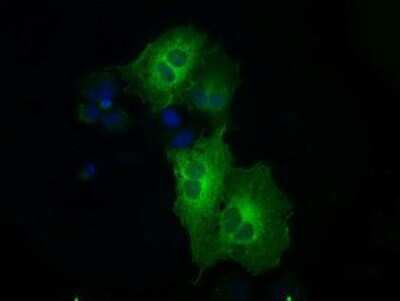
Immunocytochemistry/Immunofluorescence: MEK1 Antibody (1F5) [NBP1-47833] Staining of COS7 cells transiently transfected by pCMV6-ENTRY MEK1.
![Immunohistochemistry-Paraffin: MEK1 Antibody (OTI1F5) [NBP1-47833]](https://resources.rndsystems.com/images/products/MEK1-Antibody-1F5-Immunohistochemistry-Paraffin-NBP1-47833-img0008.jpg "Immunohistochemistry-Paraffin: MEK1 Antibody (OTI1F5) [NBP1-47833]")
Immunohistochemistry-Paraffin: MEK1 Antibody (OTI1F5) [NBP1-47833]
Immunohistochemistry-Paraffin: MEK1 Antibody (1F5) [NBP1-47833] - Staining of paraffin-embedded prostate tissue using anti-MEK1 mouse monoclonal antibody.![Immunohistochemistry-Paraffin: MEK1 Antibody (OTI1F5) [NBP1-47833]](https://resources.rndsystems.com/images/products/MEK1-Antibody-1F5-Immunohistochemistry-Paraffin-NBP1-47833-img0002.jpg "Immunohistochemistry-Paraffin: MEK1 Antibody (OTI1F5) [NBP1-47833]")
Immunohistochemistry-Paraffin: MEK1 Antibody (OTI1F5) [NBP1-47833]
Immunohistochemistry-Paraffin: MEK1 Antibody (1F5) [NBP1-47833] - Staining of paraffin-embedded Adenocarcinoma of colon tissue using anti-MEK1 mouse monoclonal antibody.![Immunohistochemistry-Paraffin: MEK1 Antibody (OTI1F5) [NBP1-47833]](https://resources.rndsystems.com/images/products/MEK1-Antibody-1F5-Immunohistochemistry-Paraffin-NBP1-47833-img0003.jpg "Immunohistochemistry-Paraffin: MEK1 Antibody (OTI1F5) [NBP1-47833]")
Immunohistochemistry-Paraffin: MEK1 Antibody (OTI1F5) [NBP1-47833]
Immunohistochemistry-Paraffin: MEK1 Antibody (1F5) [NBP1-47833] - Staining of paraffin-embedded Carcinoma of bladder tissue using anti-MEK1 mouse monoclonal antibody.![Immunohistochemistry-Paraffin: MEK1 Antibody (OTI1F5) [NBP1-47833]](https://resources.rndsystems.com/images/products/MEK1-Antibody-1F5-Immunohistochemistry-Paraffin-NBP1-47833-img0004.jpg "Immunohistochemistry-Paraffin: MEK1 Antibody (OTI1F5) [NBP1-47833]")
Immunohistochemistry-Paraffin: MEK1 Antibody (OTI1F5) [NBP1-47833]
Immunohistochemistry-Paraffin: MEK1 Antibody (1F5) [NBP1-47833] - Staining of paraffin-embedded colon tissue using anti-MEK1 mouse monoclonal antibody.![Immunohistochemistry-Paraffin: MEK1 Antibody (OTI1F5) [NBP1-47833]](https://resources.rndsystems.com/images/products/MEK1-Antibody-1F5-Immunohistochemistry-Paraffin-NBP1-47833-img0005.jpg "Immunohistochemistry-Paraffin: MEK1 Antibody (OTI1F5) [NBP1-47833]")
Immunohistochemistry-Paraffin: MEK1 Antibody (OTI1F5) [NBP1-47833]
Immunohistochemistry-Paraffin: MEK1 Antibody (1F5) [NBP1-47833] - Staining of paraffin-embedded endometrium tissue using anti-MEK1 mouse monoclonal antibody.![Immunohistochemistry-Paraffin: MEK1 Antibody (OTI1F5) [NBP1-47833]](https://resources.rndsystems.com/images/products/MEK1-Antibody-1F5-Immunohistochemistry-Paraffin-NBP1-47833-img0006.jpg "Immunohistochemistry-Paraffin: MEK1 Antibody (OTI1F5) [NBP1-47833]")
Immunohistochemistry-Paraffin: MEK1 Antibody (OTI1F5) [NBP1-47833]
Immunohistochemistry-Paraffin: MEK1 Antibody (1F5) [NBP1-47833] - Staining of paraffin-embedded Ovary tissue using anti-MEK1 mouse monoclonal antibody.![Immunohistochemistry-Paraffin: MEK1 Antibody (OTI1F5) [NBP1-47833]](https://resources.rndsystems.com/images/products/MEK1-Antibody-1F5-Immunohistochemistry-Paraffin-NBP1-47833-img0007.jpg "Immunohistochemistry-Paraffin: MEK1 Antibody (OTI1F5) [NBP1-47833]")
Immunohistochemistry-Paraffin: MEK1 Antibody (OTI1F5) [NBP1-47833]
Immunohistochemistry-Paraffin: MEK1 Antibody (1F5) [NBP1-47833] - Staining of paraffin-embedded pancreas tissue using anti-MEK1mouse monoclonal antibody.Applications for MEK1 Antibody (OTI1F5)
Application
Recommended Usage
Immunocytochemistry/ Immunofluorescence
1:100
Immunohistochemistry
1:10-1:500
Immunohistochemistry-Paraffin
1:50
Western Blot
1:2000
Reviewed Applications
Read 1 review rated 5 using NBP1-47833 in the following applications:
Formulation, Preparation, and Storage
Purification
Immunogen affinity purified
Formulation
PBS (pH 7.3), 1.0% BSA and 50% Glycerol
Preservative
0.02% Sodium Azide
Concentration
0.99 mg/ml
Shipping
The product is shipped with polar packs. Upon receipt, store it immediately at the temperature recommended below.
Stability & Storage
Store at -20C. Avoid freeze-thaw cycles.
Background: MEK1
Long Name
Mitogen-activated Protein Kinase Kinase 1
Alternate Names
MAP2K1, MKK1
Entrez Gene IDs
5604 (Human)
Gene Symbol
MAP2K1
Additional MEK1 Products
Product Documents for MEK1 Antibody (OTI1F5)
Certificate of Analysis
To download a Certificate of Analysis, please enter a lot or batch number in the search box below.
Product Specific Notices for MEK1 Antibody (OTI1F5)
This product is for research use only and is not approved for use in humans or in clinical diagnosis. Primary Antibodies are guaranteed for 1 year from date of receipt.
Related Research Areas
Citations for MEK1 Antibody (OTI1F5)
 Powered by Bioz
Customer Reviews for MEK1 Antibody (OTI1F5) (1)
5 out of 5
1 Customer Rating
Have you used MEK1 Antibody (OTI1F5)?
Submit a review and receive an Amazon gift card!
$25/€18/£15/$25CAN/¥2500 Yen for a review with an image
$10/€7/£6/$10CAN/¥1110 Yen for a review without an image
Submit a review
Customer Images
Showing
1
-
1 of
1 review
Showing All
Filter By:
-
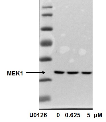
Application: Western BlotSample Tested: Human cancer cell whole cell lysateSpecies: OtherVerified Customer | Posted 10/04/2015MEK1 expression in MDA-MB-231 cells with U0126 treatment

There are no reviews that match your criteria.
Protocols
Find general support by application which include: protocols, troubleshooting, illustrated assays, videos and webinars.
- Antigen Retrieval Protocol (PIER)
- Antigen Retrieval for Frozen Sections Protocol
- Appropriate Fixation of IHC/ICC Samples
- Cellular Response to Hypoxia Protocols
- Chromogenic IHC Staining of Formalin-Fixed Paraffin-Embedded (FFPE) Tissue Protocol
- Chromogenic Immunohistochemistry Staining of Frozen Tissue
- ClariTSA™ Fluorophore Kits
- Detection & Visualization of Antibody Binding
- Fluorescent IHC Staining of Frozen Tissue Protocol
- Graphic Protocol for Heat-induced Epitope Retrieval
- Graphic Protocol for the Preparation and Fluorescent IHC Staining of Frozen Tissue Sections
- Graphic Protocol for the Preparation and Fluorescent IHC Staining of Paraffin-embedded Tissue Sections
- Graphic Protocol for the Preparation of Gelatin-coated Slides for Histological Tissue Sections
- ICC Cell Smear Protocol for Suspension Cells
- ICC Immunocytochemistry Protocol Videos
- ICC for Adherent Cells
- IHC Sample Preparation (Frozen sections vs Paraffin)
- Immunocytochemistry (ICC) Protocol
- Immunocytochemistry Troubleshooting
- Immunofluorescence of Organoids Embedded in Cultrex Basement Membrane Extract
- Immunofluorescent IHC Staining of Formalin-Fixed Paraffin-Embedded (FFPE) Tissue Protocol
- Immunohistochemistry (IHC) and Immunocytochemistry (ICC) Protocols
- Immunohistochemistry Frozen Troubleshooting
- Immunohistochemistry Paraffin Troubleshooting
- Preparing Samples for IHC/ICC Experiments
- Preventing Non-Specific Staining (Non-Specific Binding)
- Primary Antibody Selection & Optimization
- Protocol for Heat-Induced Epitope Retrieval (HIER)
- Protocol for Making a 4% Formaldehyde Solution in PBS
- Protocol for VisUCyte™ HRP Polymer Detection Reagent
- Protocol for the Fluorescent ICC Staining of Cell Smears - Graphic
- Protocol for the Fluorescent ICC Staining of Cultured Cells on Coverslips - Graphic
- Protocol for the Preparation & Fixation of Cells on Coverslips
- Protocol for the Preparation and Chromogenic IHC Staining of Frozen Tissue Sections
- Protocol for the Preparation and Chromogenic IHC Staining of Frozen Tissue Sections - Graphic
- Protocol for the Preparation and Chromogenic IHC Staining of Paraffin-embedded Tissue Sections
- Protocol for the Preparation and Chromogenic IHC Staining of Paraffin-embedded Tissue Sections - Graphic
- Protocol for the Preparation and Fluorescent ICC Staining of Cells on Coverslips
- Protocol for the Preparation and Fluorescent ICC Staining of Non-adherent Cells
- Protocol for the Preparation and Fluorescent ICC Staining of Stem Cells on Coverslips
- Protocol for the Preparation and Fluorescent IHC Staining of Frozen Tissue Sections
- Protocol for the Preparation and Fluorescent IHC Staining of Paraffin-embedded Tissue Sections
- Protocol for the Preparation of Gelatin-coated Slides for Histological Tissue Sections
- Protocol for the Preparation of a Cell Smear for Non-adherent Cell ICC - Graphic
- R&D Systems Quality Control Western Blot Protocol
- TUNEL and Active Caspase-3 Detection by IHC/ICC Protocol
- The Importance of IHC/ICC Controls
- Troubleshooting Guide: Immunohistochemistry
- Troubleshooting Guide: Western Blot Figures
- Western Blot Conditions
- Western Blot Protocol
- Western Blot Protocol for Cell Lysates
- Western Blot Troubleshooting
- Western Blot Troubleshooting Guide
- View all Protocols, Troubleshooting, Illustrated assays and Webinars
Loading...
Associated Pathways


IL-9 Signaling Pathways
 IL-15 Signaling Pathways
 IL-21 Signaling Pathways
 MAPK Signaling: Oxidative Stress Pathway
 MAPK Signaling Pathway: Mitogen Stimulation Pathway
 Pathogen or Damage-activated C-Type Lectin Receptor Signaling Pathways
 TGF-beta Signaling Pathways
 VEGF - VEGF R2 Signaling Pathways
