Several distinct genes encode alkaline phosphatases (APs) in mice with different tissue-specific expression patterns. The Alpl gene, also known as Akp2, encodes the liver/bone/kidney isozyme, also known as the tissue-nonspecific AP (TNAP) (1). The Alpl gene is a key regulator of bone mineralization in mice (2). A variety of mutations in the human ALPL gene leads to different forms of hypophosphatasia, characterized by poorly mineralized cartilage and bones (3). The native ALPL is a glycosylated homodimer attached to the membrane through a GPI-anchor. The C-terminal pro peptide (residues 504 to 524) is not present in the mature form.
Mouse Alkaline Phosphatase/ALPL Antibody
R&D Systems | Catalog # AF2910

Key Product Details
Species Reactivity
Validated:
Mouse
Cited:
Human, Mouse, Rat, Transgenic Mouse
Applications
Validated:
Immunohistochemistry, Western Blot, Flow Cytometry, Immunocytochemistry, Immunoprecipitation, CyTOF-ready
Cited:
Immunohistochemistry, Immunohistochemistry-Paraffin, Immunohistochemistry-Frozen, Western Blot, Flow Cytometry, Immunocytochemistry, CyTof
Label
Unconjugated
Antibody Source
Polyclonal Goat IgG
Loading...
Product Specifications
Immunogen
Mouse myeloma cell line NS0-derived recombinant mouse Alkaline Phosphatase/ALPL
Phe18-Gly503
Accession # P09242
Phe18-Gly503
Accession # P09242
Specificity
Detects mouse Alkaline Phosphatase/ALPL in direct ELISAs and Western blots.
Clonality
Polyclonal
Host
Goat
Isotype
IgG
Scientific Data Images for Mouse Alkaline Phosphatase/ALPL Antibody
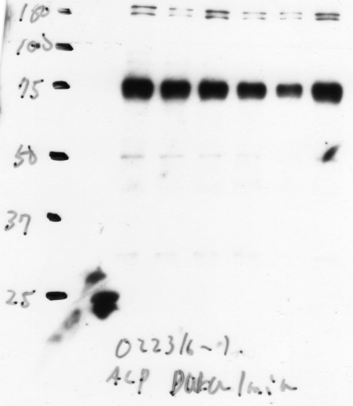
Detection of Mouse Alkaline Phosphatase/ALPL by Western Blot.
Western blot shows lysates of D3 mouse embryonic stem cell line, MEF mouse embryonic feeder cells, and mouse embryo tissue. PVDF membrane was probed with 1 µg/mL of Goat Anti-Mouse Alkaline Phosphatase/ALPL Antigen Affinity-purified Polyclonal Antibody (Catalog # AF2910) followed by HRP-conjugated Anti-Goat IgG Secondary Antibody (Catalog # HAF109). Specific bands were detected for Alkaline Phosphatase/ALPL at approximately 75-80 kDa (as indicated). This experiment was conducted under reducing conditions and using Immunoblot Buffer Group 1.
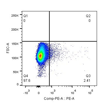
Detection of ALPL in Rat Stem Cells by Flow Cytometry.
Rat Mesenchymal Stem Cells were stained with Goat Anti-Mouse Alkaline Phosphatase/ALPL Antigen Affinity-purified Polyclonal Antibody (Catalog # AF2910, filled histogram) or isotype control antibody (Catalog # AB-108-C, open histogram), followed by Phycoerythrin-conjugated Anti-Goat IgG Secondary Antibody (Catalog # F0107).
Alkaline Phosphatase/ALPL in Rat Mesenchymal Stem Cells.
Alkaline Phosphatase/ALPL was detected in immersion fixed rat mesenchymal stem cells using Goat Anti-Mouse Alkaline Phosphatase/ALPL Antigen Affinity-purified Polyclonal Antibody (Catalog # AF2910) at 10 µg/mL for 3 hours at room temperature. Cells were stained using the NorthernLights™ 557-conjugated Anti-Goat IgG Secondary Antibody (red; Catalog # NL001) and counterstained with DAPI (blue). Specific staining was localized to cell surfaces and cytoplasm. View our protocol for Fluorescent ICC Staining of Cells on Coverslips.
Alkaline Phosphatase/ALPL in Mouse Embryo.
Alkaline Phosphatase/ALPL was detected in immersion fixed frozen sections of mouse embryonic (E13.5) developing vertebra using Goat Anti-Mouse Alkaline Phosphatase/ALPL Antigen Affinity-purified Poly-clonal Antibody (Catalog # AF2910) at 2 µg/mL overnight at 4 °C. Tissue was stained red and counterstained with DAPI (blue).Image courtesy of Paul J. Simmons, Ph.D., Director, Center for Stem Cell Research, The University of Texas Health Science Center at Houston, Houston, Texas, USA.View our protocol for Fluorescent IHC Staining of Frozen Tissue Sections.
Detection of Alkaline Phosphatase/ALPL by Western Blot
Downregulation of HDAC9 rescued lineage differentiation imbalance and ameliorated senescence in aged BMMSCs. a Alizarin Red staining was performed, and osteogenesis-related proteins were detected by western blotting in aged BMMSCs transfected with HDAC9 siRNA. b Oil Red O staining was performed, and adipogenesis-related proteins were detected by western blotting in aged BMMSCs transfected with HDAC9 siRNA. c Alizarin Red staining was performed, and osteogenic-related proteins were detected by western blotting in young BMMSCs and young BMMSCs transfected with HDAC9 siRNA. d Oil Red O staining was performed, and adipogenic-related proteins were detected by western blotting in young BMMSCs and young BMMSCs transfected with HDAC9 siRNA. e, f Expressions of the senescence-related proteins p53 and p-p53 in BMMSCs cultured in vitro from aged mice (e) and young mice (f) were examined by western blotting. The data are presented as the means ± SD of each independent experiment performed in triplicate. *P < 0.05, **P < 0.01, ***P < 0.001, one-way analysis of variance (ANOVA) Image collected and cropped by CiteAb from the following open publication (https://pubmed.ncbi.nlm.nih.gov/32620134), licensed under a CC-BY license. Not internally tested by R&D Systems.
Detection of Alkaline Phosphatase/ALPL by Western Blot
Inhibition of HDAC9 improved the lineage differentiation of endogenous BMMSCs ex vivo. The BMMSCs harvested from the mice from the control, shScr-treated, & shHDAC9-treated groups 4 weeks after bone intrainjection. e Alizarin Red staining was performed, & osteogenesis-related proteins detected in BMMSCs from the three group. Image collected & cropped by CiteAb from the following open publication (https://pubmed.ncbi.nlm.nih.gov/32620134), licensed under a CC-BY license. Not internally tested by R&D Systems.
Detection of Alkaline Phosphatase/ALPL by Western Blot
The representative images of Ki‐67 staining (A) and quantified by the positive‐stained percentage through ImageJ software (B). Scale bar, 100 μm. (C‐D) Alizarin Red staining was performed to detect mineralized nodules formed in Con, AS‐Exo and NS‐Exo 14 days after osteogenic induction and quantified with a spectrophotometer after dissolving with cetylpyridinium chloride. (E‐F) Lipid droplet formation was detected by Oil Red O staining 7 days after adipogenic induction with positive area quantified. Scale bar, 100 μm. (G‐H) The protein expression levels of Runx2, ALP and PPAR‐ gamma in Con, AS‐Exo and NS‐Exo groups were measured through Western blot and quantified by ImageJ software. n = 3 per group. Data are shown as mean ± SD; ns, not significant; *P < .05; **P < .01; ***P < .001. AS‐Exo, adult serum exosomes; Con, control; NS‐Exo, neonatal serum exosomes Image collected and cropped by CiteAb from the following open publication (https://pubmed.ncbi.nlm.nih.gov/32608556), licensed under a CC-BY license. Not internally tested by R&D Systems.
Detection of Alkaline Phosphatase/ALPL by Western Blot
Inhibition of autophagy blocked the ability of HDAC9 siRNA to rebalance BMMSC differentiation. To investigate the HDAC9-autophagy axis regulating BMMSC function, BMMSCs were respectively transfected with Nc siRNA and HDAC9 siRNA or co-transfected with HDAC9 siRNA and BECN1 siRNA. a Alizarin Red staining was performed, and osteogenesis-related proteins were analyzed in aged BMMSCs from above three groups. b Oil Red O staining was performed, and adipogenesis-related proteins were analyzed in three groups of cells described above. Scale bars = 100 μm. The data are presented as the means ± SD of each independent experiment performed in triplicate. *P < 0.05, **P < 0.01, one-way analysis of variance (ANOVA) Image collected and cropped by CiteAb from the following open publication (https://pubmed.ncbi.nlm.nih.gov/32620134), licensed under a CC-BY license. Not internally tested by R&D Systems.Applications for Mouse Alkaline Phosphatase/ALPL Antibody
Application
Recommended Usage
CyTOF-ready
Ready to be labeled using established conjugation methods. No BSA or other carrier proteins that could interfere with conjugation.
Flow Cytometry
2.5 µg/106 cells
Sample: Rat mesenchymal stem cells
Sample: Rat mesenchymal stem cells
Immunocytochemistry
5-15 µg/mL
Sample: Immersion fixed rat mesenchymal stem cells
Sample: Immersion fixed rat mesenchymal stem cells
Immunohistochemistry
5-15 µg/mL
Sample: Immersion fixed frozen sections of mouse embryonic (E13.5) developing vertebra
Sample: Immersion fixed frozen sections of mouse embryonic (E13.5) developing vertebra
Immunoprecipitation
25 µg/mL
Sample: Conditioned cell culture medium spiked with Recombinant Mouse Alkaline Phosphatase/ALPL (Catalog # 2910-AP), see our available Western blot detection antibodies
Sample: Conditioned cell culture medium spiked with Recombinant Mouse Alkaline Phosphatase/ALPL (Catalog # 2910-AP), see our available Western blot detection antibodies
Western Blot
1 µg/mL
Sample: D3 mouse embryonic stem cell line, MEF mouse embryonic feeder cells, and mouse embryo tissue
Sample: D3 mouse embryonic stem cell line, MEF mouse embryonic feeder cells, and mouse embryo tissue
Reviewed Applications
Read 3 reviews rated 5 using AF2910 in the following applications:
Flow Cytometry Panel Builder
Bio-Techne Knows Flow Cytometry
Save time and reduce costly mistakes by quickly finding compatible reagents using the Panel Builder Tool.
Advanced Features
- Spectra Viewer - Custom analysis of spectra from multiple fluorochromes
- Spillover Popups - Visualize the spectra of individual fluorochromes
- Antigen Density Selector - Match fluorochrome brightness with antigen density

Formulation, Preparation, and Storage
Purification
Antigen Affinity-purified
Reconstitution
Reconstitute at 0.2 mg/mL in sterile PBS. For liquid material, refer to CoA for concentration.
Loading...
Formulation
Lyophilized from a 0.2 μm filtered solution in PBS with Trehalose. *Small pack size (SP) is supplied either lyophilized or as a 0.2 µm filtered solution in PBS.
Shipping
Lyophilized product is shipped at ambient temperature. Liquid small pack size (-SP) is shipped with polar packs. Upon receipt, store immediately at the temperature recommended below.
Stability & Storage
Use a manual defrost freezer and avoid repeated freeze-thaw cycles.
- 12 months from date of receipt, -20 to -70 °C as supplied.
- 1 month, 2 to 8 °C under sterile conditions after reconstitution.
- 6 months, -20 to -70 °C under sterile conditions after reconstitution.
Calculators
Background: Alkaline Phosphatase/ALPL
References
- Terao, M. and B. Mintz (1987) Proc. Natl. Acad. Sci. USA 84:7051.
- Hessle, L. et al. (2002) Proc. Natl. Acad. Sci. USA 99:9445.
- Di Mauro, S. et al. (2002) J. Bone Miner. Res. 17:1383.
Long Name
Alkaline Phosphatase Liver
Alternate Names
Akp2, AP-TNAP, HOPS, TNAP, TNSALP
Gene Symbol
ALPL
UniProt
Additional Alkaline Phosphatase/ALPL Products
Product Documents for Mouse Alkaline Phosphatase/ALPL Antibody
Certificate of Analysis
To download a Certificate of Analysis, please enter a lot or batch number in the search box below.
Note: Certificate of Analysis not available for kit components.
Product Specific Notices for Mouse Alkaline Phosphatase/ALPL Antibody
For research use only
Related Research Areas
Citations for Mouse Alkaline Phosphatase/ALPL Antibody
 Powered by Bioz
Customer Reviews for Mouse Alkaline Phosphatase/ALPL Antibody (3)
5 out of 5
3 Customer Ratings
Have you used Mouse Alkaline Phosphatase/ALPL Antibody?
Submit a review and receive an Amazon gift card!
$25/€18/£15/$25CAN/¥2500 Yen for a review with an image
$10/€7/£6/$10CAN/¥1110 Yen for a review without an image
Submit a review
Customer Images
Showing
1
-
3 of
3 reviews
Showing All
Filter By:
-
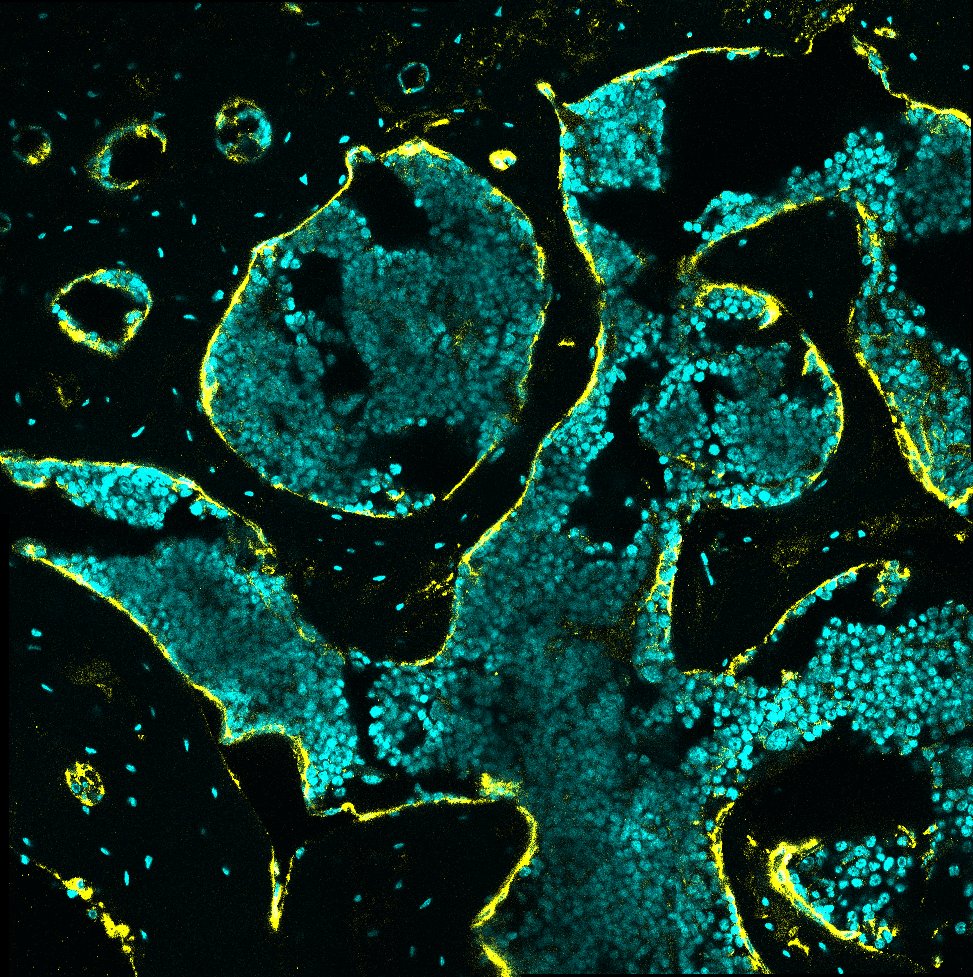
Application: Immunocytochemistry/ImmunofluorescenceSample Tested: bone marrowSpecies: MouseVerified Customer | Posted 09/15/2023Mouse tibiae slices stained with ALP (AF2910) at a 1:200 dilution + Alexa Fluor donkey-antigoat 594 1:200. DAPI (cyan) ALPL (yellow)

-
Application: ELISASample Tested: Bone marrow cellsSpecies: MouseVerified Customer | Posted 04/29/2021Mouse bone marrow cells were stained with Goat Anti-Mouse Alkaline Phosphatase/ALPL Antigen Affinity-purified Polyclonal Antibody or followed by Phycoerythrin-conjugated Anti-Goat IgG Secondary Antibody.
-
Application: Western BlotSample Tested: Differentiated osteoblastsSpecies: MouseVerified Customer | Posted 06/28/2016
There are no reviews that match your criteria.
Protocols
Find general support by application which include: protocols, troubleshooting, illustrated assays, videos and webinars.
- 7-Amino Actinomycin D (7-AAD) Cell Viability Flow Cytometry Protocol
- Antigen Retrieval Protocol (PIER)
- Antigen Retrieval for Frozen Sections Protocol
- Appropriate Fixation of IHC/ICC Samples
- Cellular Response to Hypoxia Protocols
- Chromogenic IHC Staining of Formalin-Fixed Paraffin-Embedded (FFPE) Tissue Protocol
- Chromogenic Immunohistochemistry Staining of Frozen Tissue
- ClariTSA™ Fluorophore Kits
- Detection & Visualization of Antibody Binding
- Extracellular Membrane Flow Cytometry Protocol
- Flow Cytometry Protocol for Cell Surface Markers
- Flow Cytometry Protocol for Staining Membrane Associated Proteins
- Flow Cytometry Staining Protocols
- Flow Cytometry Troubleshooting Guide
- Fluorescent IHC Staining of Frozen Tissue Protocol
- Graphic Protocol for Heat-induced Epitope Retrieval
- Graphic Protocol for the Preparation and Fluorescent IHC Staining of Frozen Tissue Sections
- Graphic Protocol for the Preparation and Fluorescent IHC Staining of Paraffin-embedded Tissue Sections
- Graphic Protocol for the Preparation of Gelatin-coated Slides for Histological Tissue Sections
- ICC Cell Smear Protocol for Suspension Cells
- ICC Immunocytochemistry Protocol Videos
- ICC for Adherent Cells
- IHC Sample Preparation (Frozen sections vs Paraffin)
- Immunocytochemistry (ICC) Protocol
- Immunocytochemistry Troubleshooting
- Immunofluorescence of Organoids Embedded in Cultrex Basement Membrane Extract
- Immunofluorescent IHC Staining of Formalin-Fixed Paraffin-Embedded (FFPE) Tissue Protocol
- Immunohistochemistry (IHC) and Immunocytochemistry (ICC) Protocols
- Immunohistochemistry Frozen Troubleshooting
- Immunohistochemistry Paraffin Troubleshooting
- Immunoprecipitation Protocol
- Intracellular Flow Cytometry Protocol Using Alcohol (Methanol)
- Intracellular Flow Cytometry Protocol Using Detergents
- Intracellular Nuclear Staining Flow Cytometry Protocol Using Detergents
- Intracellular Staining Flow Cytometry Protocol Using Alcohol Permeabilization
- Intracellular Staining Flow Cytometry Protocol Using Detergents to Permeabilize Cells
- Preparing Samples for IHC/ICC Experiments
- Preventing Non-Specific Staining (Non-Specific Binding)
- Primary Antibody Selection & Optimization
- Propidium Iodide Cell Viability Flow Cytometry Protocol
- Protocol for Heat-Induced Epitope Retrieval (HIER)
- Protocol for Liperfluo
- Protocol for Making a 4% Formaldehyde Solution in PBS
- Protocol for VisUCyte™ HRP Polymer Detection Reagent
- Protocol for the Characterization of Human Th22 Cells
- Protocol for the Characterization of Human Th9 Cells
- Protocol for the Fluorescent ICC Staining of Cell Smears - Graphic
- Protocol for the Fluorescent ICC Staining of Cultured Cells on Coverslips - Graphic
- Protocol for the Preparation & Fixation of Cells on Coverslips
- Protocol for the Preparation and Chromogenic IHC Staining of Frozen Tissue Sections
- Protocol for the Preparation and Chromogenic IHC Staining of Frozen Tissue Sections - Graphic
- Protocol for the Preparation and Chromogenic IHC Staining of Paraffin-embedded Tissue Sections
- Protocol for the Preparation and Chromogenic IHC Staining of Paraffin-embedded Tissue Sections - Graphic
- Protocol for the Preparation and Fluorescent ICC Staining of Cells on Coverslips
- Protocol for the Preparation and Fluorescent ICC Staining of Non-adherent Cells
- Protocol for the Preparation and Fluorescent ICC Staining of Stem Cells on Coverslips
- Protocol for the Preparation and Fluorescent IHC Staining of Frozen Tissue Sections
- Protocol for the Preparation and Fluorescent IHC Staining of Paraffin-embedded Tissue Sections
- Protocol for the Preparation of Gelatin-coated Slides for Histological Tissue Sections
- Protocol for the Preparation of a Cell Smear for Non-adherent Cell ICC - Graphic
- Protocol: Annexin V and PI Staining by Flow Cytometry
- Protocol: Annexin V and PI Staining for Apoptosis by Flow Cytometry
- R&D Systems Quality Control Western Blot Protocol
- TUNEL and Active Caspase-3 Detection by IHC/ICC Protocol
- The Importance of IHC/ICC Controls
- Troubleshooting Guide: Fluorokine Flow Cytometry Kits
- Troubleshooting Guide: Immunohistochemistry
- Troubleshooting Guide: Western Blot Figures
- Western Blot Conditions
- Western Blot Protocol
- Western Blot Protocol for Cell Lysates
- Western Blot Troubleshooting
- Western Blot Troubleshooting Guide
- View all Protocols, Troubleshooting, Illustrated assays and Webinars
Loading...