Mouse Caspase-11/Caspase-4 Antibody
R&D Systems | Catalog # MAB8648
Recombinant Monoclonal Antibody.

Key Product Details
Validated by
Biological Validation
Species Reactivity
Mouse
Applications
Western Blot
Label
Unconjugated
Antibody Source
Recombinant Monoclonal Rabbit IgG Clone # 1172C
Loading...
Product Specifications
Immunogen
HEK293 human embryonic kidney cell line transfected with mouse Caspase-11/Caspase-4
Accession # P70343
Accession # P70343
Specificity
Detects mouse Caspase-11/Caspase-4 in Western blots.
Clonality
Monoclonal
Host
Rabbit
Isotype
IgG
Scientific Data Images for Mouse Caspase-11/Caspase-4 Antibody
Detection of Mouse Caspase-11/Caspase-4 by Western Blot.
Western blot shows lysates of mouse splenocytes untreated (-) or treated (+) with LPS. PVDF membrane was probed with 1 µg/mL of Rabbit Anti-Mouse Caspase-11/Caspase-4 Monoclonal Antibody (Catalog # MAB8648) followed by HRP-conjugated Anti-Rabbit IgG Secondary Antibody (Catalog # HAF008). Specific bands were detected for Caspase-11/Caspase-4 at approximately 36, 44, and 46 kDa (as indicated). This experiment was conducted under reducing conditions and using Immunoblot Buffer Group 1.
Detection of Mouse Caspase-11/Caspase-4 by Western Blot
Pro-casp-1, casp-11, NLRP3, gasdermin D (GSDMD) expression were assessed by western blot. Statistical significance was determined by using Student’s t-test or one-way ANOVA. Asterisks indicate p-values *=p<0.05, **=p<0.01, and ***=p<0.001, only significant values are shown. All data depicted in this figure are provided as source data. Image collected and cropped by CiteAb from the following open publication (https://elifesciences.org/articles/88686), licensed under a CC-BY license. Not internally tested by R&D Systems.
Detection of Mouse Caspase-11/Caspase-4 by Western Blot
DENV NS1-induced inflammasome activation is NLPR3-independent.(A) WT and Nlrp3-/- BMDMs were primed with PAM3CSK4 (1μg/mL) for 17h and then treated with DENV2 NS1 at indicated concentrations, nigericin (5μM), or medium (PAM only). IL-1 beta levels in supernatant 2h (nigericin) or 24h (NS1 and PAM only) were measured by ELISA. Statistical significance was determined using two-way ANOVA with Holm-Sidak’s multiple comparisons test. (B) Representative Western blots of cell lysates from WT and Nlrp3-/- BMDMs after priming with PAM3CSK4 (1μg/mL) for 17h and treatment with DENV2 NS1 (10 or 5 μg/mL), treatment with nigericin (5μM), or no treatment for 24h. (C) BMDMs were primed with PAM3CSK4 (1μg/mL) for 17h and then pre-treated with MCC950 at the indicated concentrations before addition of DENV2 NS1 (10μg/mL), nigericin (5μM), or medium (Inhibitor only). IL-1 beta levels in the supernatant after 2h (Nigericin) or 24h (NS1 and PAM only) were measured by ELISA. (D) Representative Western blots of cell lysates from BMDMs nucleofected with Cas9-gRNA ribonuclear protein complexes to knock out the indicated genes. Two gRNAs per gene were used per nucleofection. NTG = non-targeting guide. (E) Knockout BMDMs from (D) were primed with PAM3CSK4 (1μg/mL) for 17h and treated with DENV2 NS1 (10μg/mL) or left untreated for 48h. Statistical significance was determined using two-way ANOVA followed by with Holm-Sidak’s multiple comparisons test. The data are shown as the mean ± SD of 3 biological replicates (A,C), a representative image taken from 2 biological replicates (B,D), or data pooled from 8 independent experiments with at least 3 biological replicates per guide (E). *p<0.05, **p<0.01, *** p< 0.001, ****p<0.0001, ns (not significant), p> 0.05. Image collected and cropped by CiteAb from the following open publication (https://dx.plos.org/10.1371/journal.ppat.1012167), licensed under a CC-BY license. Not internally tested by R&D Systems.
Detection of Mouse Caspase-11/Caspase-4 by Western Blot
NAD+ specifically inhibits the non-canonical inflammasome by targeting caspase-11.Bone marrow was isolated from mice and bone marrow-derived macrophages (BMDMs) were differentiated in vitro. Subsequently, BMDMs were cultured in the presence of NAD+ or PBS. BMDMs were then primed with either Pam3CSK4 or lipopolysaccharide (LPS) O111:B4. Next primed BMDMs were stimulated with ATP or LPS and cholera toxin B (CTB). (A) Pro-casp-1, pro-casp-11, casp-11, NLRP3, casp-1, IL1 beta, and gasdermin D (GSDMD) expression were determined using western blot and (B) IL-1 beta secretion and LDH release were assessed in the supernatant. Column plots display mean with standard deviation (n=5-8). Image collected and cropped by CiteAb from the following open publication (https://elifesciences.org/articles/88686), licensed under a CC-BY license. Not internally tested by R&D Systems.Applications for Mouse Caspase-11/Caspase-4 Antibody
Application
Recommended Usage
Western Blot
1 µg/mL
Sample: Mouse splenocytes treated with LPS
Sample: Mouse splenocytes treated with LPS
Reviewed Applications
Read 1 review rated 4 using MAB8648 in the following applications:
Formulation, Preparation, and Storage
Purification
Protein A or G purified from cell culture supernatant
Reconstitution
Reconstitute at 0.5 mg/mL in sterile PBS. For liquid material, refer to CoA for concentration.
Loading...
Formulation
Lyophilized from a 0.2 μm filtered solution in PBS with Trehalose. *Small pack size (SP) is supplied either lyophilized or as a 0.2 µm filtered solution in PBS.
Shipping
Lyophilized product is shipped at ambient temperature. Liquid small pack size (-SP) is shipped with polar packs. Upon receipt, store immediately at the temperature recommended below.
Stability & Storage
Use a manual defrost freezer and avoid repeated freeze-thaw cycles.
- 12 months from date of receipt, -20 to -70 °C as supplied.
- 1 month, 2 to 8 °C under sterile conditions after reconstitution.
- 6 months, -20 to -70 °C under sterile conditions after reconstitution.
Calculators
Background: Caspase-11/Caspase-4
Alternate Names
CASP11, Caspl, Ich-3
Gene Symbol
CASP4
UniProt
Additional Caspase-11/Caspase-4 Products
Product Documents for Mouse Caspase-11/Caspase-4 Antibody
Certificate of Analysis
To download a Certificate of Analysis, please enter a lot or batch number in the search box below.
Note: Certificate of Analysis not available for kit components.
Product Specific Notices for Mouse Caspase-11/Caspase-4 Antibody
For research use only
Related Research Areas
Citations for Mouse Caspase-11/Caspase-4 Antibody
 Powered by Bioz
Customer Reviews for Mouse Caspase-11/Caspase-4 Antibody (1)
4 out of 5
1 Customer Rating
Have you used Mouse Caspase-11/Caspase-4 Antibody?
Submit a review and receive an Amazon gift card!
$25/€18/£15/$25CAN/¥2500 Yen for a review with an image
$10/€7/£6/$10CAN/¥1110 Yen for a review without an image
Submit a review
Customer Images
Showing
1
-
1 of
1 review
Showing All
Filter By:
-
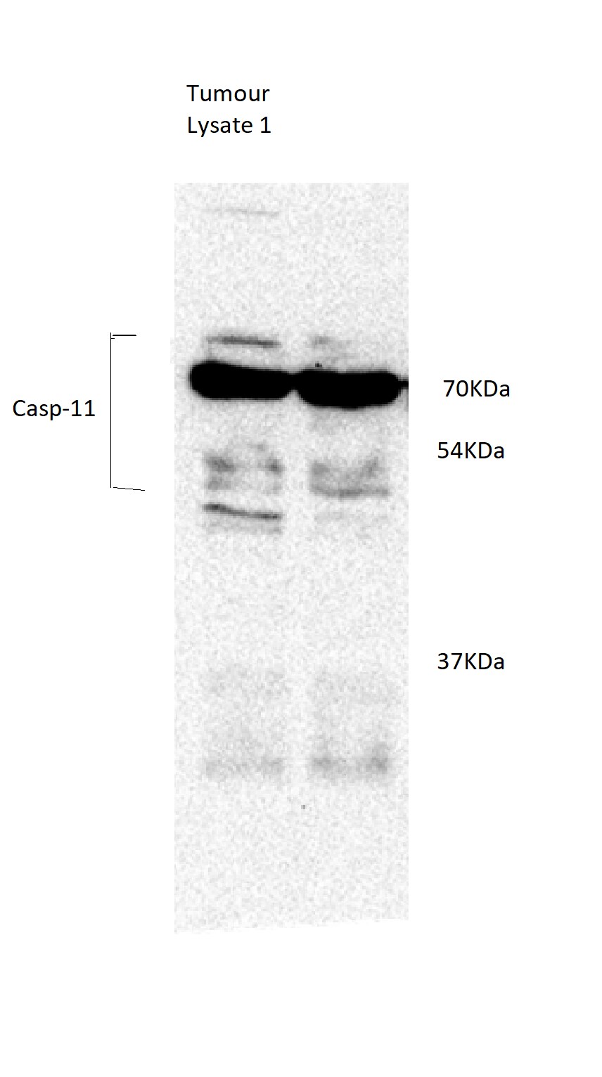
Application: Western BlotSample Tested: Tumor cell lyastesSpecies: MouseVerified Customer | Posted 04/25/201850ug protein

There are no reviews that match your criteria.
Protocols
Find general support by application which include: protocols, troubleshooting, illustrated assays, videos and webinars.
- Cellular Response to Hypoxia Protocols
- R&D Systems Quality Control Western Blot Protocol
- Troubleshooting Guide: Western Blot Figures
- Western Blot Conditions
- Western Blot Protocol
- Western Blot Protocol for Cell Lysates
- Western Blot Troubleshooting
- Western Blot Troubleshooting Guide
- View all Protocols, Troubleshooting, Illustrated assays and Webinars
Loading...