Endoglin (CD105) is a 90 kDa type I transmembrane glycoprotein of the zona pellucida (ZP) family of proteins (1-3). Endoglin and betaglycan/T beta RIII are type III receptors for TGF beta superfamily ligands, sharing 71% amino acid (aa) identity within the transmembrane (TM) and cytoplasmic domains. Endoglin is highly expressed on proliferating vascular endothelial cells, chondrocytes, and syncytiotrophoblasts of term placenta, with lower amounts on hematopoietic, mesenchymal, and neural crest stem cells, activated monocytes, and lymphoid and myeloid leukemic cells (2-5). Mouse Endoglin cDNA encodes 653 aa including a 26 aa signal sequence, a 555 aa extracellular domain (ECD) with an orphan domain and a two-part ZP domain, a TM domain, and a 47 aa cytoplasmic domain (1-3). A mouse isoform with a 35 aa cytoplasmic domain (S-endoglin) can oppose effects of long (L) Endoglin (6, 7). The mouse Endoglin ECD shares 69%, 84%, 62%, 63%, and 66% aa identity with human, rat, bovine, porcine, and canine Endoglin, respectively. Endoglin homodimers interact with TGF-beta 1 and TGF-beta 3 (but not TGF-beta 2) but only after binding T beta RII (8). Similarly, they interact with activin-A and BMP-7 via activin type IIA or B receptors, and with BMP-2 via BMPR-1A/ALK-3 or BMPR-1B/ALK-6 (9). BMP-9, however, is reported to bind Endoglin directly (10). Endoglin modifies ligand-induced signaling in multiple ways. For example, expression of Endoglin can inhibit TGF-beta 1 signals but enhance BMP-7 signals in the same myoblast cell line (11). In endothelial cells, Endoglin inhibits T beta RI/ALK5 but enhances ALK1-mediated activation (12). Deletion of mouse Endoglin causes lethal vascular and cardiovascular defects, and human Endoglin haploinsufficiency can a cause the vascular disorder, hereditary hemorrhagic telangiectasia type I (13, 14). These abnormalities confirm the essential function of Endoglin in differentiation of smooth muscle, angiogenesis, and neovascularization (2-4, 12-14). In preeclampsia of pregnancy, high levels of proteolytically generated soluble Endoglin and VEGF R1 (sFlt-1), along with low placental growth factor (PlGF), are pathogenic due to anti-angiogenic activity (15).
Mouse Endoglin/CD105 Antibody (209701)
R&D Systems | Catalog # MAB1320

Key Product Details
Species Reactivity
Validated:
Cited:
Applications
Validated:
Cited:
Label
Antibody Source
Product Specifications
Immunogen
Glu21-Gly581 (predicted)
Accession # NP_031958
Specificity
Clonality
Host
Isotype
Scientific Data Images for Mouse Endoglin/CD105 Antibody (209701)
Endoglin/CD105 in Mouse Embryo.
Endoglin/CD105 was detected in immersion fixed frozen sections of mouse embryo (13.5 d.p.c.) using Rat Anti-Mouse Endoglin/CD105 Monoclonal Antibody (Catalog # MAB1320) at 10 µg/mL overnight at 4 °C. Tissue was stained using the NorthernLights™ 557-conjugated Anti-Rat IgG Secondary Antibody (red; NL013) and counterstained with DAPI (blue). Specific staining was localized to endothelial cells of the developing hindlimb. View our protocol for Fluorescent IHC Staining of Frozen Tissue Sections.Image depicts staining at 10X magnification.
Endoglin/CD105 in Mouse Embryo.
Endoglin/CD105 was detected in immersion fixed frozen sections of mouse embryo (13.5 d.p.c.) using Rat Anti-Mouse Endoglin/CD105 Monoclonal Antibody (Catalog # MAB1320) at 10 µg/mL overnight at 4 °C. Tissue was stained using the NorthernLights™ 557-conjugated Anti-Rat IgG Secondary Antibody (red; NL013) and counterstained with DAPI (blue). Specific staining was localized to endothelial cells of the developing hindlimb. View our protocol for Fluorescent IHC Staining of Frozen Tissue Sections.Image depicts staining at 40X magnification.
Endoglin/CD105 in Mouse Mesenchymal Stem Cells.
Endoglin/CD105 was detected in immersion fixed mouse mesenchymal stem cells using Rat Anti-Mouse Endoglin/CD105 Monoclonal Antibody (Catalog # MAB1320) at 10 µg/mL for 3 hours at room temperature. Cells were stained using the NorthernLights™ 557-conjugated Anti-Rat IgG Secondary Antibody (red; NL013) and counterstained with DAPI (blue). Specific staining was localized to cytoplasmic. View our protocol for Fluorescent ICC Staining of Non-adherent Cells.
Detection of Endoglin/CD105 in MS-1 cells by Flow Cytometry
MS-1 cells were stained with Rat Anti-Mouse Endoglin/CD105 Monoclonal Antibody (Catalog # MAB1320, filled histogram) or isotype control antibody (Catalog # MAB006, open histogram) followed by Phycoerythrin-conjugated Anti-Rat IgG Secondary Antibody (Catalog # F0105B). View our protocol for Staining Membrane-associated Proteins.
Detection of Endoglin/CD105 in Mouse Embryo.
Endoglin/CD105 was detected in immersion fixed frozen sections of mouse embryo using Rat Anti-Mouse Endoglin/CD105 Monoclonal Antibody (Catalog # MAB1320) at 3 µg/ml for 1 hour at room temperature followed by incubation with the Anti-Rat IgG VisUCyte™ HRP Polymer Antibody (Catalog # VC005) or the HRP-conjugated Anti-Rat IgG Secondary Antibody (Catalog # HAF005). Tissue was stained using DAB (brown) and counterstained with hematoxylin (blue). Specific staining was localized to endothelial cells of developing limbs. View our protocol for Chromogenic IHC Staining of Frozen Tissue Sections.
Detection of Endoglin/CD105 in Mouse Embryo.
Endoglin/CD105 was detected in immersion fixed frozen sections of mouse embryo using Rat Anti-Mouse Endoglin/CD105 Monoclonal Antibody (Catalog # MAB1320) at 3 µg/ml for 1 hour at room temperature followed by incubation with the Anti-Rat IgG VisUCyte™ HRP Polymer Antibody (Catalog # VC005) or the HRP-conjugated Anti-Rat IgG Secondary Antibody (Catalog # HAF005). Tissue was stained using DAB (brown) and counterstained with hematoxylin (blue). Specific staining was localized to endothelial cells of the developing liver. View our protocol for Chromogenic IHC Staining of Frozen Tissue Sections.Applications for Mouse Endoglin/CD105 Antibody (209701)
CyTOF-ready
Flow Cytometry
Sample: MS‑1 mouse pancreatic islet endothelial cell line
Immunocytochemistry
Sample: Immersion fixed fixed mouse mesenchymal stem cells, immersion fixed D3 mouse embryonic cell line differentiated to embryoid bodies, and immersion fixed MS-1 mouse pancreatic islet endothelial cell line
Immunohistochemistry
Sample:
Immersion fixed frozen sections of mouse embryo (13.5 d.p.c.)
Western Blot
Sample: Recombinant Mouse Endoglin/CD105 Fc Chimera (Catalog # 1320-EN)
under non-reducing conditions only
Reviewed Applications
Read 4 reviews rated 4.5 using MAB1320 in the following applications:
Flow Cytometry Panel Builder
Bio-Techne Knows Flow Cytometry
Save time and reduce costly mistakes by quickly finding compatible reagents using the Panel Builder Tool.
Advanced Features
- Spectra Viewer - Custom analysis of spectra from multiple fluorochromes
- Spillover Popups - Visualize the spectra of individual fluorochromes
- Antigen Density Selector - Match fluorochrome brightness with antigen density

Formulation, Preparation, and Storage
Purification
Reconstitution
Reconstitute at 0.5 mg/mL in sterile PBS. For liquid material, refer to CoA for concentration.
Formulation
*Small pack size (-SP) is supplied either lyophilized or as a 0.2 µm filtered solution in PBS.
Shipping
Stability & Storage
- 12 months from date of receipt, -20 to -70 °C as supplied.
- 1 month, 2 to 8 °C under sterile conditions after reconstitution.
- 6 months, -20 to -70 °C under sterile conditions after reconstitution.
Calculators
Background: Endoglin/CD105
References
- Ge, A.Z. and E.C. Butcher (1994) Gene 138:201.
- ten Dijke, P. et al. (2008) Angiogenesis 11:79.
- Bernabeu, C. et al. (2007) J. Cell. Biochem. 102:1375.
- Mancini, M.L. et al. (2007) Dev. Biol. 308:520.
- Moody, J.L. et al. (2007) Stem Cells 25:2809.
- Velasco, S. et al. (2008) J. Cell Sci. 121:913.
- Perez-Gomez, E. et al. (2005) Oncogene 24:4450.
- Cheifetz, S, et al. (1992) J. Biol. Chem. 267:19027.
- Barbara, N.P. et al. (1999) J. Biol. Chem. 274:584.
- Scharpfenecker, M. et al. (2007) J. Cell Sci. 120:964.
- Scherner, O. et al. (2007) J. Biol. Chem. 282:13934.
- Pece-Barbara, N. et al. (2005) J. Biol. Chem. 280:27800.
- Arthur, H.M. et al. (2000) Dev. Biol. 217:42.
- Lebrin, F. and C.L. Mummery (2008) Trends Cardiovasc. Med. 18:25.
- Venkatesha, S. et al. (2006) Nat. Med. 12:642.
Alternate Names
Gene Symbol
UniProt
Additional Endoglin/CD105 Products
Product Documents for Mouse Endoglin/CD105 Antibody (209701)
Certificate of Analysis
To download a Certificate of Analysis, please enter a lot or batch number in the search box below.
Note: Certificate of Analysis not available for kit components.
Product Specific Notices for Mouse Endoglin/CD105 Antibody (209701)
For research use only
Citations for Mouse Endoglin/CD105 Antibody (209701)
 Powered by Bioz
Customer Reviews for Mouse Endoglin/CD105 Antibody (209701) (4)
Have you used Mouse Endoglin/CD105 Antibody (209701)?
Submit a review and receive an Amazon gift card!
$25/€18/£15/$25CAN/¥2500 Yen for a review with an image
$10/€7/£6/$10CAN/¥1110 Yen for a review without an image
Submit a review
Customer Images
-
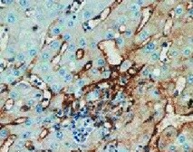
Application: ImmunohistochemistrySample Tested: Hepatocellular carcinomaSpecies: HumanVerified Customer | Posted 10/06/2021

-
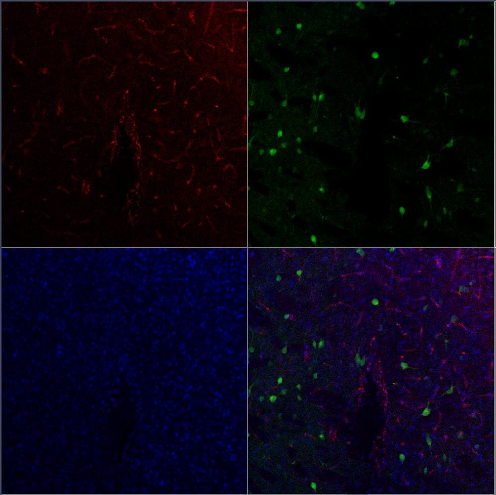
Application: Immunohistochemistry-FrozenSample Tested: brain and spinal cord, Embryonic brain and E15.5 MouseSpecies: MouseVerified Customer | Posted 01/22/2019Endoglin staining in an E12.5 mouse embryonic cortex. the tissue is contrasted with DAPI in blueThe sections did not need any antigen retrieval. Immunofluorescence-with frozen sections and immunofluorescence with 500µm thick PFA fixed embryonic brain slices.
-
Application: ImmunohistochemistrySample Tested: Mouse Brain slices fixed with 4% PFA solutionSpecies: Mus MusculusVerified Customer | Posted 08/01/2016Endoglin/CD105 was detected 4% PFA fixed mouse brain. Brain sections were cutted with an Vibratom. Endoglin detection was performed with Endoglin/CD105 Monoclonal Antibody (Catalog # MAB1320) at 20 µg/mL overnight at 4 °C. Brain sections were afterwards stained with AlexaFluor 647 coupled Anti-Rat IgG Secondary Antibody and counterstained with Hoechst 33342 (blue). Specific staining was localized to endothelial cells in Striatum of mouse brain. Transduced Neurons Express GFP.
-
Application: Immunohistochemistry-ParaffinSample Tested: See PMID 22683021Species: MouseVerified Customer | Posted 02/04/2015
There are no reviews that match your criteria.
Protocols
Find general support by application which include: protocols, troubleshooting, illustrated assays, videos and webinars.
- 7-Amino Actinomycin D (7-AAD) Cell Viability Flow Cytometry Protocol
- Antigen Retrieval Protocol (PIER)
- Antigen Retrieval for Frozen Sections Protocol
- Appropriate Fixation of IHC/ICC Samples
- Cellular Response to Hypoxia Protocols
- Chromogenic IHC Staining of Formalin-Fixed Paraffin-Embedded (FFPE) Tissue Protocol
- Chromogenic Immunohistochemistry Staining of Frozen Tissue
- ClariTSA™ Fluorophore Kits
- Detection & Visualization of Antibody Binding
- Extracellular Membrane Flow Cytometry Protocol
- Flow Cytometry Protocol for Cell Surface Markers
- Flow Cytometry Protocol for Staining Membrane Associated Proteins
- Flow Cytometry Staining Protocols
- Flow Cytometry Troubleshooting Guide
- Fluorescent IHC Staining of Frozen Tissue Protocol
- Graphic Protocol for Heat-induced Epitope Retrieval
- Graphic Protocol for the Preparation and Fluorescent IHC Staining of Frozen Tissue Sections
- Graphic Protocol for the Preparation and Fluorescent IHC Staining of Paraffin-embedded Tissue Sections
- Graphic Protocol for the Preparation of Gelatin-coated Slides for Histological Tissue Sections
- ICC Cell Smear Protocol for Suspension Cells
- ICC Immunocytochemistry Protocol Videos
- ICC for Adherent Cells
- IHC Sample Preparation (Frozen sections vs Paraffin)
- Immunocytochemistry (ICC) Protocol
- Immunocytochemistry Troubleshooting
- Immunofluorescence of Organoids Embedded in Cultrex Basement Membrane Extract
- Immunofluorescent IHC Staining of Formalin-Fixed Paraffin-Embedded (FFPE) Tissue Protocol
- Immunohistochemistry (IHC) and Immunocytochemistry (ICC) Protocols
- Immunohistochemistry Frozen Troubleshooting
- Immunohistochemistry Paraffin Troubleshooting
- Intracellular Flow Cytometry Protocol Using Alcohol (Methanol)
- Intracellular Flow Cytometry Protocol Using Detergents
- Intracellular Nuclear Staining Flow Cytometry Protocol Using Detergents
- Intracellular Staining Flow Cytometry Protocol Using Alcohol Permeabilization
- Intracellular Staining Flow Cytometry Protocol Using Detergents to Permeabilize Cells
- Preparing Samples for IHC/ICC Experiments
- Preventing Non-Specific Staining (Non-Specific Binding)
- Primary Antibody Selection & Optimization
- Propidium Iodide Cell Viability Flow Cytometry Protocol
- Protocol for Heat-Induced Epitope Retrieval (HIER)
- Protocol for Liperfluo
- Protocol for Making a 4% Formaldehyde Solution in PBS
- Protocol for VisUCyte™ HRP Polymer Detection Reagent
- Protocol for the Characterization of Human Th22 Cells
- Protocol for the Characterization of Human Th9 Cells
- Protocol for the Fluorescent ICC Staining of Cell Smears - Graphic
- Protocol for the Fluorescent ICC Staining of Cultured Cells on Coverslips - Graphic
- Protocol for the Preparation & Fixation of Cells on Coverslips
- Protocol for the Preparation and Chromogenic IHC Staining of Frozen Tissue Sections
- Protocol for the Preparation and Chromogenic IHC Staining of Frozen Tissue Sections - Graphic
- Protocol for the Preparation and Chromogenic IHC Staining of Paraffin-embedded Tissue Sections
- Protocol for the Preparation and Chromogenic IHC Staining of Paraffin-embedded Tissue Sections - Graphic
- Protocol for the Preparation and Fluorescent ICC Staining of Cells on Coverslips
- Protocol for the Preparation and Fluorescent ICC Staining of Non-adherent Cells
- Protocol for the Preparation and Fluorescent ICC Staining of Stem Cells on Coverslips
- Protocol for the Preparation and Fluorescent IHC Staining of Frozen Tissue Sections
- Protocol for the Preparation and Fluorescent IHC Staining of Paraffin-embedded Tissue Sections
- Protocol for the Preparation of Gelatin-coated Slides for Histological Tissue Sections
- Protocol for the Preparation of a Cell Smear for Non-adherent Cell ICC - Graphic
- Protocol: Annexin V and PI Staining by Flow Cytometry
- Protocol: Annexin V and PI Staining for Apoptosis by Flow Cytometry
- R&D Systems Quality Control Western Blot Protocol
- TUNEL and Active Caspase-3 Detection by IHC/ICC Protocol
- The Importance of IHC/ICC Controls
- Troubleshooting Guide: Fluorokine Flow Cytometry Kits
- Troubleshooting Guide: Immunohistochemistry
- Troubleshooting Guide: Western Blot Figures
- Western Blot Conditions
- Western Blot Protocol
- Western Blot Protocol for Cell Lysates
- Western Blot Troubleshooting
- Western Blot Troubleshooting Guide
- View all Protocols, Troubleshooting, Illustrated assays and Webinars


