Mouse Ly-6G/Ly-6C (Gr-1) Antibody (RB6-8C5)
R&D Systems | Catalog # MAB1037

Key Product Details
Species Reactivity
Validated:
Mouse
Cited:
Human, Mouse, Bovine, Transgenic Mouse, Xenograft
Applications
Validated:
Immunohistochemistry, Flow Cytometry, Immunocytochemistry, Immunoprecipitation, COMET, CyTOF-ready
Cited:
Immunohistochemistry, Immunohistochemistry-Paraffin, Immunohistochemistry-Frozen, Western Blot, Neutralization, Flow Cytometry, Immunocytochemistry, Immunodepletion
Label
Unconjugated
Antibody Source
Monoclonal Rat IgG2B Clone # RB6-8C5
Loading...
Product Specifications
Immunogen
The immunogen for this antibody is Ly-6G (Gr-1, Gr1).
Specificity
Detects Ly-6G/Ly-6C (Gr-1). Weak cross-reactivity with Ly-6C is observed.
Clonality
Monoclonal
Host
Rat
Isotype
IgG2B
Scientific Data Images for Mouse Ly-6G/Ly-6C (Gr-1) Antibody (RB6-8C5)
Detection of Ly-6G/Ly-6C in Frozen Mouse Spleen viaseqIF™ staining on COMET™
Ly-6G/Ly-6C was detected in frozen sections of mouse spleen using Rat Anti-Mouse Ly-6G/Ly-6C Monoclonal Antibody (Catalog # MAB1037) at 7ug/mL at 37 ° Celsius for 4 minutes. Before incubation with the primary antibody, tissue underwent preprocessing by incubating tissue with Multi Staining Buffer (Lunaphore Catalog # BU06) for 5 minutes at room temperature followed by a 20 minute incubation in Tris-Buffered Saline + 0.2% Triton at room temperature. Tissue was stained using the Alexa Fluor™ 555 Goat anti-Rat IgG Secondary Antibody at 1:100 at 37 ° Celsius for 2 minutes. (Yellow; Lunaphore Catalog # DR555RT) and counterstained with DAPI (blue; Lunaphore Catalog # DR100). Specific staining was localized to the cell membrane. Protocol available in COMET™ Panel Builder. in Mouse Spleno-cytes.")
Ly-6G/Ly-6C (Gr-1) in Mouse Spleno-cytes.
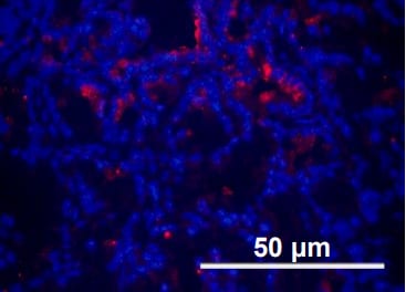
Ly-6G/Ly-6C (Gr-1) was detected in immersion fixed mouse splenocytes using Mouse Ly-6G/Ly-6C (Gr-1) Monoclonal Anti-body (Catalog # MAB1037) at 10 µg/mL for 3 hours at room temperature. Cells were stained using the NorthernLights™ 557-conjugated Anti-Rat IgG Secondary Antibody (red; Catalog # NL013) and counter-stained with DAPI (blue). View our protocol for Fluorescent ICC Staining of Non-adherent Cells. in Mouse Spleen.")
Ly-6G/Ly-6C (Gr-1) in Mouse Spleen.
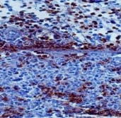
Ly-6G/Ly-6C (Gr-1) was detected in perfusion fixed frozen sections of mouse spleen using Rat Anti-Mouse Ly-6G/Ly-6C (Gr-1)Monoclonal Antibody (Catalog # MAB1037) at 3 µg/mL overnight at 4 °C. Tissue was stained using the Anti-Rat HRP-DAB Cell & Tissue Staining Kit (brown; Catalog # CTS017) and counterstained with hematoxylin (blue). Specific staining was localized to cytoplasm in splenocytes. View our protocol for Chromogenic IHC Staining of Frozen Tissue Sections. Antibody by Immunohistochemistry")
Detection of Mouse Mouse Ly-6G/Ly-6C (Gr-1) Antibody by Immunohistochemistry
Lymphocyte/neutrophil ratio increases upon NHE1 inhibition in tumor sections of KPfC mice.(A) H&E (left) and PAS-stained KPfC mouse tissue sections after vehicle and gemcitabine + cariporide (GEM+CARI) therapy. Cells of innate immunity, such as neutrophils (arrows), utilize glycogen and are thus PAS+ (purple), in contrast to, for example, lymphocytes. Scale bar: 50 μm. (B) Representative IHC images stained for Ly6G+ neutrophils (magenta, arrows on left image), CD3+ lymphocytes (yellow, arrows on the right image), and nuclei with DAPI (cyan). Scale bar: 50 μm. (C) CD3/Ly6G ratio was assessed by dividing the number of CD3+ cells by the number of Ly6G+ cells in every tumor node. Data points depict the mean CD3/Ly6G ratio derived from each tumor node in individual mice; NVehicle = 10, NGEM = 9, NCARI = 10, NGEM+CARI = 11 mice. (D) To obtain the CD3/Ly6G ratio per tumor node, the number of CD3+ cells was divided by the respective number of Ly6G+ cells in each tumor node. Data points depict individual tumor nodes; nVehicle = 386, nGEM = 276, nCARI = 301, nGEM+CARI = 398. Data and statistical comparison in D and E are represented as median ± 95% CI using Kruskal-Wallis statistical test with Dunn’s post hoc test. Image collected and cropped by CiteAb from the following publication (https://pubmed.ncbi.nlm.nih.gov/37643024), licensed under a CC-BY license. Not internally tested by R&D Systems.
Detection of Mouse Gr-1/Ly-6G by Immunohistochemistry
Lymphocyte/neutrophil ratio increases upon NHE1 inhibition in tumor sections of KPfC mice.(A) H&E (left) and PAS-stained KPfC mouse tissue sections after vehicle and gemcitabine + cariporide (GEM+CARI) therapy. Cells of innate immunity, such as neutrophils (arrows), utilize glycogen and are thus PAS+ (purple), in contrast to, for example, lymphocytes. Scale bar: 50 μm. (B) Representative IHC images stained for Ly6G+ neutrophils (magenta, arrows on left image), CD3+ lymphocytes (yellow, arrows on the right image), and nuclei with DAPI (cyan). Scale bar: 50 μm. (C) CD3/Ly6G ratio was assessed by dividing the number of CD3+ cells by the number of Ly6G+ cells in every tumor node. Data points depict the mean CD3/Ly6G ratio derived from each tumor node in individual mice; NVehicle = 10, NGEM = 9, NCARI = 10, NGEM+CARI = 11 mice. (D) To obtain the CD3/Ly6G ratio per tumor node, the number of CD3+ cells was divided by the respective number of Ly6G+ cells in each tumor node. Data points depict individual tumor nodes; nVehicle = 386, nGEM = 276, nCARI = 301, nGEM+CARI = 398. Data and statistical comparison in D and E are represented as median ± 95% CI using Kruskal-Wallis statistical test with Dunn’s post hoc test. Image collected and cropped by CiteAb from the following open publication (https://pubmed.ncbi.nlm.nih.gov/37643024), licensed under a CC-BY license. Not internally tested by R&D Systems. by Immunohistochemistry")
Detection of Mouse Ly-6G/Ly-6C (Gr-1) by Immunohistochemistry
Lymphocyte/neutrophil ratio increases upon NHE1 inhibition in tumor sections of KPfC mice.(A) H&E (left) and PAS-stained KPfC mouse tissue sections after vehicle and gemcitabine + cariporide (GEM+CARI) therapy. Cells of innate immunity, such as neutrophils (arrows), utilize glycogen and are thus PAS+ (purple), in contrast to, for example, lymphocytes. Scale bar: 50 μm. (B) Representative IHC images stained for Ly6G+ neutrophils (magenta, arrows on left image), CD3+ lymphocytes (yellow, arrows on the right image), and nuclei with DAPI (cyan). Scale bar: 50 μm. (C) CD3/Ly6G ratio was assessed by dividing the number of CD3+ cells by the number of Ly6G+ cells in every tumor node. Data points depict the mean CD3/Ly6G ratio derived from each tumor node in individual mice; NVehicle = 10, NGEM = 9, NCARI = 10, NGEM+CARI = 11 mice. (D) To obtain the CD3/Ly6G ratio per tumor node, the number of CD3+ cells was divided by the respective number of Ly6G+ cells in each tumor node. Data points depict individual tumor nodes; nVehicle = 386, nGEM = 276, nCARI = 301, nGEM+CARI = 398. Data and statistical comparison in D and E are represented as median ± 95% CI using Kruskal-Wallis statistical test with Dunn’s post hoc test. Image collected and cropped by CiteAb from the following open publication (https://pubmed.ncbi.nlm.nih.gov/37643024), licensed under a CC-BY license. Not internally tested by R&D Systems.Applications for Mouse Ly-6G/Ly-6C (Gr-1) Antibody (RB6-8C5)
Application
Recommended Usage
COMET
7 µg/mL
Sample: Perfusion fixed frozen sections of mouse spleen
Sample: Perfusion fixed frozen sections of mouse spleen
CyTOF-ready
Ready to be labeled using established conjugation methods. No BSA or other carrier proteins that could interfere with conjugation.
Flow Cytometry
0.25 µg/106 cells
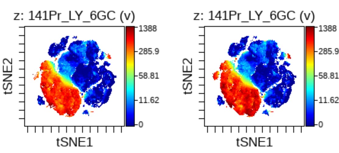
Sample: Mouse splenocytes or peripheral blood cells
Sample: Mouse splenocytes or peripheral blood cells
Immunocytochemistry
8-25 µg/mL
Sample: Immersion fixed mouse splenocytes
Sample: Immersion fixed mouse splenocytes
Immunohistochemistry
3-25 µg/mL
Sample: Perfusion fixed frozen sections of mouse spleen
Sample: Perfusion fixed frozen sections of mouse spleen
Immunoprecipitation
Conlan, J.W. and R.J. North (1994) J. Exp. Med. 179:259.
Reviewed Applications
Read 8 reviews rated 4.6 using MAB1037 in the following applications:
Flow Cytometry Panel Builder
Bio-Techne Knows Flow Cytometry
Save time and reduce costly mistakes by quickly finding compatible reagents using the Panel Builder Tool.
Advanced Features
- Spectra Viewer - Custom analysis of spectra from multiple fluorochromes
- Spillover Popups - Visualize the spectra of individual fluorochromes
- Antigen Density Selector - Match fluorochrome brightness with antigen density

Formulation, Preparation, and Storage
Purification
Protein A or G purified from hybridoma culture supernatant
Reconstitution
Reconstitute at 0.5 mg/mL in sterile PBS. For liquid material, refer to CoA for concentration.
Loading...
Formulation
Lyophilized from a 0.2 μm filtered solution in PBS with Trehalose. See Certificate of Analysis for details.
*Small pack size (-SP) is supplied either lyophilized or as a 0.2 µm filtered solution in PBS.
*Small pack size (-SP) is supplied either lyophilized or as a 0.2 µm filtered solution in PBS.
Shipping
Lyophilized product is shipped at ambient temperature. Liquid small pack size (-SP) is shipped with polar packs. Upon receipt, store immediately at the temperature recommended below.
Stability & Storage
Use a manual defrost freezer and avoid repeated freeze-thaw cycles.
- 12 months from date of receipt, -20 to -70 °C as supplied.
- 1 month, 2 to 8 °C under sterile conditions after reconstitution.
- 6 months, -20 to -70 °C under sterile conditions after reconstitution.
Calculators
Background: Ly-6G/Ly-6C (Gr-1)
References
- Spangrude, G.J. et al. (1988) Science 241:58.
- Fleming, T.J. et al. (1993) J. Immunol. 151:2399.
- Lewinsohn, D.M. et al.(1987) J. Immunol. 147:22.
- Lagasse, E. and I.L. Weissman (1996) J. Immunol. Methods 197:139.
Long Name
A Myeloid Differentiation Antigen
Alternate Names
Ly-6C, Ly-6G, Ly6G
Entrez Gene IDs
546644 (Mouse)
Gene Symbol
LY6G
Additional Ly-6G/Ly-6C (Gr-1) Products
Product Documents for Mouse Ly-6G/Ly-6C (Gr-1) Antibody (RB6-8C5)
Certificate of Analysis
To download a Certificate of Analysis, please enter a lot or batch number in the search box below.
Note: Certificate of Analysis not available for kit components.
Product Specific Notices for Mouse Ly-6G/Ly-6C (Gr-1) Antibody (RB6-8C5)
For research use only
Citations for Mouse Ly-6G/Ly-6C (Gr-1) Antibody (RB6-8C5)
 Powered by Bioz
Customer Reviews for Mouse Ly-6G/Ly-6C (Gr-1) Antibody (RB6-8C5) (8)
4.6 out of 5
8 Customer Ratings
Have you used Mouse Ly-6G/Ly-6C (Gr-1) Antibody (RB6-8C5)?
Submit a review and receive an Amazon gift card!
$25/€18/£15/$25CAN/¥2500 Yen for a review with an image
$10/€7/£6/$10CAN/¥1110 Yen for a review without an image
Submit a review
Customer Images
Showing
1
-
5 of
8 reviews
Showing All
Filter By:
-
Application: ImmunohistochemistrySample Tested: Colon tissueSpecies: MouseVerified Customer | Posted 08/17/2021

-
Application: Immunocytochemistry/ImmunofluorescenceSample Tested: GranulocytesSpecies: MouseVerified Customer | Posted 06/02/2021
-
Application: Flow CytometrySample Tested: Pancreas tissueSpecies: MouseVerified Customer | Posted 01/15/2021
-
Application: Immunocytochemistry/ImmunofluorescenceSample Tested: Lung cancer tissueSpecies: MouseVerified Customer | Posted 04/18/2018
-
Application: Flow CytometrySample Tested: Pancreatic cancer cellsSpecies: pancreatic cancer cell line and MouseVerified Customer | Posted 04/11/2018
-
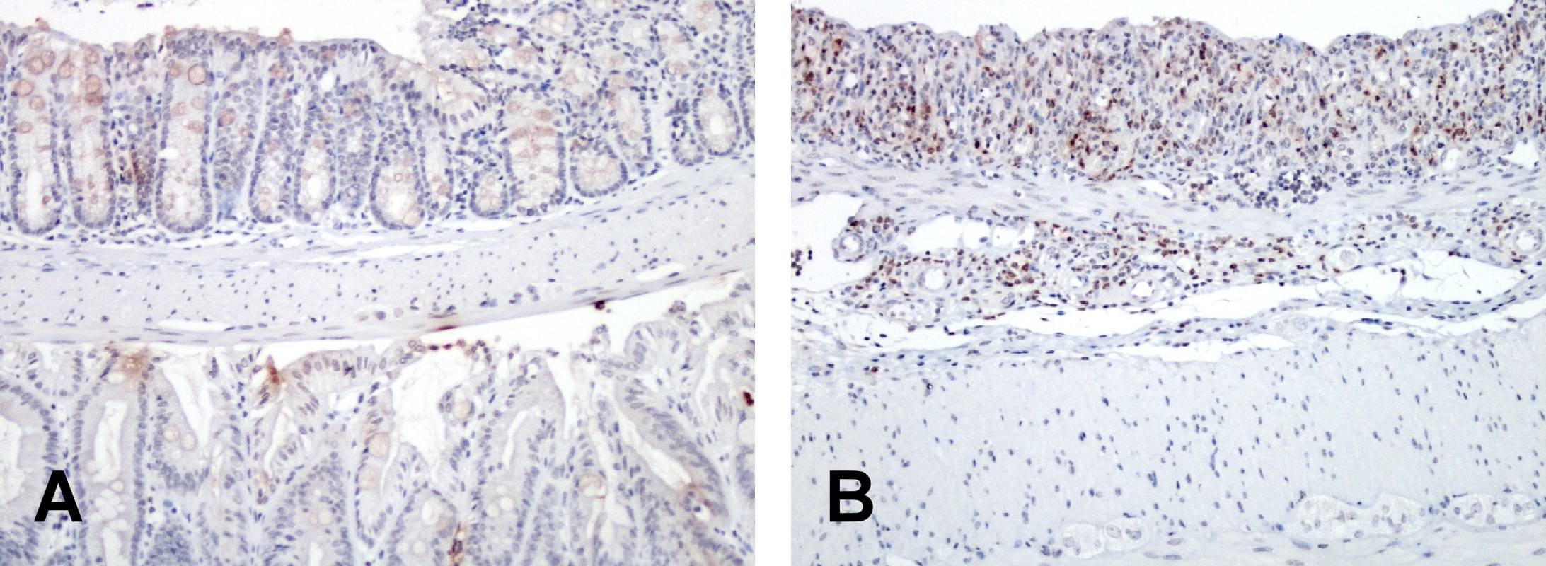
Application: ImmunohistochemistrySample Tested: colonSpecies: MouseVerified Customer | Posted 12/01/2017A. Normal mouse colon; B. DSS induced mouse colitis colon. The staining only work at 1:20 dilution instead of the recommended ~1:50 dilution. We tried 4 difference sources and this is the best one.
-
Application: Flow CytometrySample Tested: GranulocytesSpecies: MouseVerified Customer | Posted 07/17/2017
-
Application: Flow CytometrySample Tested: NeutrophilsSpecies: MouseVerified Customer | Posted 05/30/2017
There are no reviews that match your criteria.
Protocols
Find general support by application which include: protocols, troubleshooting, illustrated assays, videos and webinars.
- 7-Amino Actinomycin D (7-AAD) Cell Viability Flow Cytometry Protocol
- Antigen Retrieval Protocol (PIER)
- Antigen Retrieval for Frozen Sections Protocol
- Appropriate Fixation of IHC/ICC Samples
- Cellular Response to Hypoxia Protocols
- Chromogenic IHC Staining of Formalin-Fixed Paraffin-Embedded (FFPE) Tissue Protocol
- Chromogenic Immunohistochemistry Staining of Frozen Tissue
- ClariTSA™ Fluorophore Kits
- Detection & Visualization of Antibody Binding
- Extracellular Membrane Flow Cytometry Protocol
- Flow Cytometry Protocol for Cell Surface Markers
- Flow Cytometry Protocol for Staining Membrane Associated Proteins
- Flow Cytometry Staining Protocols
- Flow Cytometry Troubleshooting Guide
- Fluorescent IHC Staining of Frozen Tissue Protocol
- Graphic Protocol for Heat-induced Epitope Retrieval
- Graphic Protocol for the Preparation and Fluorescent IHC Staining of Frozen Tissue Sections
- Graphic Protocol for the Preparation and Fluorescent IHC Staining of Paraffin-embedded Tissue Sections
- Graphic Protocol for the Preparation of Gelatin-coated Slides for Histological Tissue Sections
- ICC Cell Smear Protocol for Suspension Cells
- ICC Immunocytochemistry Protocol Videos
- ICC for Adherent Cells
- IHC Sample Preparation (Frozen sections vs Paraffin)
- Immunocytochemistry (ICC) Protocol
- Immunocytochemistry Troubleshooting
- Immunofluorescence of Organoids Embedded in Cultrex Basement Membrane Extract
- Immunofluorescent IHC Staining of Formalin-Fixed Paraffin-Embedded (FFPE) Tissue Protocol
- Immunohistochemistry (IHC) and Immunocytochemistry (ICC) Protocols
- Immunohistochemistry Frozen Troubleshooting
- Immunohistochemistry Paraffin Troubleshooting
- Immunoprecipitation Protocol
- Intracellular Flow Cytometry Protocol Using Alcohol (Methanol)
- Intracellular Flow Cytometry Protocol Using Detergents
- Intracellular Nuclear Staining Flow Cytometry Protocol Using Detergents
- Intracellular Staining Flow Cytometry Protocol Using Alcohol Permeabilization
- Intracellular Staining Flow Cytometry Protocol Using Detergents to Permeabilize Cells
- Preparing Samples for IHC/ICC Experiments
- Preventing Non-Specific Staining (Non-Specific Binding)
- Primary Antibody Selection & Optimization
- Propidium Iodide Cell Viability Flow Cytometry Protocol
- Protocol for Heat-Induced Epitope Retrieval (HIER)
- Protocol for Liperfluo
- Protocol for Making a 4% Formaldehyde Solution in PBS
- Protocol for VisUCyte™ HRP Polymer Detection Reagent
- Protocol for the Characterization of Human Th22 Cells
- Protocol for the Characterization of Human Th9 Cells
- Protocol for the Fluorescent ICC Staining of Cell Smears - Graphic
- Protocol for the Fluorescent ICC Staining of Cultured Cells on Coverslips - Graphic
- Protocol for the Preparation & Fixation of Cells on Coverslips
- Protocol for the Preparation and Chromogenic IHC Staining of Frozen Tissue Sections
- Protocol for the Preparation and Chromogenic IHC Staining of Frozen Tissue Sections - Graphic
- Protocol for the Preparation and Chromogenic IHC Staining of Paraffin-embedded Tissue Sections
- Protocol for the Preparation and Chromogenic IHC Staining of Paraffin-embedded Tissue Sections - Graphic
- Protocol for the Preparation and Fluorescent ICC Staining of Cells on Coverslips
- Protocol for the Preparation and Fluorescent ICC Staining of Non-adherent Cells
- Protocol for the Preparation and Fluorescent ICC Staining of Stem Cells on Coverslips
- Protocol for the Preparation and Fluorescent IHC Staining of Frozen Tissue Sections
- Protocol for the Preparation and Fluorescent IHC Staining of Paraffin-embedded Tissue Sections
- Protocol for the Preparation of Gelatin-coated Slides for Histological Tissue Sections
- Protocol for the Preparation of a Cell Smear for Non-adherent Cell ICC - Graphic
- Protocol: Annexin V and PI Staining by Flow Cytometry
- Protocol: Annexin V and PI Staining for Apoptosis by Flow Cytometry
- TUNEL and Active Caspase-3 Detection by IHC/ICC Protocol
- The Importance of IHC/ICC Controls
- Troubleshooting Guide: Fluorokine Flow Cytometry Kits
- Troubleshooting Guide: Immunohistochemistry
- View all Protocols, Troubleshooting, Illustrated assays and Webinars
Loading...