Interleukin 17 (also known as CTLA-8) is a T cell-expressed pleiotropic cytokine that exhibits a high degree of homology to a protein encoded by the ORF13 gene of herpes virus Saimiri. cDNA clones encoding IL-17 have been isolated from activated rat, mouse and human T cells. Mouse IL-17 cDNA encodes a 158 amino acid (aa) residue precursor protein with a 21 amino acid residue signal peptide that is cleaved to yield the 137 aa residue mature IL-17. Both recombinant and natural IL-17 have been shown to exist as disulfide linked homodimers. At the amino acid level, mIL-17 shows 57% and 87% sequence identity with herpesvirus and rat IL-17, respectively. An IL-17 specific mouse cell surface receptor (IL-17 R) has been cloned. While the expression of IL-17 mRNA is restricted to activated alpha beta TCR+CD4-CD8-T cells, the expression of mIL-17 R mRNA has been detected in virtually all cells and tissues tested. IL-17 exhibits multiple biological activities on a variety of cells including: the induction of IL-6 and IL-8 production in fibroblasts; the enhancement of surface expression of ICAM-1 in fibroblasts; activation of NF-kappa B and costimulation of T cell proliferation.
Mouse IL-17/IL-17A Antibody (50104)
R&D Systems | Catalog # MAB421

Key Product Details
Validated by
Species Reactivity
Validated:
Cited:
Applications
Validated:
Cited:
Label
Antibody Source
Product Specifications
Immunogen
Thr22-Ala158
Accession # Q62386
Specificity
Clonality
Host
Isotype
Endotoxin Level
Scientific Data Images for Mouse IL-17/IL-17A Antibody (50104)
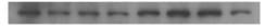
Detection of Recombinant Mouse IL‑17/IL‑17A by Western Blot.
Western blot shows 25 ng of Recombinant Mouse IL-17/ IL-17A (Catalog # 421-ML), Recombinant Human IL-17/IL-17A (Catalog # 317-ILB), Recombinant Rat IL-17/IL-17A (Catalog # 8410-IL), and Recombinant Mouse IL-17F (Catalog # 2057-IL). PVDF Membrane was probed with 1 µg/mL of Rat Anti-Mouse IL-17/ IL-17A Monoclonal Antibody (Catalog # MAB421) followed by HRP-conjugated Anti-Rat IgG Secondary Antibody (Catalog # HAF005). A specific band was detected for IL-17/IL-17A at approximately 15 kDa (as indicated). This experiment was conducted under reducing conditions and using Immunoblot Buffer Group 3.
IL‑6 Secretion Induced by IL‑17 and Neutralization by Mouse IL‑17 Antibody.
Recombinant Mouse IL-17 (Catalog # 421-ML) stimulates IL-6 secretion in the NIH-3T3 mouse embryonic fibroblast cell line in a dose-dependent manner (orange line), as measured by the Mouse IL-6 Quantikine ELISA Kit (Catalog # M6000B). IL-6 secretion elicited by Recombinant Mouse IL-17 (10 ng/mL) is neutralized (green line) by increasing concentrations of Rat Anti-Mouse IL-17 Monoclonal Antibody (Catalog # MAB421). The ND50 is typically 0.05-0.15 µg/mL.
Detection of Mouse IL-17/IL-17A by In vivo assay
Ear histology in WT and Pglyrp-deficient mice in atopic dermatitis and contact dermatitis models of skin inflammation.(A) Oxazolone model of atopic dermatitis: sensitization and 10 applications of oxazolone to the ears every other day induced acanthosis (Ac), parakeratosis (Pk), marked thickening of the sub-epidermal layer with spongiosis (Sp) and dense cellular infiltrates of primarily mononuclear and some polymorphonuclear cells (high magnification insets), that were all highly prominent in Pglyrp3−/− mice and Pglyrp4−/− mice and much less severe in WT mice. (B) Oxazolone model of contact dermatitis: sensitization and a single application of oxazolone to the ears induced strong inflammatory response in WT mice with marked spongiosis of the sub-epidermal layer (Sp) and cellular infiltrates of epidermal and sub-epidermal layers, composed of mononuclear and polymorphonuclear cells; Pglyrp1−/− and Pglyrp1−/−Pglyrp2−/− mice still had cellular infiltrates, but had substantially reduced swelling, compared to WT mice, mostly due to reduced edema. H&E stained cross-sections; bar = 200 µm for all panels, except high magnification insets (the magnified areas are shown by rectangles). Image collected and cropped by CiteAb from the following publication (https://pubmed.ncbi.nlm.nih.gov/21949809), licensed under a CC-BY license. Not internally tested by R&D Systems.
Detection of Mouse IL-17/IL-17A by In vivo assay
IL-17 is required for enhanced response to oxazolone in Pglyrp3−/− mice.(A) The level of IL-17-induced chemokine, CXCL-1, is higher in the ears of Pglyrp3−/− mice than WT mice after sensitization and application of oxazolone for 20 days. (B) Ear swelling in Pglyrp3−/− mice sensitized and treated 7 times with oxazolone every other day and also treated with neutralizing anti-IL-17 mAbs is lower than in Pglyrp3−/−mice similarly treated with oxazolone and isotype control IgG. Means ± SEM; N = 6 mice/group; significance of differences between Pglyrp3−/− and WT mice (A) or IgG control and anti-IL-17 mAbs-treated mice (B): *, P<0.05; **, P<0.005. Image collected and cropped by CiteAb from the following publication (https://pubmed.ncbi.nlm.nih.gov/21949809), licensed under a CC-BY license. Not internally tested by R&D Systems.
Detection of Mouse IL-17/IL-17A by In vivo assay
Ear histology in WT and Pglyrp-deficient mice in atopic dermatitis and contact dermatitis models of skin inflammation.(A) Oxazolone model of atopic dermatitis: sensitization and 10 applications of oxazolone to the ears every other day induced acanthosis (Ac), parakeratosis (Pk), marked thickening of the sub-epidermal layer with spongiosis (Sp) and dense cellular infiltrates of primarily mononuclear and some polymorphonuclear cells (high magnification insets), that were all highly prominent in Pglyrp3−/− mice and Pglyrp4−/− mice and much less severe in WT mice. (B) Oxazolone model of contact dermatitis: sensitization and a single application of oxazolone to the ears induced strong inflammatory response in WT mice with marked spongiosis of the sub-epidermal layer (Sp) and cellular infiltrates of epidermal and sub-epidermal layers, composed of mononuclear and polymorphonuclear cells; Pglyrp1−/− and Pglyrp1−/−Pglyrp2−/− mice still had cellular infiltrates, but had substantially reduced swelling, compared to WT mice, mostly due to reduced edema. H&E stained cross-sections; bar = 200 µm for all panels, except high magnification insets (the magnified areas are shown by rectangles). Image collected and cropped by CiteAb from the following publication (https://pubmed.ncbi.nlm.nih.gov/21949809), licensed under a CC-BY license. Not internally tested by R&D Systems.
Detection of Mouse IL-17/IL-17A by Flow Cytometry
Immunogenicity and protective efficacy of BCG + CT – MVA85A.Balb/c mice received BCG±CT i.n. followed by 1×10⁶ CFU MVA85A 10 weeks later. Lungs (A) and spleen (B) were taken at 10 (black circles), 11 (dark grey circles) and 14 (light grey circles) weeks post-BCG and cytokine-producing cells responding to an Ag85A peptide pool quantified using ICS. Responses from animals receiving BCG – MVA85A (closed circles) were compared with those receiving BCG + CT followed by MVA85A (open circles). Statistical analysis was performed using a Mann Whitney test. n = 10, five each from two experiments. (C) Balb/c mice were vaccinated as above. Control groups included unvaccinated and BCG i.d. A group receiving BCG i.n. was included to compare BCG – MVA85A i.n. to BCG i.n. Animals were exposed to ∼100 CFU M.tb via aerosol four weeks post-MVA85A. Four weeks post-challenge, lungs and spleen were homogenised and plated for CFU quantitation. (D) Balb/c mice were vaccinated and challenged as described above. Groups receiving BCG – MVA85A and BCG + CT – MVA85A received an anti-IL-17 blocking antibody (MAB421; R&D Systems) administered i.p. every three days post-challenge. One group receiving BCG – MVA85A received an IgG2a isotype control antibody (MAB006; R&D Systems) on the same regimen. Mice were culled four weeks post-challenge and lung CFU quantitated as described above. Statistical analysis was performed using a one way ANOVA and post-hoc tests on the vaccinated groups (n = 8–16). Image collected and cropped by CiteAb from the following open publication (https://pubmed.ncbi.nlm.nih.gov/24194918), licensed under a CC-BY license. Not internally tested by R&D Systems.Applications for Mouse IL-17/IL-17A Antibody (50104)
Western Blot
Sample: Recombinant Mouse IL‑17 (Catalog # 421-ML)
Neutralization
Reviewed Applications
Read 2 reviews rated 5 using MAB421 in the following applications:
Formulation, Preparation, and Storage
Purification
Reconstitution
Reconstitute at 0.5 mg/mL in sterile PBS. For liquid material, refer to CoA for concentration.
Formulation
Shipping
Stability & Storage
- 12 months from date of receipt, -20 to -70 °C as supplied.
- 1 month, 2 to 8 °C under sterile conditions after reconstitution.
- 6 months, -20 to -70 °C under sterile conditions after reconstitution.
Calculators
Background: IL-17/IL-17A
References
- Kennedy, J. et al. (1996) J. Interferon Cytokine Res. 16:611.
- Yao, Z. et al. (1995) J. Immunol. 155:5483.
- Yao, Z. et al. (1995) Immunity 3:811.
- Rouvier, E. et al. (1993) J. Immunol. 150:5445.
Long Name
Alternate Names
Entrez Gene IDs
Gene Symbol
UniProt
Additional IL-17/IL-17A Products
Product Documents for Mouse IL-17/IL-17A Antibody (50104)
Certificate of Analysis
To download a Certificate of Analysis, please enter a lot or batch number in the search box below.
Note: Certificate of Analysis not available for kit components.
Product Specific Notices for Mouse IL-17/IL-17A Antibody (50104)
For research use only
Citations for Mouse IL-17/IL-17A Antibody (50104)
 Powered by Bioz
Customer Reviews for Mouse IL-17/IL-17A Antibody (50104) (2)
Have you used Mouse IL-17/IL-17A Antibody (50104)?
Submit a review and receive an Amazon gift card!
$25/€18/£15/$25CAN/¥2500 Yen for a review with an image
$10/€7/£6/$10CAN/¥1110 Yen for a review without an image
Submit a review
Customer Images
-
Application: Western BlotSample Tested: Fibroblast-like synoviocytes and fibroblastsSpecies: MouseVerified Customer | Posted 08/09/2021

-
Application: ImmunohistochemistrySample Tested: Tumor cell lyastesSpecies: MouseVerified Customer | Posted 04/13/2018
There are no reviews that match your criteria.
Protocols
Find general support by application which include: protocols, troubleshooting, illustrated assays, videos and webinars.
- Cellular Response to Hypoxia Protocols
- R&D Systems Quality Control Western Blot Protocol
- Troubleshooting Guide: Western Blot Figures
- Western Blot Conditions
- Western Blot Protocol
- Western Blot Protocol for Cell Lysates
- Western Blot Troubleshooting
- Western Blot Troubleshooting Guide
- View all Protocols, Troubleshooting, Illustrated assays and Webinars
FAQs for Mouse IL-17/IL-17A Antibody (50104)
-
Q: Does Mouse IL-17/IL-17A antibody (catalog # MAB421) cross-react with Rat IL-17A?
A: Mouse IL-17/IL-17A antibody, catalog # MAB421, was tested against recombinant Rat IL-17A (catalog # 8410-IL ) in a Direct ELISA assay. 100% cross-rectivity was observed to recombinant Rat IL-17A.
Associated Pathways


