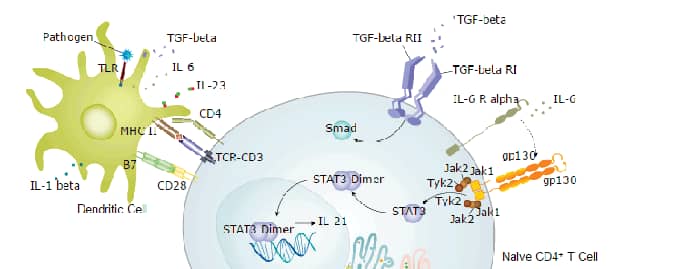
Interleukin 23 (IL-23) is a heterodimeric cytokine composed of two disulfide-linked subunits, a p19 subunit that is unique to IL-23, and a p40 subunit that is shared with IL-12 (1‑5). The functional IL-23 receptor complex consists of two receptor subunits, the IL-12 receptor beta 1 subunit (IL-12 R beta 1) and the IL-23-specific receptor subunit (IL-23 R) (3). Mouse IL-23 R cDNA encodes a 644 amino acid (aa) type I transmembrane protein with a 23 aa residue signal peptide, a 349 aa residue extracellular domain, a 23 aa residue transmembrane domain and a 249 aa residue cytoplasmic region. IL-23 R shares structural features with the IL-12 R beta 2, including an N-terminal Ig-like domain, two cytokine receptor domains and multiple glycosylation sites in the extracellular domain. IL-23 R lacks the three extracellular membrane-proximal fibronectin-type III domains present on IL-12 R beta 2. IL-23 R has a WQPWS sequence in the transmembrane-proximal cytokine receptor domain similar to the cytokine receptor signature WSXWS motif. The cytoplasmic region of IL-23 R has three potential Src homology 2 domain-binding sites and two potential Stat-binding sites. The gene for human IL-23 R is located on human chromosome 1 within 150 kb of IL-12 R beta 2. Human and mouse IL-23 R share 66% amino acid sequence identity. Mouse IL-23 R is expressed in mouse Th1 and Th2 cells, bone marrow, dendritic cells and macrophages. It is also expressed by mouse CD4+ CD45RBlow memory T cells but at much lower levels by mouse CD4+ CD45RBhigh cells. IL-23 initiates a signal transduction cascade similar to that of IL-12 and involves Jak2, Tyk2, STAT1, STAT3, STAT4, and STAT5. IL-23 has biological activities that are similar to, but distinct from, IL-12.

Scientific images may be resized, cropped, or adjusted for brightness or contrast for presentation purposes. These updates are limited to presentation and do not affect the underlying scientific interpretation of the data. Where available, additional source data may be provided upon request.
Key Product Details
Validated by
Species Reactivity
Applications
Label
Antibody Source
Product Specifications
Immunogen
Gly24-Asp372
Accession # Q8K4B4
Specificity
Clonality
Host
Isotype
Scientific Data Images for Mouse IL-23R Antibody (753317)
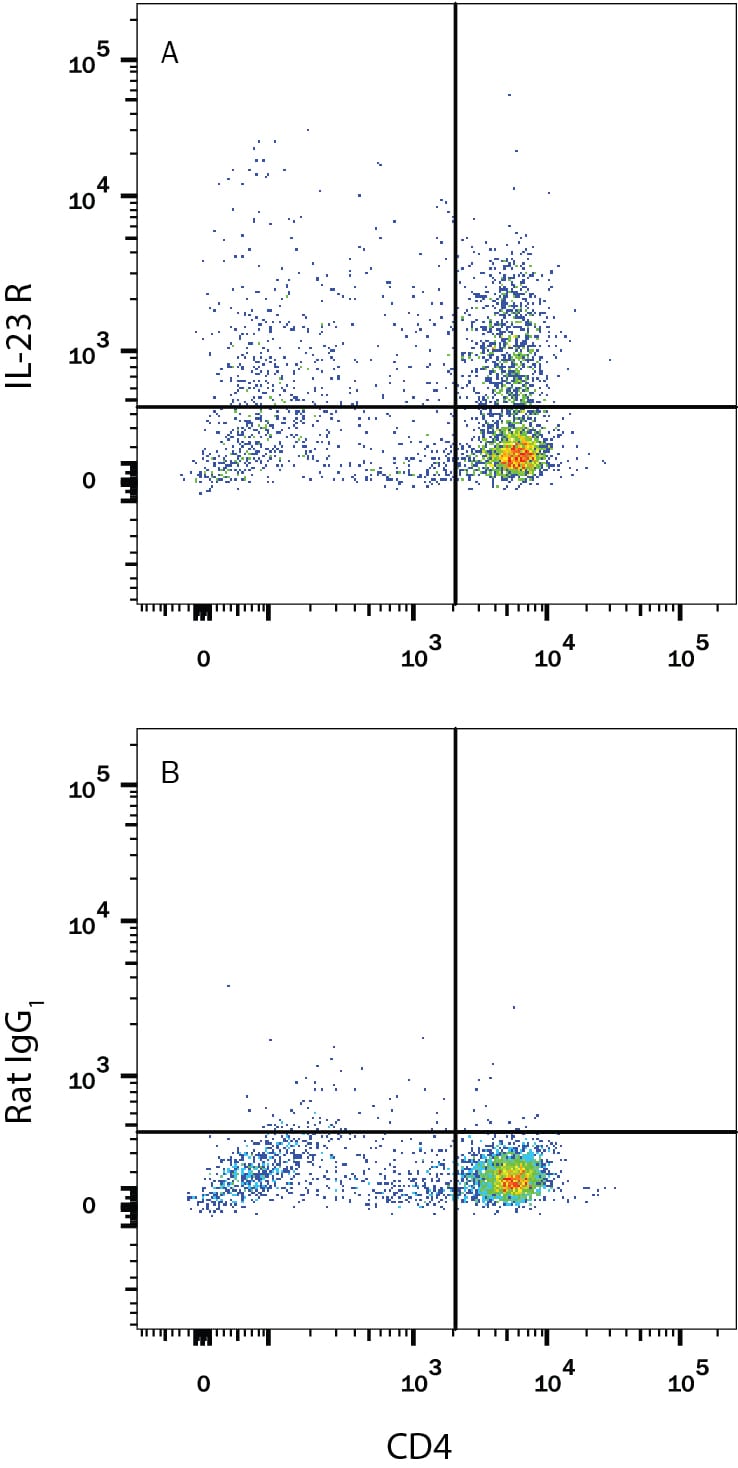
Detection of IL‑23 R in Mouse Splenocytes by Flow Cytometry.
Mouse splenocytes treated with 10 µg/mL Anti-Mouse CD3 Antibody (Catalog # MAB484), 5 µg/mL Anti-Mouse CD28 Antibody (Catalog # AF483), 10 ng/mL Recombinant Mouse TGF-beta 1 (Catalog # 100-B), 20 ng/mL Recombinant Mouse IL-23 (Catalog # 1887-ML), 40 ng/mL Recombinant Mouse IL-6 (Catalog # 406-ML), and 10 ng/mL Recombinant Mouse IL-1 beta (Catalog # 401-ML) for 5 days to induce Th17 activation were stained with Rat Anti-Mouse CD4 PE-conjugated Monoclonal Antibody (Catalog # FAB554P) and either (A) Rat Anti-Mouse IL-23 R APC-conjugated Monoclonal Antibody (Catalog # FAB16861A) or (B) Rat IgG1 APC Isotype Control (Catalog # IC005A). Data shown is from the APC-conjugated version of this clone. Conjugation to a bright fluorochrome is suggested due to the low expression level of IL-23 R. View our protocol for Staining Membrane-associated Proteins.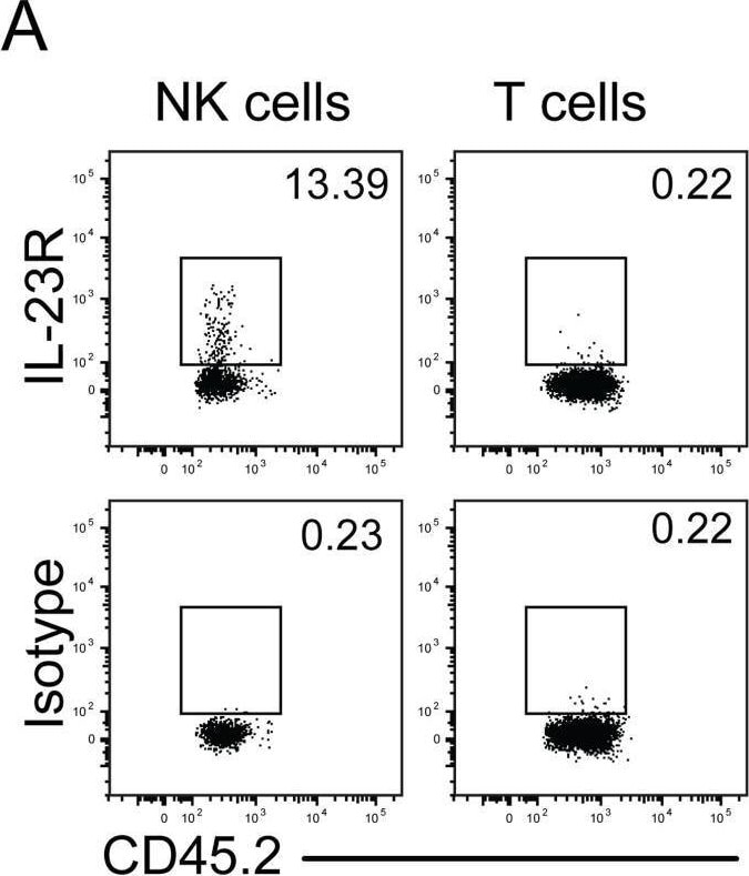
Detection of IL-23R by Flow Cytometry
Defect in IL-23p19 production by Syk-deficient kidney DCs underlies decreased NK cell GM-CSF-mediated control of C. albicans.(A) Representative staining of naïve control kidney NK cells (CD45.2+ NK1.1+ CD3−) and T cells (CD45.2+ CD3+) with anti-IL-23R vs. isotype-matched irrelevant specificity control. Image collected and cropped by CiteAb from the following open publication (https://pubmed.ncbi.nlm.nih.gov/25033445), licensed under a CC-BY license. Not internally tested by R&D Systems.Applications for Mouse IL-23R Antibody (753317)
CyTOF-ready
Flow Cytometry
Sample: Mouse splenocytes induced to Th17 cells with Anti-CD3 Antibody, Anti-CD28 Antibody, Recombinant Mouse TGF-beta 1, Recombinant Mouse IL-23, Recombinant Mouse IL-6, and Recombinant Mouse IL-1 beta for 5 days
Flow Cytometry Panel Builder
Bio-Techne Knows Flow Cytometry
Save time and reduce costly mistakes by quickly finding compatible reagents using the Panel Builder Tool.
Advanced Features
- Spectra Viewer - Custom analysis of spectra from multiple fluorochromes
- Spillover Popups - Visualize the spectra of individual fluorochromes
- Antigen Density Selector - Match fluorochrome brightness with antigen density

Formulation, Preparation, and Storage
Purification
Reconstitution
Reconstitute at 0.5 mg/mL in sterile PBS. For liquid material, refer to CoA for concentration.
Formulation
Shipping
Stability & Storage
- 12 months from date of receipt, -20 to -70 °C as supplied.
- 1 month, 2 to 8 °C under sterile conditions after reconstitution.
- 6 months, -20 to -70 °C under sterile conditions after reconstitution.
Calculators
Background: IL-23R
References
- Oppmann, B. et al. (2000) Immunity 13:715.
- Lankford, C.S. and D.M. Frucht (2003) J. Leukoc. Biol. 73:49.
- Parham, C. et al. (2002) J. Immunol. 168:5448.
- Belladonna, M.L. et al. (2002) J. Immunol. 168:5448.
- Aggarwal, S. et al. (2003) J. Biol. Chem. 278:1910.
Long Name
Alternate Names
Gene Symbol
UniProt
Additional IL-23R Products
Product Documents for Mouse IL-23R Antibody (753317)
Certificate of Analysis
To download a Certificate of Analysis, please enter a lot or batch number in the search box below.
Note: Certificate of Analysis not available for kit components.
Product Specific Notices for Mouse IL-23R Antibody (753317)
For research use only
Related Research Areas
Customer Reviews for Mouse IL-23R Antibody (753317)
There are currently no reviews for this product. Be the first to review Mouse IL-23R Antibody (753317) and earn rewards!
Have you used Mouse IL-23R Antibody (753317)?
Submit a review and receive an Amazon gift card!
$25/€18/£15/$25CAN/¥2500 for a review with an image
$10/€7/£6/$10CAN/¥1110 for a review without an image
Submit a review
Protocols
Find general support by application which include: protocols, troubleshooting, illustrated assays, videos and webinars.
- 7-Amino Actinomycin D (7-AAD) Cell Viability Flow Cytometry Protocol
- Extracellular Membrane Flow Cytometry Protocol
- Flow Cytometry Protocol for Cell Surface Markers
- Flow Cytometry Protocol for Staining Membrane Associated Proteins
- Flow Cytometry Staining Protocols
- Flow Cytometry Troubleshooting Guide
- Intracellular Flow Cytometry Protocol Using Alcohol (Methanol)
- Intracellular Flow Cytometry Protocol Using Detergents
- Intracellular Nuclear Staining Flow Cytometry Protocol Using Detergents
- Intracellular Staining Flow Cytometry Protocol Using Alcohol Permeabilization
- Intracellular Staining Flow Cytometry Protocol Using Detergents to Permeabilize Cells
- Propidium Iodide Cell Viability Flow Cytometry Protocol
- Protocol for Liperfluo
- Protocol for the Characterization of Human Th22 Cells
- Protocol for the Characterization of Human Th9 Cells
- Protocol: Annexin V and PI Staining by Flow Cytometry
- Protocol: Annexin V and PI Staining for Apoptosis by Flow Cytometry
- Troubleshooting Guide: Fluorokine Flow Cytometry Kits
- View all Protocols, Troubleshooting, Illustrated assays and Webinars