Interleukin 3 (IL-3) is a pleiotropic cytokine produced primarily by activated T cells or mast cells. IL-3 stimulates the proliferation and differentiation of hemopoietic cells including the pluripotent hematopoietic stem cells as well as various lineage-committed cells. The biological effects of IL-3 on the various cell types are mediated by the binding of IL-3 to specific cell surface receptor complexes. The functional high-affinity IL-3 receptor is a heterodimer consisting of a ligand binding alpha subunit and the beta subunit. The alpha subunit alone binds IL-3 with low affinity. The beta subunit is required for the high-affinity binding of IL-3 to the heterodimeric receptor complex. The beta subunit has also been found to be a component of the high-affinity receptor complex for IL-5 and GM-CSF and is also referred to as the beta common ( beta c) chain. In the mouse, there are two IL-3 R beta proteins. The first identified mouse IL-3 R beta was also called AIC2A and binds IL-3 with low affinity (1). The second mIL-3 R beta was referred to as AIC2B (2). AIC2B doesn’t bind IL-3 and is the homolog of the human IL-3 R beta. AIC2A was found to be the result of a gene duplication event. Both the alpha and the beta subunits are members of the cytokine receptor superfamily (3).

Key Product Details
Species Reactivity
Validated:
Mouse
Cited:
Mouse
Applications
Validated:
Western Blot, Neutralization, Flow Cytometry, CyTOF-ready
Cited:
Neutralization, Immunocytochemistry/ Immunofluorescence
Label
Unconjugated
Antibody Source
Polyclonal Goat IgG
Loading...
Product Specifications
Immunogen
Mouse myeloma cell line NS0-derived recombinant mouse IL-3 R beta
His23-Trp440
Accession # P26954
His23-Trp440
Accession # P26954
Specificity
Detects mouse IL‑3 R beta in direct ELISAs and Western blots. In direct ELISAs, less than 5% cross-reactivity with recombinant human Common beta chain is observed.
Clonality
Polyclonal
Host
Goat
Isotype
IgG
Endotoxin Level
<0.10 EU per 1 μg of the antibody by the LAL method.
Scientific Data Images for Mouse IL-3R beta Antibody
Cell Proliferation Induced by IL‑3 and Neutralization by Mouse IL‑3 R beta Antibody.
Recombinant Mouse IL-3 (403-ML) stimulates proliferation in the NFS-60 mouse myelogenous leukemia lymphoblast cell line in a dose-dependent manner (orange line). Proliferation elicited by Recombinant Mouse IL-3 (0.5 ng/mL) is neutralized (green line) by increasing concentrations of Goat Anti-Mouse IL-3 R beta Antigen Affinity-purified Polyclonal Antibody (Catalog # AF549). The ND50 is typically 5-15 µg/mL.
Detection of IL-3R beta in mouse bone marrow by Flow Cytometry
Mouse bone marrow cells were stained with Rat Anti-Mouse CD11b/Integrin alpha M PE‑conjugated Monoclonal Antibody (Catalog # FAB1124P) and either (A) Goat Anti-Mouse IL-3R beta Antigen Affinity-purified Polyclonal Antibody (Catalog # AF549) or (B) isotype control antibody (Catalog # AB-108-C) followed by Allophycocyanin-conjugated Anti-Goat IgG Secondary Antibody (Catalog # F0108). View our protocol for Staining Membrane-associated Proteins.Applications for Mouse IL-3R beta Antibody
Application
Recommended Usage
CyTOF-ready
Ready to be labeled using established conjugation methods. No BSA or other carrier proteins that could interfere with conjugation.
Flow Cytometry
0.25 µg/106 cells
Sample: Mouse bone marrow
Sample: Mouse bone marrow
Western Blot
0.1 µg/mL
Sample: Recombinant Mouse IL‑3 R beta Fc Chimera (Catalog # 549-R3)
Sample: Recombinant Mouse IL‑3 R beta Fc Chimera (Catalog # 549-R3)
Neutralization
Measured by its ability to neutralize IL‑3-induced proliferation in the NFS‑60 mouse myelogenous leukemia lymphoblast cell line. Holmes, K. L. et al. (1985) Proc. Natl. Acad. Sci. USA 82:6687. The Neutralization Dose (ND50) is typically 5-15 µg/mL in the presence of 0.5 ng/mL Recombinant Mouse IL‑3.
Reviewed Applications
Read 1 review rated 1 using AF549 in the following applications:
Flow Cytometry Panel Builder
Bio-Techne Knows Flow Cytometry
Save time and reduce costly mistakes by quickly finding compatible reagents using the Panel Builder Tool.
Advanced Features
- Spectra Viewer - Custom analysis of spectra from multiple fluorochromes
- Spillover Popups - Visualize the spectra of individual fluorochromes
- Antigen Density Selector - Match fluorochrome brightness with antigen density

Formulation, Preparation, and Storage
Purification
Antigen Affinity-purified
Reconstitution
Reconstitute at 0.2 mg/mL in sterile PBS. For liquid material, refer to CoA for concentration.
Loading...
Formulation
Lyophilized from a 0.2 μm filtered solution in PBS with Trehalose. See Certificate of Analysis for details.
*Small pack size (-SP) is supplied either lyophilized or as a 0.2 µm filtered solution in PBS.
*Small pack size (-SP) is supplied either lyophilized or as a 0.2 µm filtered solution in PBS.
Shipping
Lyophilized product is shipped at ambient temperature. Liquid small pack size (-SP) is shipped with polar packs. Upon receipt, store immediately at the temperature recommended below.
Stability & Storage
Use a manual defrost freezer and avoid repeated freeze-thaw cycles.
- 12 months from date of receipt, -20 to -70 °C as supplied.
- 1 month, 2 to 8 °C under sterile conditions after reconstitution.
- 6 months, -20 to -70 °C under sterile conditions after reconstitution.
Calculators
Background: IL-3R beta
References
- Itoh, N. et al. (1990) Science 247:324.
- Gorman, D.M. et al. (1990) Proc. Natl. Acad. Sci. USA 87:5459.
- Schrader, J.W. in Cytokine Reference, (2001) J.J. Oppenheim and M. Feldmann, eds. Academic Press, New York, p. 1899.
Long Name
Interleukin 3 Receptor beta
Alternate Names
IL-3R beta, IL-3Rb, IL3R beta
Entrez Gene IDs
12984 (Mouse)
Gene Symbol
CSF2RB2
UniProt
Additional IL-3R beta Products
Product Documents for Mouse IL-3R beta Antibody
Certificate of Analysis
To download a Certificate of Analysis, please enter a lot or batch number in the search box below.
Note: Certificate of Analysis not available for kit components.
Product Specific Notices for Mouse IL-3R beta Antibody
For research use only
Citations for Mouse IL-3R beta Antibody
 Powered by Bioz
Customer Reviews for Mouse IL-3R beta Antibody (1)
1 out of 5
1 Customer Rating
Have you used Mouse IL-3R beta Antibody?
Submit a review and receive an Amazon gift card!
$25/€18/£15/$25CAN/¥2500 Yen for a review with an image
$10/€7/£6/$10CAN/¥1110 Yen for a review without an image
Submit a review
Customer Images
Showing
1
-
1 of
1 review
Showing All
Filter By:
-
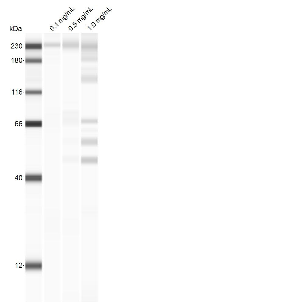
Application: Simple WesternSample Tested: Mouse SpleenSpecies: MouseVerified Customer | Posted 06/04/2019Homogenized mouse spleen probed at the indicated concentrations, using a 1:25 dil of the antibody.

There are no reviews that match your criteria.
Protocols
Find general support by application which include: protocols, troubleshooting, illustrated assays, videos and webinars.
- 7-Amino Actinomycin D (7-AAD) Cell Viability Flow Cytometry Protocol
- Cellular Response to Hypoxia Protocols
- Extracellular Membrane Flow Cytometry Protocol
- Flow Cytometry Protocol for Cell Surface Markers
- Flow Cytometry Protocol for Staining Membrane Associated Proteins
- Flow Cytometry Staining Protocols
- Flow Cytometry Troubleshooting Guide
- Intracellular Flow Cytometry Protocol Using Alcohol (Methanol)
- Intracellular Flow Cytometry Protocol Using Detergents
- Intracellular Nuclear Staining Flow Cytometry Protocol Using Detergents
- Intracellular Staining Flow Cytometry Protocol Using Alcohol Permeabilization
- Intracellular Staining Flow Cytometry Protocol Using Detergents to Permeabilize Cells
- Propidium Iodide Cell Viability Flow Cytometry Protocol
- Protocol for Liperfluo
- Protocol for the Characterization of Human Th22 Cells
- Protocol for the Characterization of Human Th9 Cells
- Protocol: Annexin V and PI Staining by Flow Cytometry
- Protocol: Annexin V and PI Staining for Apoptosis by Flow Cytometry
- R&D Systems Quality Control Western Blot Protocol
- Troubleshooting Guide: Fluorokine Flow Cytometry Kits
- Troubleshooting Guide: Western Blot Figures
- Western Blot Conditions
- Western Blot Protocol
- Western Blot Protocol for Cell Lysates
- Western Blot Troubleshooting
- Western Blot Troubleshooting Guide
- View all Protocols, Troubleshooting, Illustrated assays and Webinars
Loading...