IRF1 is a member of the interferon regulator transcription factor family, a family characterized by a helix-turn-helix DNA binding domain enriched in tryptophan repeats. IRF family members show diverse cellular regulation of interferon-stimulated gene transcription, viral-mediated gene activation, apoptosis, differentiation, and cellular growth. IRF1 knock out mice show defects in NK and T cell function, as well as, develop CD30+ lymphoproliferative disease.

Key Product Details
Species Reactivity
Validated:
Mouse
Cited:
Mouse
Applications
Validated:
Western Blot
Cited:
Western Blot, Immunocytochemistry
Label
Unconjugated
Antibody Source
Polyclonal Goat IgG
Loading...
Product Specifications
Immunogen
E. coli-derived recombinant mouse IRF1
Thr147-Pro329
Accession # P15314
Thr147-Pro329
Accession # P15314
Specificity
Detects mouse IRF1 in Western blots.
Clonality
Polyclonal
Host
Goat
Isotype
IgG
Scientific Data Images for Mouse IRF1 Antibody
Detection of Mouse IRF1 by Western Blot.
Western blot shows lysates of NIH-3T3 mouse embryonic fibroblast cell line. Gels were loaded with 30 µg of whole cell lysate (WCL), 20 µg of cytoplasmic (Cyto), and 10 µg of nuclear extracts (Nuc). PVDF membrane was probed with 1 µg/mL Mouse IRF1 Antigen Affinity-purified Polyclonal Antibody (Catalog # AF4715) followed by HRP-conjugated Anti-Goat IgG Secondary Antibody (Catalog # HAF017). A specific band for IRF1 was detected at approximately 50 kDa (as indicated). This experiment was conducted under reducing conditions and using Immunoblot Buffer Group 1.
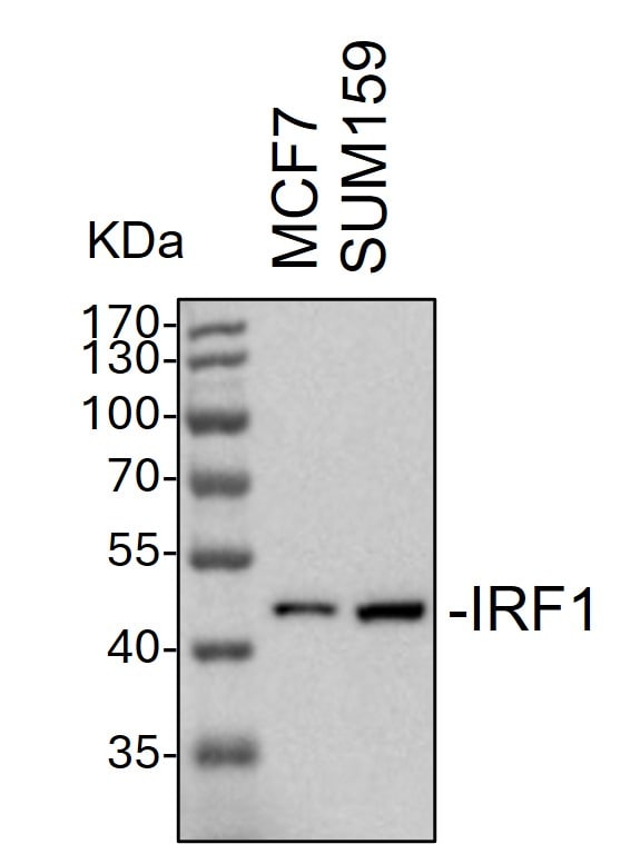
Detection of Mouse IRF1 antibody by Western Blot.
Whole cell lysates from MCF7 and SUM159 cells were loaded with 50 ug/lane.10% SDS-PAGE. IRF1 Antibody (AF4715) was used for primary antibody: 1:1000, 4℃, overnight. Image from a verified customer review.Applications for Mouse IRF1 Antibody
Application
Recommended Usage
Western Blot
1 µg/mL
Sample: NIH-3T3 mouse embryonic fibroblast cell line
Sample: NIH-3T3 mouse embryonic fibroblast cell line
Reviewed Applications
Read 2 reviews rated 4.5 using AF4715 in the following applications:
Formulation, Preparation, and Storage
Purification
Antigen Affinity-purified
Reconstitution
Reconstitute at 0.2 mg/mL in sterile PBS. For liquid material, refer to CoA for concentration.
Loading...
Formulation
Lyophilized from a 0.2 μm filtered solution in PBS with Trehalose. See Certificate of Analysis for details.
*Small pack size (-SP) is supplied either lyophilized or as a 0.2 µm filtered solution in PBS.
*Small pack size (-SP) is supplied either lyophilized or as a 0.2 µm filtered solution in PBS.
Shipping
Lyophilized product is shipped at ambient temperature. Liquid small pack size (-SP) is shipped with polar packs. Upon receipt, store immediately at the temperature recommended below.
Stability & Storage
Use a manual defrost freezer and avoid repeated freeze-thaw cycles.
- 12 months from date of receipt, -20 to -70 °C as supplied.
- 1 month, 2 to 8 °C under sterile conditions after reconstitution.
- 6 months, -20 to -70 °C under sterile conditions after reconstitution.
Calculators
Background: IRF1
Long Name
Interferon Regulatory Factor 1
Alternate Names
MAR
Gene Symbol
IRF1
UniProt
Additional IRF1 Products
Product Documents for Mouse IRF1 Antibody
Certificate of Analysis
To download a Certificate of Analysis, please enter a lot or batch number in the search box below.
Note: Certificate of Analysis not available for kit components.
Product Specific Notices for Mouse IRF1 Antibody
For research use only
Related Research Areas
Citations for Mouse IRF1 Antibody
 Powered by Bioz
Customer Reviews for Mouse IRF1 Antibody (2)
4.5 out of 5
2 Customer Ratings
Have you used Mouse IRF1 Antibody?
Submit a review and receive an Amazon gift card!
$25/€18/£15/$25CAN/¥2500 Yen for a review with an image
$10/€7/£6/$10CAN/¥1110 Yen for a review without an image
Submit a review
Customer Images
Showing
1
-
2 of
2 reviews
Showing All
Filter By:
-
Application: Western BlotSample Tested: breast cancer MCF7 cells and SUM-159PTSpecies: HumanVerified Customer | Posted 11/28/2024Western Blot: whole cell lysates from MCF7 and SUM159 cells were loaded with 50 ug/lane. 10% SDS-PAGE. IRF1 Antibody (AF4715) was used for primary antibody: 1:1000, 4℃, overnight.
 Bio-Techne ResponseThis review was submitted through the legacy Novus Innovators Program, reflecting a new species or application tested on a primary antibody.
 -
Application: Western BlotSample Tested: See PMID 22464096Species: MouseVerified Customer | Posted 01/08/2015
There are no reviews that match your criteria.
Protocols
Find general support by application which include: protocols, troubleshooting, illustrated assays, videos and webinars.
- Cellular Response to Hypoxia Protocols
- R&D Systems Quality Control Western Blot Protocol
- Troubleshooting Guide: Western Blot Figures
- Western Blot Conditions
- Western Blot Protocol
- Western Blot Protocol for Cell Lysates
- Western Blot Troubleshooting
- Western Blot Troubleshooting Guide
- View all Protocols, Troubleshooting, Illustrated assays and Webinars
Loading...