CD44 is a ubiquitously expressed protein that is the major receptor for hyaluronan and exerts control over cell growth and migration (1 - 5). Mouse CD44 has a 22 amino acid (aa) signal sequence, an extracellular domain (ECD) with a 100 aa hyaluronan-binding disulfide-stabilized link region and a 48-463 aa stem region, a 21 aa transmembrane domain, and a 72 aa cytoplasmic domain. Within the stem, ten variably spliced exons (v1-10, exons 6-15) produce multiple protein isoforms (1‑5). The standard or hematopoietic form, CD44H, does not include the variable segments (1‑5). Cancer aggressiveness and T cell activation have been correlated with expression of specific isoforms (2, 4). With variable N- and O-glycosylation and splicing within the stalk, CD44 can range from 80 to 200 kDa (1, 2). Within the N‑terminal invariant portion of the ECD (aa 23-222), mouse CD44 shares 92%, 77%, 77%, 79% and 71% identity with corresponding rat, human, equine, canine and bovine CD44, respectively. The many reported functions of CD44 fall within three categories (1, 2). First, CD44 binds hyaluronan and other ligands within the extracellular matrix and can function as a "platform" for growth factors and metalloproteinases. Second, CD44 is a co-receptor that modifies activity of receptors including MET and the ErbB family of tyrosine kinases. Third, the CD44 intracellular domain links the plasma membrane to the actin cytoskeleton via the ERM proteins, ezrin, radixin and moesin. CD44 can be synthesized in a soluble form (4) or may be cleaved at multiple sites by either membrane-type matrix metalloproteinases, or ADAM proteases to produce soluble ectodomains (6, 7). The cellular portion may then undergo gamma secretase-dependent intramembrane cleavage to form an A beta ‑like transmembrane portion and a cytoplasmic signaling portion that affects gene expression (8, 9). These cleavage events are thought to promote metastasis by enhancing tumor cell motility and growth (1, 2, 6).

Key Product Details
Species Reactivity
Validated:
Mouse, Rat, Porcine, Equine
Cited:
Human, Mouse
Applications
Validated:
Western Blot, Blockade of Receptor-ligand Interaction, Flow Cytometry, Immunocytochemistry, Simple Western, CyTOF-ready
Cited:
Immunohistochemistry, Immunohistochemistry-Paraffin, Western Blot, Neutralization
Label
Unconjugated
Antibody Source
Polyclonal Sheep IgG
Loading...
Product Specifications
Immunogen
Chinese hamster ovary cell line CHO-derived recombinant mouse CD44
Gln25-Thr224
Accession # NP_033981
Gln25-Thr224
Accession # NP_033981
Specificity
Detects mouse and rat CD44 in direct ELISAs and Western blots. In direct ELISAs, approximately 35% cross-reactivity with recombinant human CD44 is observed.
Clonality
Polyclonal
Host
Sheep
Isotype
IgG
Endotoxin Level
<0.10 EU per 1 μg of the antibody by the LAL method.
Scientific Data Images for CD44 Antibody
Detection of Mouse and Rat CD44 by Western Blot.
Western blot shows lysates of RAW 264.7 mouse monocyte/macrophage cell line, mouse spleen tissue, mouse lymph node tissue, and rat brain tissue. PVDF membrane was probed with 1 µg/mL of Sheep Anti-Mouse/Rat/Porcine/Equine CD44 Antigen Affinity-purified Polyclonal Antibody (Catalog # AF6127) followed by HRP-conjugated Anti-Sheep IgG Secondary Antibody (Catalog # HAF016). Specific bands were detected for CD44 at approximately 80 to 100 kDa (as indicated). This experiment was conducted under reducing conditions and using Immunoblot Buffer Group 1.
Detection of CD44 in Mouse Splenocytes by Flow Cytometry.
Mouse splenocytes were stained with Sheep Anti-Mouse/Rat/Porcine/Equine CD44 Antigen Affinity-purified Polyclonal Antibody (Catalog # AF6127, filled histogram) or control antibody (Catalog # 5-001-A, open histogram), followed by Allophycocyanin-conjugated Anti-Sheep IgG Secondary Antibody (Catalog # F0127).
Detection of CD44 in Rat Splenocytes by Flow Cytometry.
Rat splenocytes were stained with Sheep Anti-Mouse/Rat/Porcine/Equine CD44 Antigen Affinity-purified Polyclonal Antibody (Catalog # AF6127, filled histogram) or control antibody (Catalog # 5-001-A, open histogram), followed by Allophycocyanin-conjugated Anti-Sheep IgG Secondary Antibody (Catalog # F0127).
Detection of CD44 in Porcine Mesenchymal Stem Cells by Flow Cytometry.
Porcine mesenchymal stem cells were stained with Sheep Anti-Mouse/Rat/Porcine/Equine CD44 Antigen Affinity-purified Polyclonal Antibody (Catalog # AF6127, filled histogram) or isotype control antibody (Catalog # 5-001-A, open histogram), followed by Phycoerythrin-conjugated Anti-Sheep IgG Secondary Antibody (Catalog # F0126).
Detection of CD44 in Equine PBMCs by Flow Cytometry.
Equine peripheral blood mononuclear cells (PBMCs) were stained with Sheep Anti-Mouse/Rat/Porcine/Equine CD44 Antigen Affinity-purified Polyclonal Antibody (Catalog # AF6127, filled histogram) or isotype control antibody (Catalog # 5-001-A, open histogram), followed by Phycoerythrin-conjugated Anti-Sheep IgG Secondary Antibody (Catalog # F0126).
CD44 in Mouse Splenocytes.
CD44 was detected in immersion fixed mouse splenocytes using Sheep Anti-Mouse/Rat/Porcine/Equine CD44 Antigen Affinity-purified Polyclonal Antibody (Catalog # AF6127) at 10 µg/mL for 3 hours at room temperature. Cells were stained using the NorthernLights™ 557-conjugated Anti-Sheep IgG Secondary Antibody (red, upper panel; Catalog # NL010) and counterstained with DAPI (blue, lower panel). Specific staining was localized to cell surfaces. View our protocol for Fluorescent ICC Staining of Non-adherent Cells.
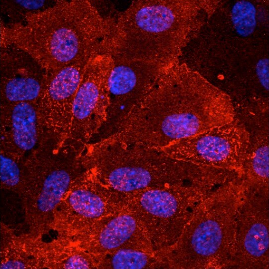
CD44 in Porcine Mesenchymal Stem Cells.
CD44 was detected in immersion fixed porcine mesenchymal stem cells using Sheep Anti-Mouse/Rat/Porcine/Equine CD44 Antigen Affinity-purified Polyclonal Antibody (Catalog # AF6127) at 10 µg/mL for 3 hours at room temperature. Cells were stained using the NorthernLights™ 557-conjugated Anti-Sheep IgG Secondary Antibody (red; Catalog # NL010) and counterstained with DAPI (blue). Specific staining was localized to cell surfaces. View our protocol for Fluorescent ICC Staining of Stem Cells on Coverslips.
Detection of Rat CD44 by Simple WesternTM.
Simple Western lane view shows lysates of NR8383 rat alveolar macrophage cell line, loaded at 0.2 mg/mL. A specific band was detected for CD44 at approximately 169 kDa (as indicated) using 10 µg/mL of Sheep Anti-Mouse/Rat/Porcine/Equine CD44 Antigen Affinity-purified Polyclonal Antibody (Catalog # AF6127) followed by 1:50 dilution of HRP-conjugated Anti-Sheep IgG Secondary Antibody (Catalog # HAF016). This experiment was conducted under reducing conditions and using the 12-230 kDa separation system.Applications for CD44 Antibody
Application
Recommended Usage
Blockade of Receptor-ligand Interaction
In a functional ELISA, 1-6 µg/mL of this antibody will block 50% of the binding of 25 ng/mL biotinylated Hyaluronan (132 kDa) to immobilized Recombinant Mouse CD44 Fc Chimera (Catalog # 6127-CD) coated at 1 µg/mL (100 μL/well). At 20 µg/mL, this antibody will block >90% of the binding.
CyTOF-ready
Ready to be labeled using established conjugation methods. No BSA or other carrier proteins that could interfere with conjugation.
Flow Cytometry
2.5 µg/106 cells
Sample: Mouse splenocytes, rat splenocytes, porcine mesenchymal stem cells, and equine peripheral blood mononuclear cells (PBMCs)
Sample: Mouse splenocytes, rat splenocytes, porcine mesenchymal stem cells, and equine peripheral blood mononuclear cells (PBMCs)
Immunocytochemistry
5-15 µg/mL
Sample: Immersion fixed mouse splenocytes and porcine mesenchymal stem cells
Sample: Immersion fixed mouse splenocytes and porcine mesenchymal stem cells
Simple Western
10 µg/mL
Sample: NR8383 rat alveolar macrophage cell line
Sample: NR8383 rat alveolar macrophage cell line
Western Blot
1 µg/mL
Sample: RAW 264.7 mouse monocyte/macrophage cell line, mouse spleen tissue, mouse lymph node tissue, and rat brain tissue
Sample: RAW 264.7 mouse monocyte/macrophage cell line, mouse spleen tissue, mouse lymph node tissue, and rat brain tissue
Reviewed Applications
Read 2 reviews rated 4.5 using AF6127 in the following applications:
Flow Cytometry Panel Builder
Bio-Techne Knows Flow Cytometry
Save time and reduce costly mistakes by quickly finding compatible reagents using the Panel Builder Tool.
Advanced Features
- Spectra Viewer - Custom analysis of spectra from multiple fluorochromes
- Spillover Popups - Visualize the spectra of individual fluorochromes
- Antigen Density Selector - Match fluorochrome brightness with antigen density

Formulation, Preparation, and Storage
Purification
Antigen Affinity-purified
Reconstitution
Sterile PBS to a final concentration of 0.2 mg/mL. For liquid material, refer to CoA for concentration.
Loading...
Formulation
Lyophilized from a 0.2 μm filtered solution in PBS with Trehalose. *Small pack size (SP) is supplied either lyophilized or as a 0.2 µm filtered solution in PBS.
Shipping
Lyophilized product is shipped at ambient temperature. Liquid small pack size (-SP) is shipped with polar packs. Upon receipt, store immediately at the temperature recommended below.
Stability & Storage
Use a manual defrost freezer and avoid repeated freeze-thaw cycles.
- 12 months from date of receipt, -20 to -70 °C as supplied.
- 1 month, 2 to 8 °C under sterile conditions after reconstitution.
- 6 months, -20 to -70 °C under sterile conditions after reconstitution.
Calculators
Background: CD44
References
- Pure, E. and R.K. Assoian (2009) Cell. Signal. 21:651.
- Ponta, H. et al. (2003) Nat. Rev. Mol. Cell Biol. 4:33.
- Screaton, G.R. et al. (1992) Proc. Natl. Acad. Sci. USA 89:12160.
- Lynch, K.W. (2004) Nat. Rev. Immunol. 4:931.
- Yu, Q. and B.P. Toole (1996) J. Biol. Chem. 271:20603.
- Nagano, O. and H. Saya (2004) Cancer Sci. 95:930.
- Nakamura, H. et al. (2004) Cancer Res. 64:876.
- Murakami, D. et al. (2003) Oncogene 22:1511.
- Lammich, S. et al. (2002) J. Biol. Chem. 277:44754.
Alternate Names
CD44, ECMR-III, HCAM, HCELL, LHR, MDU2, MDU3, MIC4, MUTCH-I, Pgp1
Gene Symbol
CD44
UniProt
Additional CD44 Products
Product Documents for CD44 Antibody
Certificate of Analysis
To download a Certificate of Analysis, please enter a lot or batch number in the search box below.
Note: Certificate of Analysis not available for kit components.
Product Specific Notices for CD44 Antibody
For research use only
Related Research Areas
Citations for CD44 Antibody
 Powered by Bioz
Customer Reviews for CD44 Antibody (2)
4.5 out of 5
2 Customer Ratings
Have you used CD44 Antibody?
Submit a review and receive an Amazon gift card!
$25/€18/£15/$25CAN/¥2500 Yen for a review with an image
$10/€7/£6/$10CAN/¥1110 Yen for a review without an image
Submit a review
Customer Images
Showing
1
-
2 of
2 reviews
Showing All
Filter By:
-
Application: Immunocytochemistry/ImmunofluorescenceSample Tested: HUVEC human umbilical vein endothelial cellsSpecies: HumanVerified Customer | Posted 09/27/2017This antibody works great for my staining.

-
Application: Western BlotSample Tested: See PMID 23645665Species: RatVerified Customer | Posted 01/08/2015
There are no reviews that match your criteria.
Protocols
Find general support by application which include: protocols, troubleshooting, illustrated assays, videos and webinars.
- 7-Amino Actinomycin D (7-AAD) Cell Viability Flow Cytometry Protocol
- Appropriate Fixation of IHC/ICC Samples
- Cellular Response to Hypoxia Protocols
- ClariTSA™ Fluorophore Kits
- Detection & Visualization of Antibody Binding
- Extracellular Membrane Flow Cytometry Protocol
- Flow Cytometry Protocol for Cell Surface Markers
- Flow Cytometry Protocol for Staining Membrane Associated Proteins
- Flow Cytometry Staining Protocols
- Flow Cytometry Troubleshooting Guide
- ICC Cell Smear Protocol for Suspension Cells
- ICC Immunocytochemistry Protocol Videos
- ICC for Adherent Cells
- Immunocytochemistry (ICC) Protocol
- Immunocytochemistry Troubleshooting
- Immunofluorescence of Organoids Embedded in Cultrex Basement Membrane Extract
- Immunohistochemistry (IHC) and Immunocytochemistry (ICC) Protocols
- Intracellular Flow Cytometry Protocol Using Alcohol (Methanol)
- Intracellular Flow Cytometry Protocol Using Detergents
- Intracellular Nuclear Staining Flow Cytometry Protocol Using Detergents
- Intracellular Staining Flow Cytometry Protocol Using Alcohol Permeabilization
- Intracellular Staining Flow Cytometry Protocol Using Detergents to Permeabilize Cells
- Preparing Samples for IHC/ICC Experiments
- Preventing Non-Specific Staining (Non-Specific Binding)
- Primary Antibody Selection & Optimization
- Propidium Iodide Cell Viability Flow Cytometry Protocol
- Protocol for Liperfluo
- Protocol for VisUCyte™ HRP Polymer Detection Reagent
- Protocol for the Characterization of Human Th22 Cells
- Protocol for the Characterization of Human Th9 Cells
- Protocol for the Fluorescent ICC Staining of Cell Smears - Graphic
- Protocol for the Fluorescent ICC Staining of Cultured Cells on Coverslips - Graphic
- Protocol for the Preparation and Fluorescent ICC Staining of Cells on Coverslips
- Protocol for the Preparation and Fluorescent ICC Staining of Non-adherent Cells
- Protocol for the Preparation and Fluorescent ICC Staining of Stem Cells on Coverslips
- Protocol for the Preparation of a Cell Smear for Non-adherent Cell ICC - Graphic
- Protocol: Annexin V and PI Staining by Flow Cytometry
- Protocol: Annexin V and PI Staining for Apoptosis by Flow Cytometry
- R&D Systems Quality Control Western Blot Protocol
- TUNEL and Active Caspase-3 Detection by IHC/ICC Protocol
- The Importance of IHC/ICC Controls
- Troubleshooting Guide: Fluorokine Flow Cytometry Kits
- Troubleshooting Guide: Western Blot Figures
- Western Blot Conditions
- Western Blot Protocol
- Western Blot Protocol for Cell Lysates
- Western Blot Troubleshooting
- Western Blot Troubleshooting Guide
- View all Protocols, Troubleshooting, Illustrated assays and Webinars
Loading...
Associated Pathways


