Serpin A3N is a serine protease inhibitor that is structurally related to alpha 1-antichymotrypsin encoded by the SERPINA3 gene (1). Serpin A3N is highly expressed in brain, testis, lung, thymus, and spleen (2). Serpin A3N secreted by Sertoli cells may regulate the activity of locally produced Granzyme B (3). Granzyme B inhibition by Serpin A3N may therefore regulate Granzyme B-mediated killing by cytotoxic lymphocytes, providing a means to disable cell-mediated immune responses.

Key Product Details
Species Reactivity
Validated:
Mouse
Cited:
Human, Mouse, Transgenic Mouse
Applications
Validated:
Western Blot, Immunoprecipitation
Cited:
Immunohistochemistry, Western Blot, Immunofluorescence, Immunocytochemistry, Immunoprecipitation, In vivo assay
Label
Unconjugated
Antibody Source
Polyclonal Goat IgG
Loading...
Product Specifications
Immunogen
Mouse myeloma cell line NS0-derived recombinant mouse Serpin A3N
Phe21-Lys418
Accession # Q91WP6
Phe21-Lys418
Accession # Q91WP6
Specificity
Detects mouse Serpin A3N in direct ELISAs and Western blots. In direct ELISAs, less than 2% cross-reactivity with recombinant mouse (rm) Serpin A1, recombinant human Serpin A3 and rmSerpin A5 is observed.
Clonality
Polyclonal
Host
Goat
Isotype
IgG
Scientific Data Images for Mouse Serpin A3N Antibody
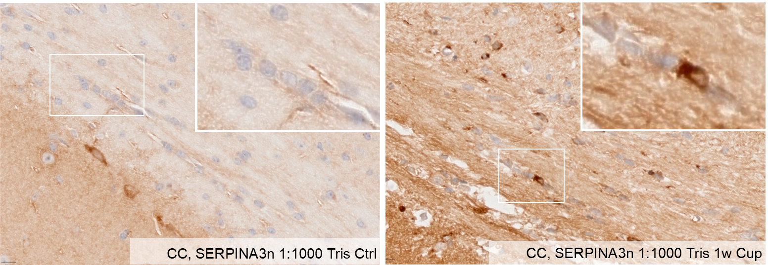
Serpin A3N in Mouse Brain Tissue.
Serpin A3N + labelled oligodendrocytes can be seen in the Corpus callosum of cuprizone-intoxicated mice. Primary antibody: Serpin A3N 1:1000, 18h incubation at 4°CSecondary antibody: rabbit anti goat, 1:200, 21°C, 1h incubation. Visualization via DAB. Image from a verified customer review.
Detection of Mouse Serpin A3N by Western Blot
HS diet increased the serpina3n expression in the hippocampus of APP/PS1 mice. (A) GSEA showed that the HS diet intervention changed the status of axon guidance, neuroactive ligand–receptor interaction, oxidoreductase activity, and blood vessel diameter. (B) A radar map of differential gene expression levels. Serpina3n is most significantly changed, and its mRNA expression is the highest among the serpin family. (C) Protein level of serpina3n was assessed using Western blotting; GAPDH was used as a control. n = 4 biologically independent animals. (D) mRNA levels of the serpin family in HS- and ND-treated APP/PS1 mice were compared; serpina3n is the most remarkably changed mRNA. Image collected and cropped by CiteAb from the following open publication (https://www.mdpi.com/1422-0067/25/21/11731), licensed under a CC-BY license. Not internally tested by R&D Systems.Applications for Mouse Serpin A3N Antibody
Application
Recommended Usage
Immunoprecipitation
25 µg/mL
Sample: Conditioned cell culture medium spiked with Recombinant Mouse Serpin A3N (Catalog # 4709-PI), see our available Western blot detection antibodies
Sample: Conditioned cell culture medium spiked with Recombinant Mouse Serpin A3N (Catalog # 4709-PI), see our available Western blot detection antibodies
Western Blot
0.1 µg/mL
Sample: Recombinant Mouse Serpin A3N (Catalog # 4709-PI)
Sample: Recombinant Mouse Serpin A3N (Catalog # 4709-PI)
Reviewed Applications
Read 2 reviews rated 5 using AF4709 in the following applications:
Formulation, Preparation, and Storage
Purification
Antigen Affinity-purified
Reconstitution
Reconstitute at 0.2 mg/mL in sterile PBS. For liquid material, refer to CoA for concentration.
Loading...
Formulation
Lyophilized from a 0.2 μm filtered solution in PBS with Trehalose. *Small pack size (SP) is supplied either lyophilized or as a 0.2 µm filtered solution in PBS.
Shipping
Lyophilized product is shipped at ambient temperature. Liquid small pack size (-SP) is shipped with polar packs. Upon receipt, store immediately at the temperature recommended below.
Stability & Storage
Use a manual defrost freezer and avoid repeated freeze-thaw cycles.
- 12 months from date of receipt, -20 to -70 °C as supplied.
- 1 month, 2 to 8 °C under sterile conditions after reconstitution.
- 6 months, -20 to -70 °C under sterile conditions after reconstitution.
Calculators
Background: Serpin A3N
References
- Forsyth, S. et al. (2003) Genomics 81:336.
- Horvath, A. J. et al. (2004) J. Mol. Evol. 59:488.
- Hirst, C. E. et al. (2001) Mol. Hum. Reprod. 7:1133.
Alternate Names
Spi2
Gene Symbol
SERPINA3N
UniProt
Additional Serpin A3N Products
Product Documents for Mouse Serpin A3N Antibody
Certificate of Analysis
To download a Certificate of Analysis, please enter a lot or batch number in the search box below.
Note: Certificate of Analysis not available for kit components.
Product Specific Notices for Mouse Serpin A3N Antibody
For research use only
Related Research Areas
Citations for Mouse Serpin A3N Antibody
 Powered by Bioz
Customer Reviews for Mouse Serpin A3N Antibody (2)
5 out of 5
2 Customer Ratings
Have you used Mouse Serpin A3N Antibody?
Submit a review and receive an Amazon gift card!
$25/€18/£15/$25CAN/¥2500 Yen for a review with an image
$10/€7/£6/$10CAN/¥1110 Yen for a review without an image
Submit a review
Customer Images
Showing
1
-
2 of
2 reviews
Showing All
Filter By:
-
Application: Immunohistochemistry-ParaffinSample Tested: Brain tissueSpecies: MouseVerified Customer | Posted 06/17/2025Serpin A3N + labelled oligodendrocytes can be seen in the Corpus callosum of cuprizone-intoxicated micePrimary antibody: Serpina3n 1:1000, 18h incubation at 4°C Secondary antibody: rabbit anti goat, 1:200, 21°C, 1h incubation Visualisation via DAB
 Bio-Techne ResponseThis review reflects a new species or application tested on a primary antibody.
 -
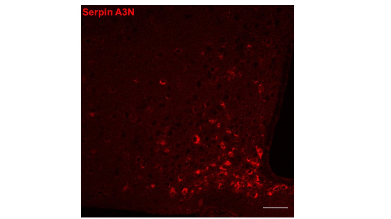
Application: Immunocytochemistry/ImmunofluorescenceSample Tested: Adult brainSpecies: MouseVerified Customer | Posted 07/08/2021The red showed serpin A3N in the ARC of hypothalamus. Scale bar: 50 um. Primary dilution: 1 : 250 Secondary donkey anti goat 555, dilution 1 : 1000
There are no reviews that match your criteria.
Protocols
Find general support by application which include: protocols, troubleshooting, illustrated assays, videos and webinars.
- Cellular Response to Hypoxia Protocols
- Immunoprecipitation Protocol
- R&D Systems Quality Control Western Blot Protocol
- Troubleshooting Guide: Western Blot Figures
- Western Blot Conditions
- Western Blot Protocol
- Western Blot Protocol for Cell Lysates
- Western Blot Troubleshooting
- Western Blot Troubleshooting Guide
- View all Protocols, Troubleshooting, Illustrated assays and Webinars
Loading...