SOST, also known as sclerostin, is a member of the cerberus/DAN family, a group of secreted glycoproteins characterized by a cysteine-knot motif. Cerberus/DAN family members are putative BMP antagonists, and include Dan, Cerberus, Gremlin, PRDC, and Caronte. While the overall sequence identity between members of the family is low, they have conserved spacing of six cysteine residues. Cerberus and Dan have an additional cysteine residue used for dimerization; however, SOST does not and is secreted as a monomer. SOST was originally identified as an important regulator of bone homeostasis. Positional cloning studies identified that mutations in the SOST gene can cause sclerosteosis and van Buchem disease, bone dysplasia disorders characterized by progressive skeletal overgrowth. Significant levels of SOST expression are detected in bone, cartilage, kidney, and liver. SOST is expressed by osteoclasts in developing bones of mouse embryos, including both intramembranously forming skull bones and endochondrally forming long bones. SOST plays a physiological role as a negative regulator of bone formation by repressing BMP-induced osteogenesis. SOST has been shown to have unique ligand specificity, binding BMP-5, -6, and -7 with high affinity and BMP-2 and -4 with low affinity. This seems to be the first example of a BMP antagonist being localized to osteoclasts, cells derived from the hematopoietic lineage, that function to degrade bone matrix. Human and mouse SOST share 88% amino acid identity (1-3).

Key Product Details
Species Reactivity
Validated:
Mouse
Cited:
Human, Mouse, Rat, Transgenic Mouse, Xenograft
Applications
Validated:
Immunohistochemistry, Western Blot
Cited:
Immunohistochemistry, Immunohistochemistry-Paraffin, Immunohistochemistry-Frozen, Western Blot, Neutralization, Flow Cytometry, Immunocytochemistry
Label
Unconjugated
Antibody Source
Polyclonal Goat IgG
Loading...
Product Specifications
Immunogen
Mouse myeloma cell line NS0-derived recombinant mouse SOST/Sclerostin
Gln24-Tyr211
Accession # NP_077769
Gln24-Tyr211
Accession # NP_077769
Specificity
Detects mouse SOST/Sclerostin in direct ELISAs and Western blots. In direct ELISAs, less than 5% cross-reactivity with recombinant human SOST is observed.
Clonality
Polyclonal
Host
Goat
Isotype
IgG
Scientific Data Images for Mouse SOST/Sclerostin Antibody
Detection of Human and Mouse SOST/Sclerostin by Western Blot.
Western blot shows lysates of human bone marrow and mouse bone marrow. PVDF membrane was probed with 2 µg/mL of Goat Anti-Mouse SOST/Sclerostin Antigen Affinity-purified Polyclonal Antibody (Catalog # AF1589) followed by HRP-conjugated Anti-Goat IgG Secondary Antibody (Catalog # HAF017). A specific band was detected for SOST/Sclerostin at approximately 28 kDa (as indicated). This experiment was conducted under reducing conditions and using Immunoblot Buffer Group 1.
SOST/Sclerostin in Mouse Embryo.
SOST/Sclerostin was detected in immersion fixed frozen sections of mouse embryo (adrenal gland) using Goat Anti-Mouse SOST/Sclerostin Antigen Affinity-purified Polyclonal Antibody (Catalog # AF1589) at 15 µg/mL overnight at 4 °C. Tissue was stained using the Anti-Goat HRP-DAB Cell & Tissue Staining Kit (brown; Catalog # CTS008) and counterstained with hematoxylin (blue). View our protocol for Chromogenic IHC Staining of Frozen Tissue Sections.
SOST/Sclerostin in Mouse Embryo.
SOST/Sclerostin was detected in immersion fixed frozen sections of mouse embryo (15 d.p.c.) using Goat Anti-Mouse SOST/Sclerostin Antigen Affinity-purified Polyclonal Antibody (Catalog # AF1589) at 15 µg/mL overnight at 4 °C. Tissue was stained using the Anti-Goat HRP-DAB Cell & Tissue Staining Kit (brown; Catalog # CTS008) and counterstained with hematoxylin (blue). View our protocol for Chromogenic IHC Staining of Frozen Tissue Sections.
Detection of Mouse Mouse SOST/Sclerostin Antibody by Immunohistochemistry
Mice were treated with vehicle or 50 μg/kg/day for 14 days and the effect on bone architecture examined using μCT (a). Further sections of bone were stained with H&E (b) or immunostained for cathepsin K (c) or sclerostin (d). Large images are 10 times and insert images are 40 times magnification. Scale bars in large images equals 100 μm and in insert images 50 μm. Osteoclasts were defined as large, strongly cathepsin K stained cells on a bone surface and are indicated by arrows in figure c. Examples of sclerostin positive osteocytes are indicated by arrows in figure d. Image collected and cropped by CiteAb from the following publication (https://pubmed.ncbi.nlm.nih.gov/28249797), licensed under a CC-BY license. Not internally tested by R&D Systems.
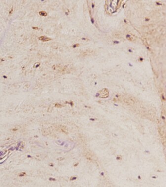
Detection of Mouse Mouse SOST/Sclerostin Antibody by Immunohistochemistry
Mice were treated with vehicle or 50 μg/kg/day for 28 days. The fibula was sectioned and immunostained for sclerostin. Scale bars equals 100 μm. Image collected and cropped by CiteAb from the following publication (https://pubmed.ncbi.nlm.nih.gov/28249797), licensed under a CC-BY license. Not internally tested by R&D Systems.
Detection of Mouse SOST/Sclerostin by Immunohistochemistry
Loading-induced increased PGE2 secretion with a decrease in SOST expression was inhibited in cKO mice. a, b ELISA analysis of PGE2 level in bone marrow-flushed tibial diaphysis (a) and serum (b) after 5-day mechanical loading. n = 6 per group. c Representative COX-2 immunohistostaining (black arrows) and d, e quantification of COX-2-positive osteocytes in diaphyseal 37% cortical bone. Scale bar, 30 μm. n = 6 per group. f Representative SOST immunohistostaining (black arrows) and g, h quantification of SOST-positive osteocytes in diaphyseal 37% cortical bone. Scale bar, 30 μm. n = 6 per group. i Representative beta -catenin immunohistostaining (black arrows) and j, k quantification of beta -catenin-positive osteoblasts on endosteal surface of diaphyseal 37% cortical bone. Scale bar, 30 μm. n = 5–6 per group. l–n Relative gene expression of Sost (l), COX-2 (m), and beta -catenin (n) was determined by RT-qPCR in the tibial diaphysis of WT and cKO mice. n = 5 per group. Mean ± SD. *P < 0.05; **P < 0.01; ***P < 0.001. Paired t test was done for loaded and contralateral tibias and unpaired t test was done for loaded or control tibias between WT and cKO mice Image collected and cropped by CiteAb from the following open publication (https://pubmed.ncbi.nlm.nih.gov/35851577), licensed under a CC-BY license. Not internally tested by R&D Systems.
Detection of SOST/Sclerostin by Western Blot
OCY‐EVs Transported to the Brain under Physiological & Pathological Conditions.Representative E) WB images of DMP1 & SOST expression. Scale bar: 100 µm. beta ‐actin was used as loading control. OB, Osteoblast; BMM, bone marrow macrophage. n = 3 per group. Image collected & cropped by CiteAb from the following open publication (https://pubmed.ncbi.nlm.nih.gov/35508803), licensed under a CC-BY license. Not internally tested by R&D Systems.
Detection of SOST/Sclerostin by Western Blot
OCY‐EVs Transported to the Brain under Physiological & Pathological Conditions.Representative H) WB images of osteocyte markers (SOST & DMP1) & EVs classical markers (TSG101 & Flotillin‐1). Scale bar: 100 nm. Image collected & cropped by CiteAb from the following open publication (https://pubmed.ncbi.nlm.nih.gov/35508803), licensed under a CC-BY license. Not internally tested by R&D Systems.
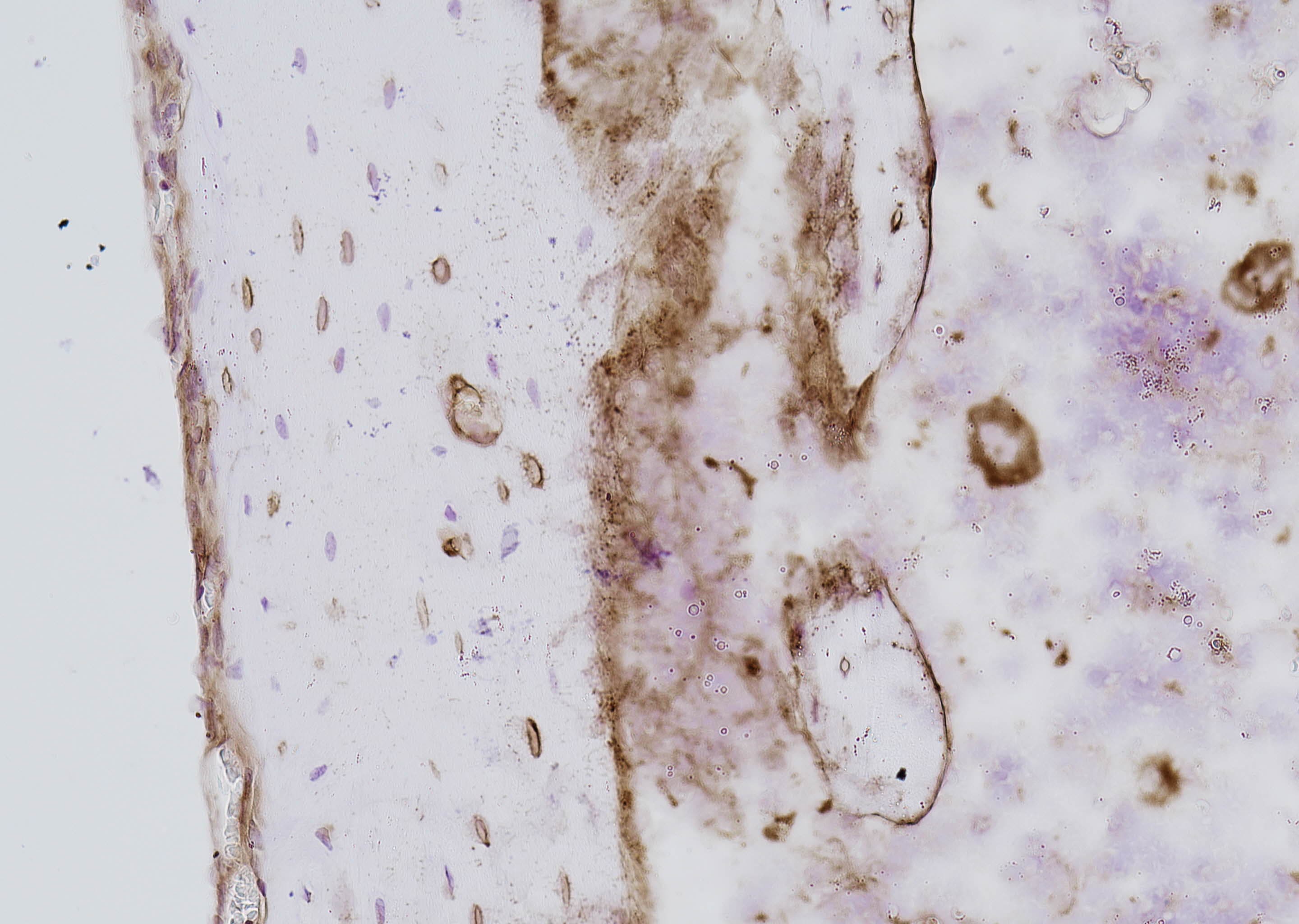
Detection of SOST/Sclerostin by Immunohistochemistry
Localized sclerostin accumulation and increased expression of PLR-related proteins in 20-week-old male db/db diabetic mice cortical bone. Immunohistochemical staining shows increased expression of CTSK (A), MMP-13 (B), and sclerostin (C) within the osteocyte LCS of db/db mice compared to WT mice. Linear regression analysis (D) reveals a significant positive correlation between sclerostin expression and CTSK and MMP-13 using combined data from both groups. Data are presented as mean ± SD. Statistical analysis was performed using an unpaired Student’s t-test for (A–C) (n = 5 mice per group) and Pearson correlation for (D). *p < 0.05, **p < 0.01, ***p < 0.001. Image collected and cropped by CiteAb from the following open publication (https://pubmed.ncbi.nlm.nih.gov/40352664), licensed under a CC-BY license. Not internally tested by R&D Systems.Applications for Mouse SOST/Sclerostin Antibody
Application
Recommended Usage
Immunohistochemistry
5-15 µg/mL
Sample: Immersion fixed frozen sections of mouse embryo (adrenal gland) and immersion fixed frozen sections of mouse embryo (kidney)
Sample: Immersion fixed frozen sections of mouse embryo (adrenal gland) and immersion fixed frozen sections of mouse embryo (kidney)
Western Blot
2 µg/mL
Sample: Human bone marrow and mouse bone marrow
Sample: Human bone marrow and mouse bone marrow
Reviewed Applications
Read 7 reviews rated 4.6 using AF1589 in the following applications:
Formulation, Preparation, and Storage
Purification
Antigen Affinity-purified
Reconstitution
Reconstitute at 0.2 mg/mL in sterile PBS. For liquid material, refer to CoA for concentration.
Loading...
Formulation
Lyophilized from a 0.2 μm filtered solution in PBS with Trehalose. *Small pack size (SP) is supplied either lyophilized or as a 0.2 µm filtered solution in PBS.
Shipping
Lyophilized product is shipped at ambient temperature. Liquid small pack size (-SP) is shipped with polar packs. Upon receipt, store immediately at the temperature recommended below.
Stability & Storage
Use a manual defrost freezer and avoid repeated freeze-thaw cycles.
- 12 months from date of receipt, -20 to -70 °C as supplied.
- 1 month, 2 to 8 °C under sterile conditions after reconstitution.
- 6 months, -20 to -70 °C under sterile conditions after reconstitution.
Calculators
Background: SOST/Sclerostin
References
- Kusu, N. et al. (2003) J. Biol. Chem. 278:24113.
- Balemans, W. et al. (2001) Hum. Mol. Genet. 10:537.
- Brunkow, M.E. et al. (2001) Am. J. Hum. Genet. 68:577.
Alternate Names
sclerostin, VBCHsclerosteosis
Gene Symbol
SOST
UniProt
Additional SOST/Sclerostin Products
Product Documents for Mouse SOST/Sclerostin Antibody
Certificate of Analysis
To download a Certificate of Analysis, please enter a lot or batch number in the search box below.
Note: Certificate of Analysis not available for kit components.
Product Specific Notices for Mouse SOST/Sclerostin Antibody
For research use only
Related Research Areas
Citations for Mouse SOST/Sclerostin Antibody
 Powered by Bioz
Customer Reviews for Mouse SOST/Sclerostin Antibody (7)
4.6 out of 5
7 Customer Ratings
Have you used Mouse SOST/Sclerostin Antibody?
Submit a review and receive an Amazon gift card!
$25/€18/£15/$25CAN/¥2500 Yen for a review with an image
$10/€7/£6/$10CAN/¥1110 Yen for a review without an image
Submit a review
Customer Images

Showing
1
-
5 of
7 reviews
Showing All
Filter By:
-
Application: ELISASample Tested: Recombinant proteinSpecies: MouseVerified Customer | Posted 08/31/2023

-
Application: ImmunohistochemistrySample Tested: boneSpecies: MouseVerified Customer | Posted 07/07/2021
-
Application: ImmunohistochemistrySample Tested: BONESpecies: MouseVerified Customer | Posted 06/27/2021
-
Application: Flow CytometrySample Tested: Recombinant proteinSpecies: MouseVerified Customer | Posted 06/25/2021
-
Application: Western BlotSample Tested: Bone Extracts, bone marrow, Bone marrow cells and Liver tissueSpecies: MouseVerified Customer | Posted 04/21/2017
-
Application: Western BlotSample Tested: See PMID 23494985Species: MouseVerified Customer | Posted 01/05/2015
-
Application: Western BlotSample Tested: See PMID 20336693Species: OtherVerified Customer | Posted 01/05/2015
There are no reviews that match your criteria.
Protocols
Find general support by application which include: protocols, troubleshooting, illustrated assays, videos and webinars.
- Antigen Retrieval Protocol (PIER)
- Antigen Retrieval for Frozen Sections Protocol
- Appropriate Fixation of IHC/ICC Samples
- Cellular Response to Hypoxia Protocols
- Chromogenic IHC Staining of Formalin-Fixed Paraffin-Embedded (FFPE) Tissue Protocol
- Chromogenic Immunohistochemistry Staining of Frozen Tissue
- ClariTSA™ Fluorophore Kits
- Detection & Visualization of Antibody Binding
- Fluorescent IHC Staining of Frozen Tissue Protocol
- Graphic Protocol for Heat-induced Epitope Retrieval
- Graphic Protocol for the Preparation and Fluorescent IHC Staining of Frozen Tissue Sections
- Graphic Protocol for the Preparation and Fluorescent IHC Staining of Paraffin-embedded Tissue Sections
- Graphic Protocol for the Preparation of Gelatin-coated Slides for Histological Tissue Sections
- IHC Sample Preparation (Frozen sections vs Paraffin)
- Immunofluorescent IHC Staining of Formalin-Fixed Paraffin-Embedded (FFPE) Tissue Protocol
- Immunohistochemistry (IHC) and Immunocytochemistry (ICC) Protocols
- Immunohistochemistry Frozen Troubleshooting
- Immunohistochemistry Paraffin Troubleshooting
- Preparing Samples for IHC/ICC Experiments
- Preventing Non-Specific Staining (Non-Specific Binding)
- Primary Antibody Selection & Optimization
- Protocol for Heat-Induced Epitope Retrieval (HIER)
- Protocol for Making a 4% Formaldehyde Solution in PBS
- Protocol for VisUCyte™ HRP Polymer Detection Reagent
- Protocol for the Preparation & Fixation of Cells on Coverslips
- Protocol for the Preparation and Chromogenic IHC Staining of Frozen Tissue Sections
- Protocol for the Preparation and Chromogenic IHC Staining of Frozen Tissue Sections - Graphic
- Protocol for the Preparation and Chromogenic IHC Staining of Paraffin-embedded Tissue Sections
- Protocol for the Preparation and Chromogenic IHC Staining of Paraffin-embedded Tissue Sections - Graphic
- Protocol for the Preparation and Fluorescent IHC Staining of Frozen Tissue Sections
- Protocol for the Preparation and Fluorescent IHC Staining of Paraffin-embedded Tissue Sections
- Protocol for the Preparation of Gelatin-coated Slides for Histological Tissue Sections
- R&D Systems Quality Control Western Blot Protocol
- TUNEL and Active Caspase-3 Detection by IHC/ICC Protocol
- The Importance of IHC/ICC Controls
- Troubleshooting Guide: Immunohistochemistry
- Troubleshooting Guide: Western Blot Figures
- Western Blot Conditions
- Western Blot Protocol
- Western Blot Protocol for Cell Lysates
- Western Blot Troubleshooting
- Western Blot Troubleshooting Guide
- View all Protocols, Troubleshooting, Illustrated assays and Webinars
Loading...