
Tryptase beta -1 is a serine protease with trypsin-like activity, which is sometimes also referred to as Mast Cell Protease 7 (1). It is stored in the secretory granules of mouse mast cells (2). It exhibits anticoagulant activity due to its ability to degrade fibrinogen in the presence of the diverse array of protease inhibitors in plasma (3). The deduced amino acid sequence for mouse Tryptase beta -1 consists of a signal peptide (residues 1‑18), a pro region (residues 19‑28) and the mature chain (residue 29‑273). The mature chain was expressed and purified.
Mouse Tryptase beta-1/MCP-7 Antibody
R&D Systems | Catalog # AF1937

Scientific images may be resized, cropped, or adjusted for brightness or contrast for presentation purposes. These updates are limited to presentation and do not affect the underlying scientific interpretation of the data. Where available, additional source data may be provided upon request.
Key Product Details
Species Reactivity
Validated:
Mouse
Cited:
Mouse
Applications
Validated:
Western Blot, Immunoprecipitation
Cited:
Immunohistochemistry, Immunohistochemistry-Paraffin, Western Blot, Immunocytochemistry, ELISA Development (Capture)
Label
Unconjugated
Antibody Source
Polyclonal Goat IgG
Loading...
Product Specifications
Immunogen
Mouse myeloma cell line NS0-derived recombinant mouse Tryptase beta ‑1/MCP‑7/Mcpt7
Ile29-Phe273
Accession # Q02844
Ile29-Phe273
Accession # Q02844
Specificity
Detects mouse Tryptase beta ‑1/MCP‑7/Mcpt7 in direct ELISAs and Western blots. In direct ELISAs, approximately 10% cross‑reactivity with recombinant human (rh) TPSB2 is observed, 5% cross-reactivity with rhTPS1 is observed, and less than 1% cross-reactivity with rhTPSG1 and recombinant mouse MCPT1 is observed.
Clonality
Polyclonal
Host
Goat
Isotype
IgG
Scientific Data Images for Mouse Tryptase beta-1/MCP-7 Antibody
Detection of Tryptase beta-1/Mcpt7 by Immunohistochemistry
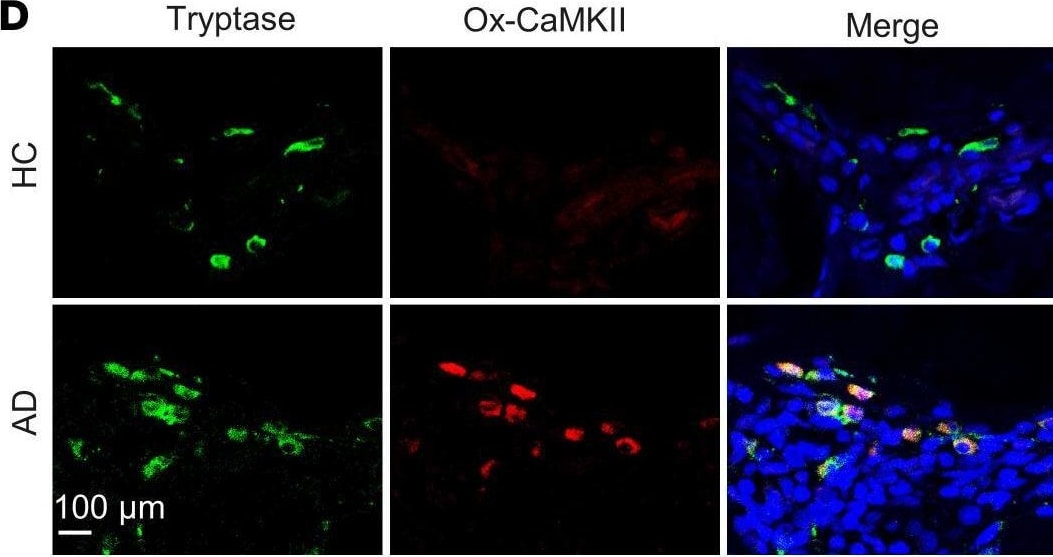
ROS-resistant CaMKII protects against allergen-induced mast cell activation.(A–C) Analyses of beta -hexosaminidase (A, n = 6), histamine release (B, n = 4), and cytokines and chemokines (C, n = 6) in supernatants of DNP-activated BMMCs derived from WT and CaMKII MM-VV delta mice. (D) Representative immunofluorescence images of dorsal skin sections and fluorescence analysis of ox-CaMKII staining in the skin mast cells of patients with AD and controls. Scale bar: 100 μm. (E) Quantitative analyses for total number of ox-CaMKII+ mast cells (n = 10). Data represent mean ± SEM of 2 independent experiments. Data were compared using a 2-tailed Student’s t test. *P < 0.05, **P < 0.01, ***P < 0.001. Image collected and cropped by CiteAb from the following open publication (https://insight.jci.org/articles/view/152559), licensed under a CC-BY license. Not internally tested by R&D Systems.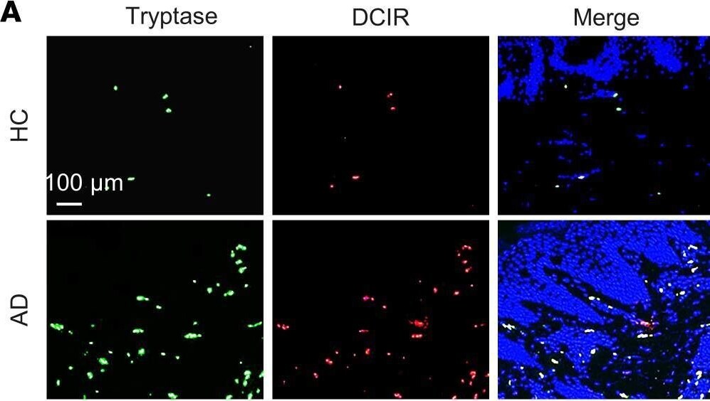
Detection of Tryptase beta-1/Mcpt7 by Immunohistochemistry
DCIR mediates cockroach allergen binding and uptake.(A and B) Representative immunofluorescence images of dorsal skin sections and fluorescence analysis of DCIR staining in the skin tissues of patients with AD and controls (n = 8). Scale bar: 100 μm. (C) Quantification analysis of DCIR+tryptase+ cells in the lesion skin of patients with AD and controls. (D) Flow cytometry analysis of DCIR expression in human mast cell line cKit+Fc epsilon RI+ LAD2 cells. (E) Scheme of experimental protocol for the direct bindings of human recombinant DCIR (hrDCIR) to BSA, CRE, and Man-BSA. (F) Direct binding of different doses of hrDCIR (0–5.0 μg/mL) to BSA, CRE, or Man-BSA (n = 3). (G) Representative immunofluorescence images of FITC-CRE uptake by LAD2. Scale bar: 15 μm. (H and I) Flow cytometry analysis (H) and quantification (I) of FITC-CRE uptake at different doses (1–500 ng/mL) by LAD2 cells (n = 3). (J) Inhibition of FITC-CRE uptake in LAD2 cells pretreated with DCIR neutralizing antibody ( alpha -DCIR) or IgG isotype (n = 4). Data represent mean ± SEM of 2 independent experiments. Data in B, C, and I were compared using a 2-tailed Student’s t test. Data in F and J were compared by 2-way ANOVA. *P < 0.05, **P < 0.01, ***P < 0.001. Image collected and cropped by CiteAb from the following open publication (https://insight.jci.org/articles/view/152559), licensed under a CC-BY license. Not internally tested by R&D Systems.Applications for Mouse Tryptase beta-1/MCP-7 Antibody
Application
Recommended Usage
Immunoprecipitation
25 µg/mL
Sample: Conditioned cell culture medium spiked with Recombinant Mouse Tryptase beta ‑1/MCP‑7/Mcpt7 (Catalog # 1937-SE), see our available Western blot detection antibodies
Sample: Conditioned cell culture medium spiked with Recombinant Mouse Tryptase beta ‑1/MCP‑7/Mcpt7 (Catalog # 1937-SE), see our available Western blot detection antibodies
Western Blot
0.1 µg/mL
Sample: Recombinant Mouse Tryptase beta ‑1/MCP‑7/Mcpt7 (Catalog # 1937-SE)
Sample: Recombinant Mouse Tryptase beta ‑1/MCP‑7/Mcpt7 (Catalog # 1937-SE)
Formulation, Preparation, and Storage
Purification
Antigen Affinity-purified
Reconstitution
Reconstitute at 0.2 mg/mL in sterile PBS. For liquid material, refer to CoA for concentration.
Loading...
Formulation
Lyophilized from a 0.2 μm filtered solution in PBS with Trehalose. *Small pack size (SP) is supplied either lyophilized or as a 0.2 µm filtered solution in PBS.
Shipping
Lyophilized product is shipped at ambient temperature. Liquid small pack size (-SP) is shipped with polar packs. Upon receipt, store immediately at the temperature recommended below.
Stability & Storage
Use a manual defrost freezer and avoid repeated freeze-thaw cycles.
- 12 months from date of receipt, -20 to -70 °C as supplied.
- 1 month, 2 to 8 °C under sterile conditions after reconstitution.
- 6 months, -20 to -70 °C under sterile conditions after reconstitution.
Calculators
Background: Tryptase beta-1/MCP-7
References
- McNeil, H.P. et al. (1992) Proc. Natl. Acad. Sci. USA 89:11174.
- Matsumoto, R et al. (1995) J. Biol. Chem. 270:19524.
- Huang, C. et al. (1997) J. Biol. Chem. 272:31885.
Long Name
Mast Cell Protease 7
Alternate Names
Mast Cell Tryptase, MCP-7, Mcp7, Mcpt7, TPSB1, TRYB1, Tryptase beta1
Entrez Gene IDs
100503895 (Mouse)
Gene Symbol
TPSAB1
UniProt
Additional Tryptase beta-1/MCP-7 Products
Product Documents for Mouse Tryptase beta-1/MCP-7 Antibody
Certificate of Analysis
To download a Certificate of Analysis, please enter a lot or batch number in the search box below.
Note: Certificate of Analysis not available for kit components.
Product Specific Notices for Mouse Tryptase beta-1/MCP-7 Antibody
For research use only
Related Research Areas
Citations for Mouse Tryptase beta-1/MCP-7 Antibody
 Powered by Bioz
Customer Reviews for Mouse Tryptase beta-1/MCP-7 Antibody
There are currently no reviews for this product. Be the first to review Mouse Tryptase beta-1/MCP-7 Antibody and earn rewards!
Have you used Mouse Tryptase beta-1/MCP-7 Antibody?
Submit a review and receive an Amazon gift card!
$25/€18/£15/$25CAN/¥2500 for a review with an image
$10/€7/£6/$10CAN/¥1110 for a review without an image
Submit a review
Protocols
Find general support by application which include: protocols, troubleshooting, illustrated assays, videos and webinars.
- Cellular Response to Hypoxia Protocols
- Immunoprecipitation Protocol
- R&D Systems Quality Control Western Blot Protocol
- Troubleshooting Guide: Western Blot Figures
- Western Blot Conditions
- Western Blot Protocol
- Western Blot Protocol for Cell Lysates
- Western Blot Troubleshooting
- Western Blot Troubleshooting Guide
- View all Protocols, Troubleshooting, Illustrated assays and Webinars
Loading...