Notch-1 Antibody - (Cleaved N terminal) - BSA Free
Novus Biologicals | Catalog # NB300-251
![Western Blot: Notch-1 Antibody(Cleaved N terminal) [NB300-251]](https://resources.rndsystems.com/images/products/Notch-1-Antibody-Cleaved-N-terminal-Western-Blot-NB300-251-img0010.jpg "Western Blot: Notch-1 Antibody(Cleaved N terminal) [NB300-251]")
Key Product Details
Species Reactivity
Validated:
Cited:
Applications
Validated:
Cited:
Label
Antibody Source
Format
Product Specifications
Immunogen
Specificity
Clonality
Host
Isotype
Description
Store vial at -20C prior to opening. Aliquot contents and freeze at -20C or below for extended storage. Avoid cycles of freezing and thawing. Centrifuge product if not completely clear after standing at room temperature. This product is stable for several weeks at 4C as an undiluted liquid. Dilute only prior to immediate use.
Scientific Data Images for Notch-1 Antibody - (Cleaved N terminal) - BSA Free
Western Blot: Notch-1 Antibody(Cleaved N terminal) [NB300-251]
Western Blot: Notch-1 Antibody - (Cleaved N terminal) [NB300-251] - Lane 1: MCF-7 control lysate. Lane 2: MCF-7 +1 nM 17beta-estradiol. Lane 3: MCF-7 + 10 uM gamma secretase inhibitor. Load: 35 ug per lane. Primary antibody: Notch1 antibody at 1:500 for overnight at 4C. Secondary antibody: IRDye800 rabbit secondary antibody at 1:10,000 for 45 min at RT. Block: 5% BLOTTO overnight at 4C. Predicted/Observed size: 80 kDa for Notch1.![Immunohistochemistry: Notch-1 Antibody - (Cleaved N terminal) [NB300-251]](https://resources.rndsystems.com/images/products/Notch-1-Antibody-Cleaved-N-terminal-Immunohistochemistry-NB300-251-img0007.jpg "Immunohistochemistry: Notch-1 Antibody - (Cleaved N terminal) [NB300-251]")
Immunohistochemistry: Notch-1 Antibody - (Cleaved N terminal) [NB300-251]
Immunohistochemistry: Notch-1 Antibody - (Cleaved N terminal) [NB300-251] - Analysis of Tissue: Exocrine glands of human pancreas Fixation: FFPE Primary antibody: Notch1 antibody at 1:200 Staining: moderate to strong membranous staining and faint to moderate cytoplasmic staining. Islets showed faint staining.![Western Blot: Notch-1 Antibody(Cleaved N terminal) [NB300-251]](https://resources.rndsystems.com/images/products/Notch-1-Antibody-Cleaved-N-terminal-Western-Blot-NB300-251-img0003.jpg "Western Blot: Notch-1 Antibody(Cleaved N terminal) [NB300-251]")
Western Blot: Notch-1 Antibody(Cleaved N terminal) [NB300-251]
Western Blot: Notch-1 Antibody - (Cleaved N terminal) [NB300-251] - Notch 1 Antibody [NB 300-251]- at 1:500, against myc-tagged transiently transfected mouse Notch constructs in 293 cells.![Western Blot: Notch-1 Antibody(Cleaved N terminal) [NB300-251]](https://resources.rndsystems.com/images/products/Notch-1-Antibody-Cleaved-N-terminal-Western-Blot-NB300-251-img0009.jpg "Western Blot: Notch-1 Antibody(Cleaved N terminal) [NB300-251]")
Western Blot: Notch-1 Antibody(Cleaved N terminal) [NB300-251]
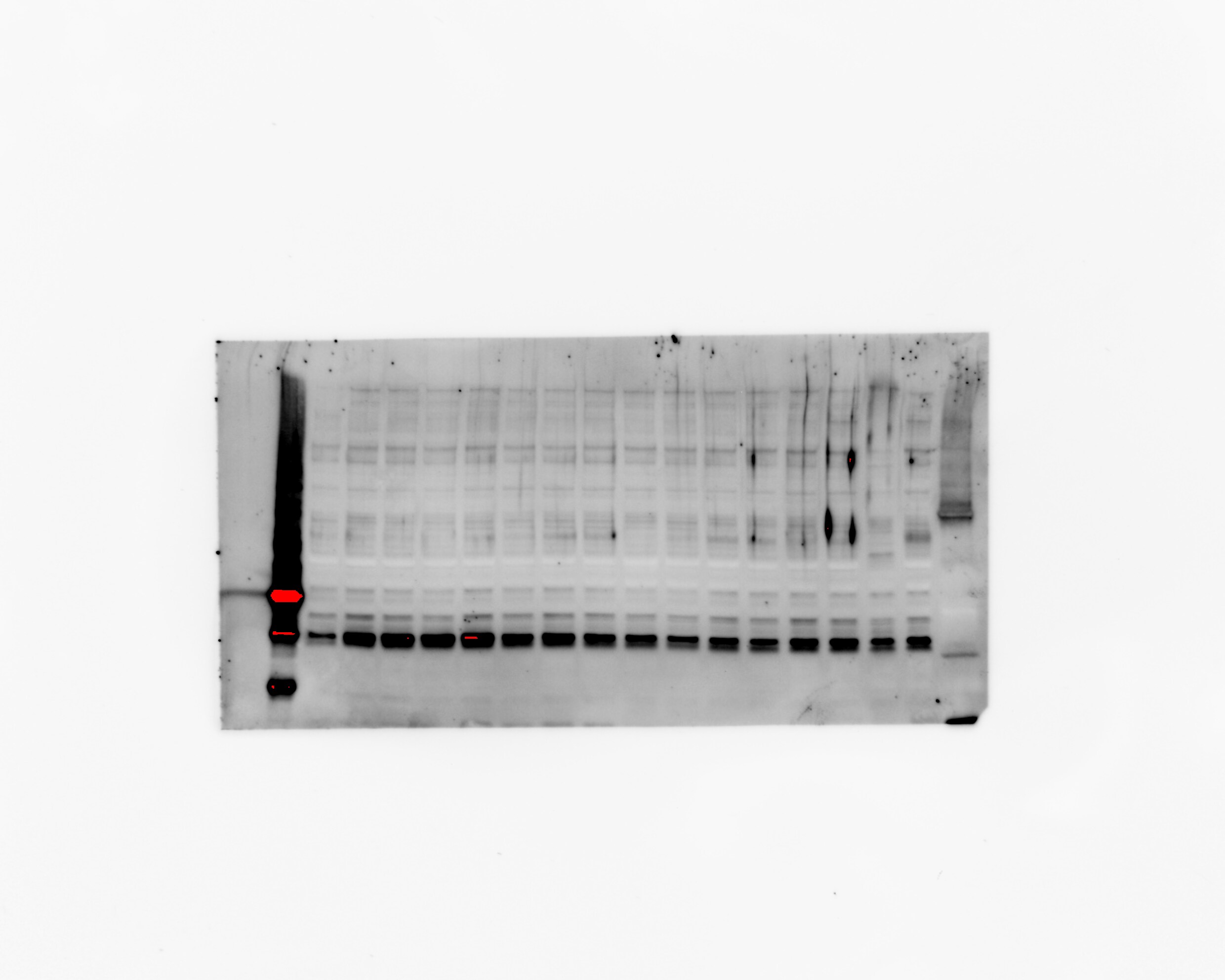
Western Blot: Notch-1 Antibody - (Cleaved N terminal) [NB300-251] - Lane 1: No transfection. Lane 2: N1 (mouse deleted extracellular domain)-myc. Lane 3: N1 (mouse intracellular domain)-myc. Lane 4: N2 (mouse deleted extracellular domain)-myc. Lane 5: N2 (mouse intracellular domain)-myc. Lane 6: N3 (mouse deleted extracellular domain)-myc. Lane 7: N3 (mouse intracellular domain)-myc. Lane 8: N4 (mouse deleted extracellular domain)-myc. Lane 9: N4 (mouse intracellular domain)-myc. Lane 10: N1 (mouse deleted extracellular domain)(V to G)-myc.![Western Blot: Notch-1 Antibody(Cleaved N terminal) [NB300-251]](https://resources.rndsystems.com/images/products/Notch-1-Antibody-Cleaved-N-terminal-Western-Blot-NB300-251-img0011.jpg "Western Blot: Notch-1 Antibody(Cleaved N terminal) [NB300-251]")
Western Blot: Notch-1 Antibody(Cleaved N terminal) [NB300-251]
Western Blot: Notch-1 Antibody - (Cleaved N terminal) [NB300-251] - Lane 1: No transfection. Lane 2: N1 (mouse deleted extracellular domain)-myc. Lane 3: N1 (mouse intracellular domain)-myc. Lane 4: N2 (mouse deleted extracellular domain)-myc. Lane 5: N2 (mouse intracellular domain)-myc. Lane 6: N3 (mouse deleted extracellular domain)-myc. Lane 7: N3 (mouse intracellular domain)-myc. Lane 8: N4 (mouse deleted extracellular domain)-myc. Lane 9: N4 (mouse intracellular domain)-myc. Lane 10: N1 (mouse deleted extracellular domain)(V to G)-myc.")
Notch-1 Antibody - (Cleaved N terminal)
Immunohistochemistry of Rabbit Notch-1 Antibody - (Cleaved N terminal). Tissue: Exocrine glands of human pancreas. Fixation: FFPE. Primary antibody: Notch1 antibody at 1:200. Staining: moderate to strong membranous staining and faint to moderate cytoplasmic staining. Islets showed faint staining.")
Notch-1 Antibody - (Cleaved N terminal)
Dot Blot of Rabbit anti-Notch 1 (Cleaved N Terminal) (Human Specific) Antibody. Antigen: Row 1 - Notch 1 Peptide (Cleaved N Terminal) Row 2 - Notch 1 (Intra) Peptide. Load: Lane 1 - 200 ng Lane 2 - 66.67 ng Lane 3 - 22.22 ng Lane 4 - 7.41 ng Lane 5 - 2.47 ng. Primary antibody: Rabbit anti-Notch-1 Antibody - (Cleaved N terminal)at 1:1,000 for 60 min at RT. Secondary antibody: HRP Rabbit Secondary at 1:40,000 for 30 min at RT. Block for 1 HR at RT.Applications for Notch-1 Antibody - (Cleaved N terminal) - BSA Free
ELISA
Immunohistochemistry
Immunohistochemistry-Paraffin
Western Blot
Reviewed Applications
Read 1 review rated 1 using NB300-251 in the following applications:
Formulation, Preparation, and Storage
Purification
Formulation
Format
Preservative
Concentration
Shipping
Stability & Storage
Background: Notch-1
Alternate Names
Gene Symbol
UniProt
Additional Notch-1 Products
Product Documents for Notch-1 Antibody - (Cleaved N terminal) - BSA Free
Certificate of Analysis
To download a Certificate of Analysis, please enter a lot or batch number in the search box below.
Product Specific Notices for Notch-1 Antibody - (Cleaved N terminal) - BSA Free
This product is for research use only and is not approved for use in humans or in clinical diagnosis. Primary Antibodies are guaranteed for 1 year from date of receipt.
Related Research Areas
Citations for Notch-1 Antibody - (Cleaved N terminal) - BSA Free
 Powered by Bioz
Customer Reviews for Notch-1 Antibody - (Cleaved N terminal) - BSA Free (1)
Have you used Notch-1 Antibody - (Cleaved N terminal) - BSA Free?
Submit a review and receive an Amazon gift card!
$25/€18/£15/$25CAN/¥2500 Yen for a review with an image
$10/€7/£6/$10CAN/¥1110 Yen for a review without an image
Submit a review
Customer Images
-
Application: Western BlotSample Tested: Rat brain tissueSpecies: RatVerified Customer | Posted 10/23/2020The bands appeared in 20KD.
 Bio-Techne ResponseThank you for reviewing our product. We are sorry to hear that this product did not perform as expected. We have been in touch with the customer to resolve this issue according to our Product Guarantee and to the customer’s satisfaction.
There are no reviews that match your criteria.
Protocols
Find general support by application which include: protocols, troubleshooting, illustrated assays, videos and webinars.
- Antigen Retrieval Protocol (PIER)
- Antigen Retrieval for Frozen Sections Protocol
- Appropriate Fixation of IHC/ICC Samples
- Cellular Response to Hypoxia Protocols
- Chromogenic IHC Staining of Formalin-Fixed Paraffin-Embedded (FFPE) Tissue Protocol
- Chromogenic Immunohistochemistry Staining of Frozen Tissue
- ClariTSA™ Fluorophore Kits
- Detection & Visualization of Antibody Binding
- ELISA Sample Preparation & Collection Guide
- ELISA Troubleshooting Guide
- Fluorescent IHC Staining of Frozen Tissue Protocol
- Graphic Protocol for Heat-induced Epitope Retrieval
- Graphic Protocol for the Preparation and Fluorescent IHC Staining of Frozen Tissue Sections
- Graphic Protocol for the Preparation and Fluorescent IHC Staining of Paraffin-embedded Tissue Sections
- Graphic Protocol for the Preparation of Gelatin-coated Slides for Histological Tissue Sections
- How to Run an R&D Systems DuoSet ELISA
- How to Run an R&D Systems Quantikine ELISA
- How to Run an R&D Systems Quantikine™ QuicKit™ ELISA
- ICC Cell Smear Protocol for Suspension Cells
- ICC Immunocytochemistry Protocol Videos
- ICC for Adherent Cells
- IHC Sample Preparation (Frozen sections vs Paraffin)
- Immunocytochemistry (ICC) Protocol
- Immunocytochemistry Troubleshooting
- Immunofluorescence of Organoids Embedded in Cultrex Basement Membrane Extract
- Immunofluorescent IHC Staining of Formalin-Fixed Paraffin-Embedded (FFPE) Tissue Protocol
- Immunohistochemistry (IHC) and Immunocytochemistry (ICC) Protocols
- Immunohistochemistry Frozen Troubleshooting
- Immunohistochemistry Paraffin Troubleshooting
- Immunoprecipitation Protocol
- Preparing Samples for IHC/ICC Experiments
- Preventing Non-Specific Staining (Non-Specific Binding)
- Primary Antibody Selection & Optimization
- Protocol for Heat-Induced Epitope Retrieval (HIER)
- Protocol for Making a 4% Formaldehyde Solution in PBS
- Protocol for VisUCyte™ HRP Polymer Detection Reagent
- Protocol for the Fluorescent ICC Staining of Cell Smears - Graphic
- Protocol for the Fluorescent ICC Staining of Cultured Cells on Coverslips - Graphic
- Protocol for the Preparation & Fixation of Cells on Coverslips
- Protocol for the Preparation and Chromogenic IHC Staining of Frozen Tissue Sections
- Protocol for the Preparation and Chromogenic IHC Staining of Frozen Tissue Sections - Graphic
- Protocol for the Preparation and Chromogenic IHC Staining of Paraffin-embedded Tissue Sections
- Protocol for the Preparation and Chromogenic IHC Staining of Paraffin-embedded Tissue Sections - Graphic
- Protocol for the Preparation and Fluorescent ICC Staining of Cells on Coverslips
- Protocol for the Preparation and Fluorescent ICC Staining of Non-adherent Cells
- Protocol for the Preparation and Fluorescent ICC Staining of Stem Cells on Coverslips
- Protocol for the Preparation and Fluorescent IHC Staining of Frozen Tissue Sections
- Protocol for the Preparation and Fluorescent IHC Staining of Paraffin-embedded Tissue Sections
- Protocol for the Preparation of Gelatin-coated Slides for Histological Tissue Sections
- Protocol for the Preparation of a Cell Smear for Non-adherent Cell ICC - Graphic
- Quantikine HS ELISA Kit Assay Principle, Alkaline Phosphatase
- Quantikine HS ELISA Kit Principle, Streptavidin-HRP Polymer
- R&D Systems Quality Control Western Blot Protocol
- Sandwich ELISA (Colorimetric) – Biotin/Streptavidin Detection Protocol
- Sandwich ELISA (Colorimetric) – Direct Detection Protocol
- TUNEL and Active Caspase-3 Detection by IHC/ICC Protocol
- The Importance of IHC/ICC Controls
- Troubleshooting Guide: ELISA
- Troubleshooting Guide: Immunohistochemistry
- Troubleshooting Guide: Western Blot Figures
- Western Blot Conditions
- Western Blot Protocol
- Western Blot Protocol for Cell Lysates
- Western Blot Troubleshooting
- Western Blot Troubleshooting Guide
- View all Protocols, Troubleshooting, Illustrated assays and Webinars
FAQs for Notch-1 Antibody - (Cleaved N terminal) - BSA Free
-
Q: Hello, does this product (NB300-251) only recognize the cleavage Notch1?
A: This antibody with recognize full length Notch1 if it is in the mix, as well as the cleaved product.
Associated Pathways


