p70 S6 Kinase/S6K [p Thr389/412] Antibody - BSA Free
Novus Biologicals | Catalog # NB600-1049
![Western Blot: p70 S6 Kinase/S6K [p Thr389/412] Antibody [NB600-1049]](https://resources.rndsystems.com/images/products/p70-S6-Kinase-S6K-[p-Thr389-412]-Antibody-Western-Blot-NB600-1049-img0005.jpg "Western Blot: p70 S6 Kinase/S6K [p Thr389/412] Antibody [NB600-1049]")
Loading...
Key Product Details
Validated by
Biological Validation
Species Reactivity
Validated:
Human, Mouse, Rat
Cited:
Human, Mouse, Rat
Applications
Validated:
Immunohistochemistry, Immunohistochemistry-Paraffin, Western Blot, Immunocytochemistry/ Immunofluorescence
Cited:
Immunohistochemistry-Paraffin, Western Blot, IF/IHC
Label
Unconjugated
Antibody Source
Polyclonal Rabbit IgG
Format
BSA Free
Loading...
Product Specifications
Immunogen
Synthetic phospho-peptide surrounding amino acids Thr389/412 of human p70 S6 Kinase/S6K
Modification
p Thr389/412
Specificity
p70 S6 Kinase/S6K [p Thr389/412] Antibody detects endogenous levels of p70 S6 Kinase only when phosphorylated at Threonine 389/412
Clonality
Polyclonal
Host
Rabbit
Isotype
IgG
Description
This product is manufactured by Abcam and distributed by Novus Biologicals. (Abcam Catalog Number: ab2571)
Scientific Data Images for p70 S6 Kinase/S6K [p Thr389/412] Antibody - BSA Free
![Immunocytochemistry/ Immunofluorescence: p70 S6 Kinase/S6K [p Thr389/412] Antibody [NB600-1049]](https://resources.rndsystems.com/images/products/p70-S6-Kinase-S6K-[p-Thr389]-Antibody-Immunocytochemistry-Immunofluorescence-NB600-1049-img0002.jpg "Immunocytochemistry/ Immunofluorescence: p70 S6 Kinase/S6K [p Thr389/412] Antibody [NB600-1049]")
Immunocytochemistry/ Immunofluorescence: p70 S6 Kinase/S6K [p Thr389/412] Antibody [NB600-1049]
Immunocytochemistry/Immunofluorescence: p70 S6 Kinase/S6K [p Thr389/412] Antibody [NB600-1049] - Analysis of Phospho-p70 S6 Kinase in 293 cells.![Immunohistochemistry: p70 S6 Kinase/S6K [p Thr389/412] Antibody [NB600-1049]](https://resources.rndsystems.com/images/products/p70-S6-Kinase-S6K-[p-Thr389]-Antibody-Immunohistochemistry-NB600-1049-img0003.jpg "Immunohistochemistry: p70 S6 Kinase/S6K [p Thr389/412] Antibody [NB600-1049]")
Immunohistochemistry: p70 S6 Kinase/S6K [p Thr389/412] Antibody [NB600-1049]
Immunohistochemistry: p70 S6 Kinase/S6K [p Thr389/412] Antibody [NB600-1049] - Human liver cancer tissue sections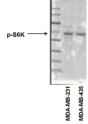
Western Blot: p70 S6 Kinase/S6K [p Thr389/412] Antibody [NB600-1049] - Analysis of phospho-p70 S6 Kinase in human breast cancer cell lines (MDA-MB-231 and MDA-MB-435) using p70 S6 Kinase/S6K [p Thr390, p Thr412] Antibody. Image from verified customer review.
![Western Blot: p70 S6 Kinase/S6K [p Thr389/412] Antibody [NB600-1049]](https://resources.rndsystems.com/images/products/p70-S6-Kinase-S6K-[p-Thr389]-Antibody-Western-Blot-NB600-1049-img0004.jpg "Western Blot: p70 S6 Kinase/S6K [p Thr389/412] Antibody [NB600-1049]")
Western Blot: p70 S6 Kinase/S6K [p Thr389/412] Antibody [NB600-1049]
Western Blot: p70 S6 Kinase/S6K [p Thr389/412] Antibody [NB600-1049] - Analysis of p70 S6 Kinase phosphorylation expression in Insulin treatedJurkat whole cell lysates. The lane on the left is treated with the antigen-specific peptide.Applications for p70 S6 Kinase/S6K [p Thr389/412] Antibody - BSA Free
Application
Recommended Usage
Immunocytochemistry/ Immunofluorescence
1:100 - 1:500
Immunohistochemistry
1:50 - 1:200
Western Blot
1:500 - 1:2000
Application Notes
Use in Immunohistochemistry-Paraffin reported in scientific literature (PMID:32339950).
Reviewed Applications
Read 1 review rated 5 using NB600-1049 in the following applications:
Formulation, Preparation, and Storage
Purification
Affinity purified
Formulation
PBS (pH 7.4), 150 mM EDTA, 50% glycerol
Format
BSA Free
Preservative
0.02% Sodium Azide
Concentration
Please see the vial label for concentration. If unlisted please contact technical services.
Shipping
The product is shipped with polar packs. Upon receipt, store it immediately at the temperature recommended below.
Stability & Storage
Aliquot and store at -20C or -80C. Avoid freeze-thaw cycles.
Background: p70 S6 Kinase
Alternate Names
p70-alpha, RPS6KB1, S6K1, STK14A
Gene Symbol
RPS6KB1
Additional p70 S6 Kinase Products
Product Documents for p70 S6 Kinase/S6K [p Thr389/412] Antibody - BSA Free
Certificate of Analysis
To download a Certificate of Analysis, please enter a lot or batch number in the search box below.
Product Specific Notices for p70 S6 Kinase/S6K [p Thr389/412] Antibody - BSA Free
This product is for research use only and is not approved for use in humans or in clinical diagnosis. Primary Antibodies are guaranteed for 1 year from date of receipt.
⚠ WARNING: This product can expose you to chemicals including mercury, which is known to the State of California to cause reproductive toxicity with developmental effects. For more information go to www.P65Warnings.ca.gov.Related Research Areas
Citations for p70 S6 Kinase/S6K [p Thr389/412] Antibody - BSA Free
 Powered by Bioz
Customer Reviews for p70 S6 Kinase/S6K [p Thr389/412] Antibody - BSA Free (1)
5 out of 5
1 Customer Rating
Have you used p70 S6 Kinase/S6K [p Thr389/412] Antibody - BSA Free?
Submit a review and receive an Amazon gift card!
$25/€18/£15/$25CAN/¥2500 Yen for a review with an image
$10/€7/£6/$10CAN/¥1110 Yen for a review without an image
Submit a review
Customer Images
![p70 S6 Kinase/S6K [p Thr389/412] Antibody - BSA Free NB600-1049](https://resources.rndsystems.com/images/reviews/Western-Blot__NB600-1049_20821.jpg)
Showing
1
-
1 of
1 review
Showing All
Filter By:
-
Application: Western BlotSample Tested: Human cancer cell whole cell lysateSpecies: HumanVerified Customer | Posted 08/16/2015phospho-p70 S6 Kinase expression in human breast cancer cell lines. NB600-1049
![p70 S6 Kinase/S6K [p Thr389/412] Antibody - BSA Free NB600-1049](data:image/png;base64,R0lGODlhAQABAAD/ACwAAAAAAQABAAACADs=)
There are no reviews that match your criteria.
Protocols
View specific protocols for p70 S6 Kinase/S6K [p Thr389/412] Antibody - BSA Free (NB600-1049):
Western Blot Protocol for S6K Antibody (NB600-1049):
Western Blot Protocol
Solutions and Reagents:
Transfer Buffer:
25 mM Tris-base (pH 8.5), 0.2 M Glycin, 20% methanol
Cell Extract Buffer:
50 mM Pipes/NaOH (pH 6.5), 2 mM EDTA, 0.1% Chaps, 5 mM DTT, 20 ug/ml Leupeptin, 10 ug/ml Pepstatin, 10 ug/ml aprotinin, and 1 mM PMSF.
SDS-PAGE Loading Buffer:
62.5 mM Tris-HCl, (pH 6.8), 2% w/v SDS, 10% glycerol, 50 mM DTT, 0.01% w/v bromphenol blue
10X TBS (Tris-Buffered Saline):
To prepare 1 liter of 10X TBS: 24.2 g Tris-base, 80 g NaCl, adjust pH to 7.6 with HCl (use at 1X).
TBS/T Washing Buffer:
1X TBS, 0.1% Tween-20
Blocking Buffer:
1X TBS/T with 5% BSA
Primary Antibody Dilution Buffer:
1X TBS/T with 5% BSA
Western Blot Detection:
Protein marker, secondary anti-rabbit antibody conjugated to HRP, chemiluminescent reagent, peroxide.
Protein Blotting:
1. Treat cells by adding fresh media containing regulator for desired time.
2. Aspirate media from cultures, wash cells with 1X PBS, aspirate. Scrape cells into PBS and spin down to pellet.
3. Lyse cells by adding Cell Extract Buffer (one volume of cell pellet, or 100 ul per well of 6-well plate or 500 ul per plate of 10 cm2 plate). Freeze and thaw 3 times. Centrifuge lysate at microcentrifuge using top speed. (~14000 rpm). Keep the supernatant and discard the pelleted cell debris.
4. Add SDS Loading Buffer and heat to 95-100oC for 5 minutes, cool on ice.
5. Microcentrifuge for 5 minutes.
6. Load 5-20 ul onto SDS-PAGE gel (10 cm x 10 cm).
Note: We recommend loading prestained molecular weight markers to verify electrotransfer.
7. Electrotransfer to nitrocellulose membrane.
Membrane Blocking & Antibody Incubations:
Note: Volumes are for 10 cm x 10 cm of membrane. For different sized membranes, adjust volumes accordingly
1. Incubate membrane in 25 ml of Blocking Buffer for 1 hour at room temperature.
2. Wash 3 times for 5 min each with 15 ml of TBS/T.
3. Incubate membrane and NB600-1049 1-2 ug/ml in 10 ml Primary Antibody Dilution Buffer with gentle agitation overnight at 4C.
4. Wash 3 times for 5 minutes each with 15 ml of TBS/T.
5. Incubate membrane with HRP-conjugated secondary antibody in 10 ml of Blocking Buffer with gentle agitation for 1 hour at room temperature.
6. Wash membrane as in step 4.
7. Proceed with detection.
Detection of Proteins:
1. Remove the wash buffer and place the blot in a plastic bag or clean tray containing chemiluminescent working solution (0.125 ml/cm2) and peroxide (ECL detection method).
2. Rotate the bag or tray to allow the solution to cover the surface of the membrane for 1-5 minutes.
3. Remove blot from bag or tray and place it between two pieces of write-on acetate transparency film. Smooth over covered blot to remove air bubbles and excess substrate.
Expose to X-ray film. An initial exposure of 10-60 seconds is recommended for film.
Find general support by application which include: protocols, troubleshooting, illustrated assays, videos and webinars.
- Antigen Retrieval Protocol (PIER)
- Antigen Retrieval for Frozen Sections Protocol
- Appropriate Fixation of IHC/ICC Samples
- Cellular Response to Hypoxia Protocols
- Chromogenic IHC Staining of Formalin-Fixed Paraffin-Embedded (FFPE) Tissue Protocol
- Chromogenic Immunohistochemistry Staining of Frozen Tissue
- ClariTSA™ Fluorophore Kits
- Detection & Visualization of Antibody Binding
- Fluorescent IHC Staining of Frozen Tissue Protocol
- Graphic Protocol for Heat-induced Epitope Retrieval
- Graphic Protocol for the Preparation and Fluorescent IHC Staining of Frozen Tissue Sections
- Graphic Protocol for the Preparation and Fluorescent IHC Staining of Paraffin-embedded Tissue Sections
- Graphic Protocol for the Preparation of Gelatin-coated Slides for Histological Tissue Sections
- ICC Cell Smear Protocol for Suspension Cells
- ICC Immunocytochemistry Protocol Videos
- ICC for Adherent Cells
- IHC Sample Preparation (Frozen sections vs Paraffin)
- Immunocytochemistry (ICC) Protocol
- Immunocytochemistry Troubleshooting
- Immunofluorescence of Organoids Embedded in Cultrex Basement Membrane Extract
- Immunofluorescent IHC Staining of Formalin-Fixed Paraffin-Embedded (FFPE) Tissue Protocol
- Immunohistochemistry (IHC) and Immunocytochemistry (ICC) Protocols
- Immunohistochemistry Frozen Troubleshooting
- Immunohistochemistry Paraffin Troubleshooting
- Preparing Samples for IHC/ICC Experiments
- Preventing Non-Specific Staining (Non-Specific Binding)
- Primary Antibody Selection & Optimization
- Protocol for Heat-Induced Epitope Retrieval (HIER)
- Protocol for Making a 4% Formaldehyde Solution in PBS
- Protocol for VisUCyte™ HRP Polymer Detection Reagent
- Protocol for the Fluorescent ICC Staining of Cell Smears - Graphic
- Protocol for the Fluorescent ICC Staining of Cultured Cells on Coverslips - Graphic
- Protocol for the Preparation & Fixation of Cells on Coverslips
- Protocol for the Preparation and Chromogenic IHC Staining of Frozen Tissue Sections
- Protocol for the Preparation and Chromogenic IHC Staining of Frozen Tissue Sections - Graphic
- Protocol for the Preparation and Chromogenic IHC Staining of Paraffin-embedded Tissue Sections
- Protocol for the Preparation and Chromogenic IHC Staining of Paraffin-embedded Tissue Sections - Graphic
- Protocol for the Preparation and Fluorescent ICC Staining of Cells on Coverslips
- Protocol for the Preparation and Fluorescent ICC Staining of Non-adherent Cells
- Protocol for the Preparation and Fluorescent ICC Staining of Stem Cells on Coverslips
- Protocol for the Preparation and Fluorescent IHC Staining of Frozen Tissue Sections
- Protocol for the Preparation and Fluorescent IHC Staining of Paraffin-embedded Tissue Sections
- Protocol for the Preparation of Gelatin-coated Slides for Histological Tissue Sections
- Protocol for the Preparation of a Cell Smear for Non-adherent Cell ICC - Graphic
- R&D Systems Quality Control Western Blot Protocol
- TUNEL and Active Caspase-3 Detection by IHC/ICC Protocol
- The Importance of IHC/ICC Controls
- Troubleshooting Guide: Immunohistochemistry
- Troubleshooting Guide: Western Blot Figures
- Western Blot Conditions
- Western Blot Protocol
- Western Blot Protocol for Cell Lysates
- Western Blot Troubleshooting
- Western Blot Troubleshooting Guide
- View all Protocols, Troubleshooting, Illustrated assays and Webinars
Loading...
Associated Pathways


IL-7 Signaling Pathways
 IL-9 Signaling Pathways
 IL-15 Signaling Pathways
 IL-21 Signaling Pathways
 MAPK Signaling Pathway: Mitogen Stimulation Pathway
 mTOR Signaling Pathway
 TGF-beta Signaling Pathways
