Tumor Necrosis Factor Alpha (TNF-alpha ) also known as Cachectin, is the prototypic ligand of the TNF superfamily. It is a pleiotropic molecule that plays a central role in inflammation, apoptosis, and immune system development. TNF-alpha is produced by a wide variety of immune and epithelial cell types (1, 2). Rat TNF-alpha consisits of a 35 amino acid (aa) cytoplasmic domain, a 21 aa transmembrane segment, and a 179 aa extracellular domain (ECD) (3). Within the ECD, rat TNF-alpha shares 94% aa sequence identity with mouse and 69-76% with bovine, canine, cotton rat, equine, feline, human, porcine, and rhesus macaque TNF-alpha. The 26 kDa type 2 transmembrane protein is assembled intracellularly to form a noncovalently linked homotrimer (4). Ligation of this complex induces reverse signaling that promotes lymphocyte co-stimulation but diminishes monocyte responsiveness (5). Cleavage of membrane bound TNF-alpha by TACE/ADAM17 releases a 55 kDa soluble trimeric form of TNF-alpha (6, 7). TNF-alpha trimers bind the ubiquitous TNF RI and the hematopoietic cell-restricted TNF RII, both of which are also expressed as homotrimers (1, 8). TNF-alpha regulates lymphoid tissue development through control of apoptosis (2). It also promotes inflammatory responses by inducing the activation of vascular endothelial cells and macrophages (2). TNF-alpha is a key cytokine in the development of several inflammatory disorders (9). It contributes to the development of type 2 diabetes through its effects on insulin resistance and fatty acid metabolism (10, 11).

Key Product Details
Validated by
Biological Validation
Species Reactivity
Validated:
Rat
Cited:
Rat
Applications
Validated:
Western Blot, ELISA Capture (Matched Antibody Pair), Neutralization
Cited:
Immunohistochemistry, Immunohistochemistry-Paraffin, Western Blot, Neutralization, Binding Assay, ELISA Development, ELISA Development (Capture), In vivo assay
Label
Unconjugated
Antibody Source
Monoclonal Mouse IgG1 Clone # 45418
Loading...
Product Specifications
Immunogen
E. coli-derived recombinant rat TNF-alpha
Accession # P16599
Accession # P16599
Specificity
Detects rat TNF-alpha in ELISAs and Western blots. In ELISAs, this antibody shows less than 3% cross-reactivity with recombinant mouse (rm) TNF‑ alpha and less than 0.2% cross-reactivity with rhTNF‑ alpha, rpTNF‑ alpha, and rhTNF‑ beta. In Western blots, this antibody shows 100% cross-reactivity with rmTNF‑ alpha and no cross-reactivity with rfeTNF‑ alpha.
Clonality
Monoclonal
Host
Mouse
Isotype
IgG1
Endotoxin Level
<0.10 EU per 1 μg of the antibody by the LAL method.
Scientific Data Images for Rat TNF‑ alpha Antibody
Cytotoxicity Induced by TNF‑ alpha and Neutralization by Rat TNF‑ alpha Antibody.
Recombinant Rat TNF‑a (Catalog # 510-RT) induces cytotoxicity in the the L‑929 mouse fibroblast cell line in a dose-dependent manner (orange line), as measured by crystal violet staining. Cytotoxicity elicited by Recombinant Rat TNF‑a (0.025 ng/mL) is neutralized (green line) by increasing concentrations of Rat TNF‑a Monoclonal Antibody (Catalog # MAB510). The ND50 is typically 10-40 µg/mL in the presence of the metabolic inhibitor actinomycin D (1 µg/mL).
Detection of Rat TNF-alpha by Block/Neutralize
IL-10 receptors expressed on hippocampal neurons.(A) Expression of IL-10 receptor mRNAs in hippocampal neurons. The expression of the IL-10 receptor was identified using RT-PCR. The mRNAs of IL-10 receptor alpha and beta were expressed in the hippocampal neurons. IL-10 receptor alpha was expressed mainly in hippocampal neurons of DIV 7. (1, cultured hippocampal neurons at DIV 7; 2, cultured hippocampal neurons at DIV 15; 3, mixed glial culture at DIV 7; 4, mixed glial culture at DIV 15; 5, microglia) Quantification (DIV 15 neuron/ DIV 7 neuron): IL-10 receptor alpha, 0.61; IL-10 receptor beta, 1.06. (B) Expression of IL-10 receptor proteins in cultured hippocampal neurons. Similar to the expression of mRNA, the IL-10 receptor alpha protein was expressed mainly in neurons of DIV 7. Anti-IL-10 receptor alpha antibodies (0.5 μg/ml, Santa Cruz, sc-985) were used for western blotting [27]. Quantification (DIV 15 neuron/ DIV 7 neurons): IL-10 receptor alpha, 0.73.(C) Expression of IL-10 receptor proteins in the developing rat brains. The IL-10 receptor alpha proteins were expressed mainly in the developing brains of embryonic and early postnatal days (E18~P3). Quantification of IL-10 receptor alpha : E18, 0.30; P1, 0.27; P3, 1.0; P7, 0.22; P14, 0.20; P3W, 0.17; P6W, 0.15 (E, embryonic days; P, postnatal days).(D) Images of IL-10 receptor expressions in cultured hippocampal neurons. Hippocampal neurons of DIV 6 were stained with antibodies of IL-10 receptor alpha (5 μg/ml, Santa Cruz, sc-985) (red) and MAP2 (the neuronal marker, green) after treatment with 4% formaldehyde and then -20 °C methanol. Hippocampal neurons expressed IL-10 receptor proteins comparable to spinal neurons or cortical neurons.(E) The induction of synaptic formation by microglia was antagonized by the neutralizing antibody of IL-10 receptor alpha. When anti-mouse IL-10 receptor alpha antibody was applied to the co-culture of mouse microglia and mouse hippocampal neurons, the density of dendritic spines was significantly decreased compared with the conApplications for Rat TNF‑ alpha Antibody
Application
Recommended Usage
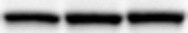
Western Blot
1 µg/mL
Sample: Recombinant Rat TNF‑ alpha (Catalog # 510-RT)
Sample: Recombinant Rat TNF‑ alpha (Catalog # 510-RT)
Neutralization
Measured by its ability to neutralize TNF‑ alpha -induced cytotoxicity in the L‑929 mouse fibroblast cell line. Matthews, N. and M.L. Neale (1987) in Lymphokines and Interferons, A Practical Approach. Clemens, M.J. et al. (eds): IRL Press. 221. The Neutralization Dose (ND50) is typically 10-40 µg/mL in the presence of 0.025 ng/mL Recombinant Rat TNF‑ alpha.
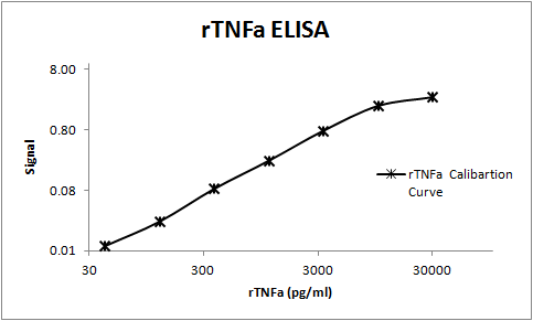
Rat TNF-alpha Sandwich Immunoassay
Please Note: Optimal dilutions of this antibody should be experimentally determined.
Reviewed Applications
Read 2 reviews rated 5 using MAB510 in the following applications:
Formulation, Preparation, and Storage
Purification
Protein A or G purified from hybridoma culture supernatant
Reconstitution
Reconstitute at 0.5 mg/mL in sterile PBS. For liquid material, refer to CoA for concentration.
Loading...
Formulation
Lyophilized from a 0.2 μm filtered solution in PBS with Trehalose. *Small pack size (SP) is supplied either lyophilized or as a 0.2 µm filtered solution in PBS.
Shipping
Lyophilized product is shipped at ambient temperature. Liquid small pack size (-SP) is shipped with polar packs. Upon receipt, store immediately at the temperature recommended below.
Stability & Storage
Use a manual defrost freezer and avoid repeated freeze-thaw cycles.
- 12 months from date of receipt, -20 to -70 °C as supplied.
- 1 month, 2 to 8 °C under sterile conditions after reconstitution.
- 6 months, -20 to -70 °C under sterile conditions after reconstitution.
Calculators
Background: TNF-alpha
References
- Idriss, H.T. and J.H. Naismith (2000) Microsc. Res. Tech. 50:184.
- Hehlgans, T. and K. Pfeffer (2005) Immunology 115:1.
- Estler, H.C. et al. (1992) Biol. Chem. Hoppe-Seyler 373:271.
- Tang, P. et al. (1996) Biochemistry 35:8216.
- Eissner G. et al. (2004) Cytokine Growth Factor Rev. 15:353.
- Black, R.A. et al. (1997) Nature 385:729.
- Moss, M.L. et al. (1997) Nature 385:733.
- Loetscher, H. et al. (1991) J. Biol. Chem. 266:18324.
- Clark, I.A. (2007) Cytokine Growth Factor Rev. 18:335.
- Romanatto, T. et al. (2007) Peptides 28:1050.
- Hector, J. et al. (2007) Horm. Metab. Res. 39:250.
Long Name
Tumor Necrosis Factor alpha
Alternate Names
Cachetin, DIF, TNF, TNF-A, TNFA, TNFalpha, TNFG1F, TNFSF1A, TNFSF2
Entrez Gene IDs
Gene Symbol
TNF
UniProt
Additional TNF-alpha Products
Product Documents for Rat TNF‑ alpha Antibody
Certificate of Analysis
To download a Certificate of Analysis, please enter a lot or batch number in the search box below.
Note: Certificate of Analysis not available for kit components.
Product Specific Notices for Rat TNF‑ alpha Antibody
For research use only
Related Research Areas
Citations for Rat TNF‑ alpha Antibody
 Powered by Bioz
Customer Reviews for Rat TNF‑ alpha Antibody (2)
5 out of 5
2 Customer Ratings
Have you used Rat TNF‑ alpha Antibody?
Submit a review and receive an Amazon gift card!
$25/€18/£15/$25CAN/¥2500 Yen for a review with an image
$10/€7/£6/$10CAN/¥1110 Yen for a review without an image
Submit a review
Customer Images
Showing
1
-
2 of
2 reviews
Showing All
Filter By:
-
Application: Western BlotSample Tested: SerumSpecies: RatVerified Customer | Posted 10/18/2021

-
Application: ELISASample Tested: Serum and PlasmaSpecies: RatVerified Customer | Posted 11/08/2017An ELISA was build targeting rat TNFa using this antibody as a capture and BAF510 as the detection. The assay was used to measure TNFa in rat serum and plasma samples.
There are no reviews that match your criteria.
Protocols
Find general support by application which include: protocols, troubleshooting, illustrated assays, videos and webinars.
- Cellular Response to Hypoxia Protocols
- R&D Systems Quality Control Western Blot Protocol
- Troubleshooting Guide: Western Blot Figures
- Western Blot Conditions
- Western Blot Protocol
- Western Blot Protocol for Cell Lysates
- Western Blot Troubleshooting
- Western Blot Troubleshooting Guide
- View all Protocols, Troubleshooting, Illustrated assays and Webinars
Loading...
Associated Pathways


IL-15 Signaling Pathways and their Primary Biological Effects in Different Immune Cell Types
 Innate Lymphoid Cell Differentiation Pathways
 mTOR Signaling Pathway
 NOD-like Receptor Signaling Pathways
 Th1 Differentiation Pathway
 TNF Superfamily Pathway: Human Ligand-Receptor Interactions & their Associated Functions
 Toll-Like Receptor Signaling Pathways
