TGF-beta 1 Antibody - BSA Free
Novus Biologicals | Catalog # NBP1-80289
![Western Blot: TGF-beta 1 Antibody [NBP1-80289]](https://resources.rndsystems.com/images/products/TGF-beta-1-Antibody-Western-Blot-NBP1-80289-img0013.jpg "Western Blot: TGF-beta 1 Antibody [NBP1-80289]")
Key Product Details
Species Reactivity
Validated:
Cited:
Applications
Validated:
Cited:
Label
Antibody Source
Format
Product Specifications
Immunogen
Reactivity Notes
Clonality
Host
Isotype
Theoretical MW
Disclaimer note: The observed molecular weight of the protein may vary from the listed predicted molecular weight due to post translational modifications, post translation cleavages, relative charges, and other experimental factors.
Description
Scientific Data Images for TGF-beta 1 Antibody - BSA Free
Western Blot: TGF-beta 1 Antibody [NBP1-80289]
Western Blot: TGF-beta 1 Antibody [NBP1-80289] - Sample Tissue: Human Fetal Heart. Antibody Dilution: 1.0ug/ml![Immunocytochemistry/ Immunofluorescence: TGF-beta 1 Antibody [NBP1-80289]](https://resources.rndsystems.com/images/products/TGF-beta-1-Antibody-Immunocytochemistry-Immunofluorescence-NBP1-80289-img0008.jpg "Immunocytochemistry/ Immunofluorescence: TGF-beta 1 Antibody [NBP1-80289]")
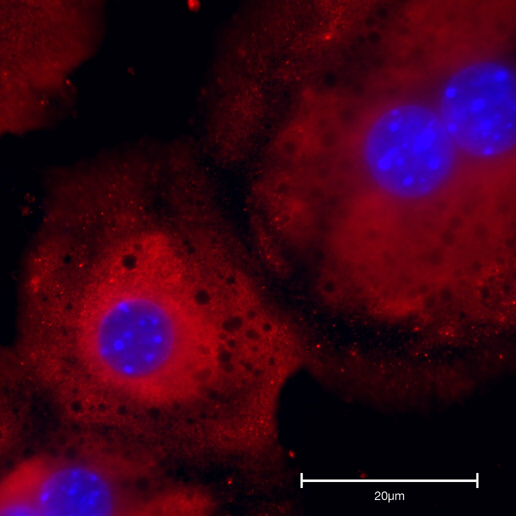
Immunocytochemistry/ Immunofluorescence: TGF-beta 1 Antibody [NBP1-80289]
Immunocytochemistry/Immunofluorescence: TGF-beta 1 Antibody [NBP1-80289] - Mouse kidney (red fluorescence). Nuclei were stained with DAPI (blue fluorescence). Working dilutions: 5-10 ug/ml![Immunohistochemistry-Paraffin: TGF-beta 1 Antibody [NBP1-80289]](https://resources.rndsystems.com/images/products/TGF-beta-1-Antibody-Immunohistochemistry-Paraffin-NBP1-80289-img0011.jpg "Immunohistochemistry-Paraffin: TGF-beta 1 Antibody [NBP1-80289]")
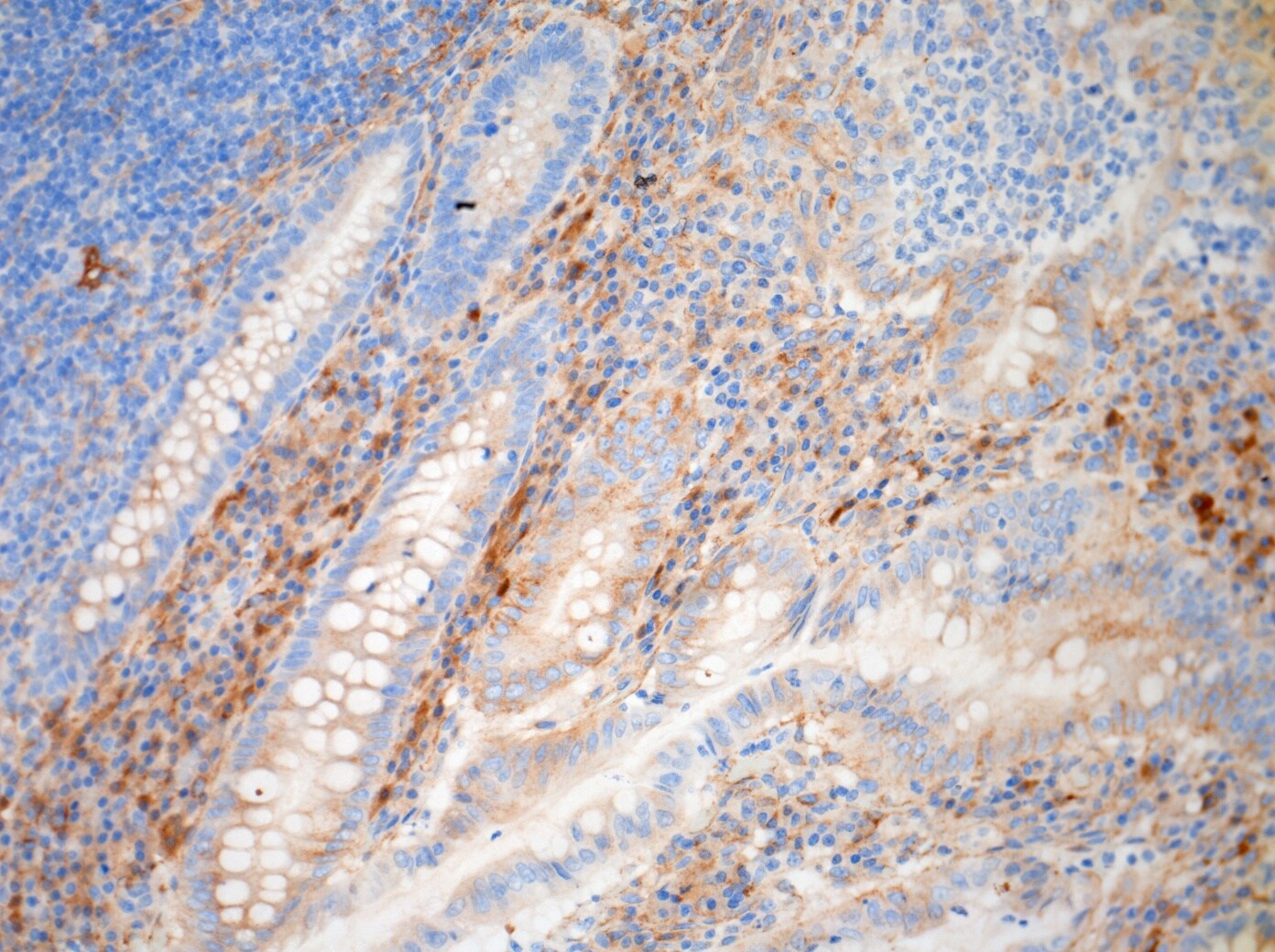
Immunohistochemistry-Paraffin: TGF-beta 1 Antibody [NBP1-80289]
Immunohistochemistry-Paraffin: TGF-beta 1 Antibody [NBP1-80289] - Human Appendix FFPE tissue section stained with TGF-beta 1 antibody. IHC-P image submitted by a verified customer review.![Western Blot: TGF-beta 1 Antibody [NBP1-80289]](https://resources.rndsystems.com/images/products/TGF-beta-1-Antibody-Western-Blot-NBP1-80289-img0010.jpg "Western Blot: TGF-beta 1 Antibody [NBP1-80289]")
Western Blot: TGF-beta 1 Antibody [NBP1-80289]
Western Blot: TGF-beta 1 Antibody [NBP1-80289] - Antibody Titration: 0.2-1 ug/ml or 1:5000 to 1:1000 Dilution. SP2/0 cell lysate.![Western Blot: TGF-beta 1 Antibody [NBP1-80289]](https://resources.rndsystems.com/images/products/TGF-beta-1-Antibody-Western-Blot-NBP1-80289-img0012.jpg "Western Blot: TGF-beta 1 Antibody [NBP1-80289]")
Western Blot: TGF-beta 1 Antibody [NBP1-80289]
Western Blot: TGF-beta 1 Antibody [NBP1-80289] - Sample Tissue: Mouse Spleen. Antibody Dilution: 1ug/ml![Western Blot: TGF-beta 1 Antibody [NBP1-80289]](https://resources.rndsystems.com/images/products/TGF-beta-1-Antibody-Western-Blot-NBP1-80289-img0014.jpg "Western Blot: TGF-beta 1 Antibody [NBP1-80289]")
Western Blot: TGF-beta 1 Antibody [NBP1-80289]
Western Blot: TGF-beta 1 Antibody [NBP1-80289] - Sample Tissue: Mouse Pancreas. Antibody Dilution: 1ug/ml![Immunohistochemistry-Paraffin: TGF-beta 1 Antibody [NBP1-80289]](https://resources.rndsystems.com/images/products/TGF-beta-1-Antibody-Immunohistochemistry-Paraffin-NBP1-80289-img0007.jpg "Immunohistochemistry-Paraffin: TGF-beta 1 Antibody [NBP1-80289]")
Immunohistochemistry-Paraffin: TGF-beta 1 Antibody [NBP1-80289]
Immunohistochemistry-Paraffin: TGF-beta 1 Antibody [NBP1-80289] - Human Spleen Tissue, 5 ug/ml.![Immunohistochemistry-Paraffin: TGF-beta 1 Antibody [NBP1-80289]](https://resources.rndsystems.com/images/products/TGF-beta-1-Antibody-Immunohistochemistry-Paraffin-NBP1-80289-img0009.jpg "Immunohistochemistry-Paraffin: TGF-beta 1 Antibody [NBP1-80289]")
Immunohistochemistry-Paraffin: TGF-beta 1 Antibody [NBP1-80289]
Immunohistochemistry-Paraffin: TGF-beta 1 Antibody [NBP1-80289] - Human prostate cancer tissue was detected using RP/AEC red color stain. Working dilutions: 5-10 ug/ml.![Imaging Mass Cytometry: TGF-beta 1 Antibody [NBP1-80289]](https://resources.rndsystems.com/images/products/TGF-beta-1-Antibody-Imaging-Mass-Cytometry-NBP1-80289-img0015.jpg "Imaging Mass Cytometry: TGF-beta 1 Antibody [NBP1-80289]")
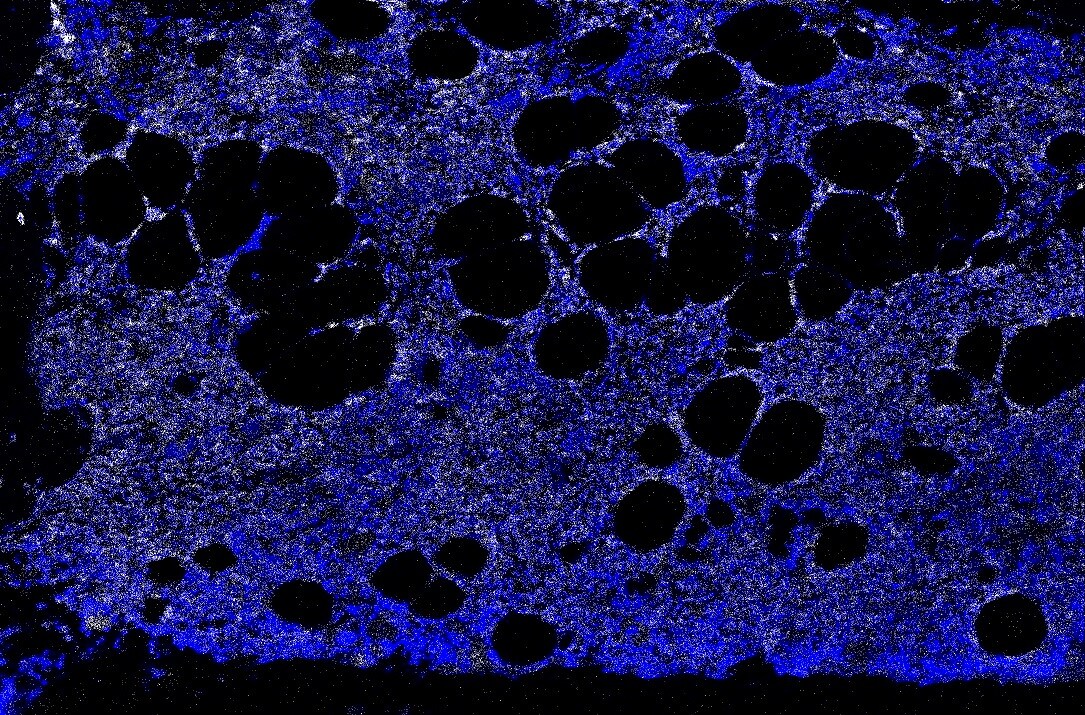
Imaging Mass Cytometry: TGF-beta 1 Antibody [NBP1-80289]
Imaging Mass Cytometry: TGF-beta 1 Antibody [NBP1-80289] - Human bone marrow FFPE tissue sections. DNA in blue, TGF-beta 1 antibody staining in white. IMC image submitted by a verified customer review.
Immunocytochemistry/Immunofluorescence: Rabbit Polyclonal TGF-beta 1 Antibody [NBP1-80289]
Immunocytochemistry/Immunofluorescence: Rabbit Polyclonal TGF-beta 1 Antibody [NBP1-80289] - Mice hepatocytes stained with TGF-beta 1 Antibody. Image from a verified customer review.
Western Blot: TGF-beta 1 Antibody - BSA Free [NBP1-80289] -
Effects of MICT and HIIT on the liver. (A) Representative histological images stained with Picro Sirius Red at 10× and 20× for (A) MICT group and (B) HIIT group. Arrows indicate collagen accumulation points. (C) Collagen 1 and (D) transforming growth factor (TGF) beta 1 and their representative blots below its respective graph with its membrane stained with Ponceau S used as loading control. Data are presented as mean +/- SD. *: p < 0.05. Each dot represents a participant. Image collected and cropped by CiteAb from the following open publication (https://www.mdpi.com/2077-0383/13/11/3273), licensed under a CC-BY license. Not internally tested by Novus Biologicals.Applications for TGF-beta 1 Antibody - BSA Free
Immunocytochemistry/ Immunofluorescence
Immunohistochemistry
Immunohistochemistry-Paraffin
Western Blot
Reviewed Applications
Read 3 reviews rated 4.7 using NBP1-80289 in the following applications:
Formulation, Preparation, and Storage
Purification
Formulation
Format
Preservative
Concentration
Shipping
Stability & Storage
Background: TGF-beta 1
Long Name
Alternate Names
Gene Symbol
UniProt
Additional TGF-beta 1 Products
Product Documents for TGF-beta 1 Antibody - BSA Free
Certificate of Analysis
To download a Certificate of Analysis, please enter a lot or batch number in the search box below.
Product Specific Notices for TGF-beta 1 Antibody - BSA Free
This product is for research use only and is not approved for use in humans or in clinical diagnosis. Primary Antibodies are guaranteed for 1 year from date of receipt.
Related Research Areas
Citations for TGF-beta 1 Antibody - BSA Free
 Powered by Bioz
Customer Reviews for TGF-beta 1 Antibody - BSA Free (3)
Have you used TGF-beta 1 Antibody - BSA Free?
Submit a review and receive an Amazon gift card!
$25/€18/£15/$25CAN/¥2500 Yen for a review with an image
$10/€7/£6/$10CAN/¥1110 Yen for a review without an image
Submit a review
Customer Images
-
Application: ImmunofluorescenceSample Tested: Primary mouse hepatocytesSpecies: MouseVerified Customer | Posted 11/19/2024Mice hepatocytes stained with NBP1-80289

-
Application: Imaging Mass CytometrySample Tested: bone marrowSpecies: HumanVerified Customer | Posted 11/12/2021IMC staining of BM FFPE samples DNA- blue TGFb-white
Bio-Techne ResponseThis review was submitted through the legacy Novus Innovators Program, reflecting a new species or application tested on a primary antibody.
-
Application: Immunohistochemistry-ParaffinSample Tested: Appendix FFPE sectionSpecies: HumanVerified Customer | Posted 09/16/2016TGFb1 human appendix
There are no reviews that match your criteria.
Protocols
Find general support by application which include: protocols, troubleshooting, illustrated assays, videos and webinars.
- Antigen Retrieval Protocol (PIER)
- Antigen Retrieval for Frozen Sections Protocol
- Appropriate Fixation of IHC/ICC Samples
- Cellular Response to Hypoxia Protocols
- Chromogenic IHC Staining of Formalin-Fixed Paraffin-Embedded (FFPE) Tissue Protocol
- Chromogenic Immunohistochemistry Staining of Frozen Tissue
- ClariTSA™ Fluorophore Kits
- Detection & Visualization of Antibody Binding
- Fluorescent IHC Staining of Frozen Tissue Protocol
- Graphic Protocol for Heat-induced Epitope Retrieval
- Graphic Protocol for the Preparation and Fluorescent IHC Staining of Frozen Tissue Sections
- Graphic Protocol for the Preparation and Fluorescent IHC Staining of Paraffin-embedded Tissue Sections
- Graphic Protocol for the Preparation of Gelatin-coated Slides for Histological Tissue Sections
- ICC Cell Smear Protocol for Suspension Cells
- ICC Immunocytochemistry Protocol Videos
- ICC for Adherent Cells
- IHC Sample Preparation (Frozen sections vs Paraffin)
- Immunocytochemistry (ICC) Protocol
- Immunocytochemistry Troubleshooting
- Immunofluorescence of Organoids Embedded in Cultrex Basement Membrane Extract
- Immunofluorescent IHC Staining of Formalin-Fixed Paraffin-Embedded (FFPE) Tissue Protocol
- Immunohistochemistry (IHC) and Immunocytochemistry (ICC) Protocols
- Immunohistochemistry Frozen Troubleshooting
- Immunohistochemistry Paraffin Troubleshooting
- Preparing Samples for IHC/ICC Experiments
- Preventing Non-Specific Staining (Non-Specific Binding)
- Primary Antibody Selection & Optimization
- Protocol for Heat-Induced Epitope Retrieval (HIER)
- Protocol for Making a 4% Formaldehyde Solution in PBS
- Protocol for VisUCyte™ HRP Polymer Detection Reagent
- Protocol for the Fluorescent ICC Staining of Cell Smears - Graphic
- Protocol for the Fluorescent ICC Staining of Cultured Cells on Coverslips - Graphic
- Protocol for the Preparation & Fixation of Cells on Coverslips
- Protocol for the Preparation and Chromogenic IHC Staining of Frozen Tissue Sections
- Protocol for the Preparation and Chromogenic IHC Staining of Frozen Tissue Sections - Graphic
- Protocol for the Preparation and Chromogenic IHC Staining of Paraffin-embedded Tissue Sections
- Protocol for the Preparation and Chromogenic IHC Staining of Paraffin-embedded Tissue Sections - Graphic
- Protocol for the Preparation and Fluorescent ICC Staining of Cells on Coverslips
- Protocol for the Preparation and Fluorescent ICC Staining of Non-adherent Cells
- Protocol for the Preparation and Fluorescent ICC Staining of Stem Cells on Coverslips
- Protocol for the Preparation and Fluorescent IHC Staining of Frozen Tissue Sections
- Protocol for the Preparation and Fluorescent IHC Staining of Paraffin-embedded Tissue Sections
- Protocol for the Preparation of Gelatin-coated Slides for Histological Tissue Sections
- Protocol for the Preparation of a Cell Smear for Non-adherent Cell ICC - Graphic
- R&D Systems Quality Control Western Blot Protocol
- TUNEL and Active Caspase-3 Detection by IHC/ICC Protocol
- The Importance of IHC/ICC Controls
- Troubleshooting Guide: Immunohistochemistry
- Troubleshooting Guide: Western Blot Figures
- Western Blot Conditions
- Western Blot Protocol
- Western Blot Protocol for Cell Lysates
- Western Blot Troubleshooting
- Western Blot Troubleshooting Guide
- View all Protocols, Troubleshooting, Illustrated assays and Webinars
FAQs for TGF-beta 1 Antibody - BSA Free
-
Q: Does TGF-beta 1 react across multiple species?
A: TGF-beta 1 is a highly conserved molecule varying by just a few amino acids between most species, and thus it is extremely likely to react across species. For example, human TGF-beta 1 is active on mouse cells and vice versa.
-
Q: Will Recombinant Human TGF-beta 1 support the maintenance of either human or non-human cultured cells?
A: TGF-beta 1 performs multiple cellular functions, including cell growth, proliferation, differentiation and apoptosis. It would be necessary to determine the specific conditions for cell growth/proliferation for the cells being cultured.
-
Q: Does TGF-beta 1 react across multiple species?
A: TGF-beta 1 is a highly conserved molecule varying by just a few amino acids between most species, and thus it is extremely likely to react across species. For example, human TGF-beta 1 is active on mouse cells and vice versa.
-
Q: Will Recombinant Human TGF-beta 1 support the maintenance of either human or non-human cultured cells?
A: TGF-beta 1 performs multiple cellular functions, including cell growth, proliferation, differentiation and apoptosis. It would be necessary to determine the specific conditions for cell growth/proliferation for the cells being cultured.
Associated Pathways


