TLR9 Antibody (26C593.2) - Azide and BSA Free
Novus Biologicals | Catalog # NBP2-24863
Key Product Details
Species Reactivity
Validated:
Cited:
Applications
Validated:
Cited:
Label
Antibody Source
Format
Product Specifications
Immunogen
Reactivity Notes
Clonality
Host
Isotype
Scientific Data Images for TLR9 Antibody (26C593.2) - Azide and BSA Free
![Western Blot: TLR9 Antibody (26C593.2)Azide Free [NBP2-24863]](https://resources.rndsystems.com/images/products/TLR9-Antibody-26C593-2-Azide-Free-Western-Blot-NBP2-24863-img0006.jpg "Western Blot: TLR9 Antibody (26C593.2)Azide Free [NBP2-24863]")
Western Blot: TLR9 Antibody (26C593.2)Azide Free [NBP2-24863]
Western Blot: TLR9 Antibody (26C593.2) - Azide Free [NBP2-24863] - Analysis of TLR9 in A) human PBMC, B) human intestine, C) mouse intestine, and D) rat intestine tissue lysates using this antibody at a dilution of 3 ug/ml.![Immunohistochemistry-Paraffin: TLR9 Antibody (26C593.2) - Azide Free [NBP2-24863]](https://resources.rndsystems.com/images/products/TLR9-Antibody-26C593-2-Azide-Free-Immunohistochemistry-Paraffin-NBP2-24863-img0007.jpg "Immunohistochemistry-Paraffin: TLR9 Antibody (26C593.2) - Azide Free [NBP2-24863]")
Immunohistochemistry-Paraffin: TLR9 Antibody (26C593.2) - Azide Free [NBP2-24863]
Immunohistochemistry-Paraffin: TLR9 Antibody (26C593.2) - Azide Free [NBP2-24863] - Formalin-fixed, paraffin-embedded human spleen probed with TLR9 antibody at 5 ug/ml. Human tissue was used for this test. Staining of formalin-fixed tissues is enhanced by boiling tissue sections in 10 mM sodium citrate buffer, pH 6.0 for 10-20 min followed by cooling at RT for 20 min.![Flow Cytometry: TLR9 Antibody (26C593.2) - Azide Free [NBP2-24863]](https://resources.rndsystems.com/images/products/TLR9-Antibody-26C593-2-Azide-Free-Flow-Cytometry-NBP2-24863-img0003.jpg "Flow Cytometry: TLR9 Antibody (26C593.2) - Azide Free [NBP2-24863]")
Flow Cytometry: TLR9 Antibody (26C593.2) - Azide Free [NBP2-24863]
Flow Cytometry: TLR9 Antibody (26C593.2) - Azide Free [NBP2-24863] - Intracellular flow analysis of TLR9 in Ramos cells using 0.5 ug of this antibody. Shaded histogram represents Ramos cells without antibody; green represents isotype control; purple represents anti-TLR9 antibody.![Flow (Intracellular): TLR9 Antibody (26C593.2) - Azide Free [NBP2-24863]](https://resources.rndsystems.com/images/products/TLR9-Antibody-26C593-2-Azide-Free-Flow-Intracellular-NBP2-24863-img0008.jpg "Flow (Intracellular): TLR9 Antibody (26C593.2) - Azide Free [NBP2-24863]")
Flow (Intracellular): TLR9 Antibody (26C593.2) - Azide Free [NBP2-24863]
Flow (Intracellular): TLR9 Antibody (26C593.2) - Azide Free [NBP2-24863] - Analysis using the PE conjugate of NBP2-24729. Staining of TLR9 in human PBMCs using 0.2 ug of this antibody. Shaded histogram represents cells without antibody; green represents a mouse IgG1-PE isotype control ; red represents anti-TLR9 antibody.![Flow Cytometry: TLR9 Antibody (26C593.2) - Azide Free [NBP2-24863]](https://resources.rndsystems.com/images/products/TLR9-Antibody-26C593-2-Azide-Free-Flow-Cytometry-NBP2-24863-img0009.jpg "Flow Cytometry: TLR9 Antibody (26C593.2) - Azide Free [NBP2-24863]")
Flow Cytometry: TLR9 Antibody (26C593.2) - Azide Free [NBP2-24863]
Flow Cytometry: TLR9 Antibody (26C593.2) - Azide Free [NBP2-24863] - Flow analysis of TLR9 in human PBMC using 0.5 ug of TLR9 antibody (red) and isotype control antibody (green).![Flow Cytometry: TLR9 Antibody (26C593.2) - Azide Free [NBP2-24863]](https://resources.rndsystems.com/images/products/TLR9-Antibody-26C593-2-Azide-Free-Flow-Cytometry-NBP2-24863-img0010.jpg "Flow Cytometry: TLR9 Antibody (26C593.2) - Azide Free [NBP2-24863]")
Flow Cytometry: TLR9 Antibody (26C593.2) - Azide Free [NBP2-24863]
Flow Cytometry: TLR9 Antibody (26C593.2) - Azide Free [NBP2-24863] - Intracellular flow cytometric analysis of TLR9 FITC in human B cells using 1ug/10^6 cells of NBP2-24908. Cells were primarily stained with PE-conjugated CD19 antibody. Cells were then fixed and permeabilized and stained with 1ug of either isotype control![Flow Cytometry: TLR9 Antibody (26C593.2) - Azide Free [NBP2-24863]](https://resources.rndsystems.com/images/products/TLR9-Antibody-26C593-2-Azide-Free-Flow-Cytometry-NBP2-24863-img0011.jpg "Flow Cytometry: TLR9 Antibody (26C593.2) - Azide Free [NBP2-24863]")
Flow Cytometry: TLR9 Antibody (26C593.2) - Azide Free [NBP2-24863]
Flow Cytometry: TLR9 Antibody (26C593.2) - Azide Free [NBP2-24863] - Analysis using the Azide Free version of NBP2-24729. Staining of TLR9 in Ramos cells using 0.5 ug of this antibody. Shaded histogram represents Ramos cells without antibody; green represents isotype control; purple represents anti-TLR9 antibody.![Flow Cytometry: TLR9 Antibody (26C593.2) - Azide Free [NBP2-24863]](https://resources.rndsystems.com/images/products/TLR9-Antibody-26C593-2-Azide-Free-Flow-Cytometry-NBP2-24863-img0012.jpg "Flow Cytometry: TLR9 Antibody (26C593.2) - Azide Free [NBP2-24863]")
Flow Cytometry: TLR9 Antibody (26C593.2) - Azide Free [NBP2-24863]
Flow Cytometry: TLR9 Antibody (26C593.2) - Azide Free [NBP2-24863] - Analysis using the FITC conjugate of NBP2-24729. Staining of TLR9 in Ramos cells using 0.5 ug of this antibody. Green represents isotype control ; red represents anti-TLR9 antibody. TLR intracellular flow kit was used for this test.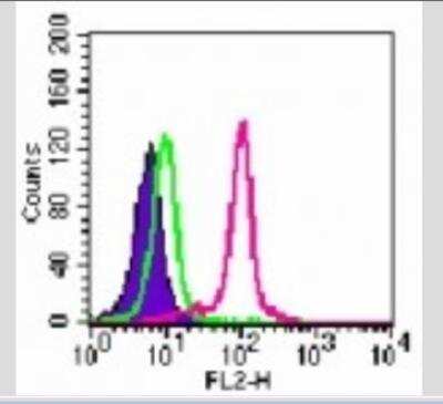
![Simple Western: TLR9 Antibody (26C593.2)Azide Free [NBP2-24863]](https://resources.rndsystems.com/images/products/TLR9-Antibody-26C593-2-Azide-Free-Simple-Western-NBP2-24863-img0014.jpg "Simple Western: TLR9 Antibody (26C593.2)Azide Free [NBP2-24863]")
Simple Western: TLR9 Antibody (26C593.2)Azide Free [NBP2-24863]
Simple Western: TLR9 Antibody (26C593.2) - Azide Free [NBP2-24863] - Simple Western lane view shows a specific band for TLR9 in 0.5 mg/ml of Ramos lysate. This experiment was performed under reducing conditions using the 66-440 kDa separation system.Applications for TLR9 Antibody (26C593.2) - Azide and BSA Free
ELISA
Flow (Intracellular)
Flow Cytometry
Immunocytochemistry/ Immunofluorescence
Immunohistochemistry-Paraffin
Immunoprecipitation
Simple Western
Western Blot
Flow Cytometry Panel Builder
Bio-Techne Knows Flow Cytometry
Save time and reduce costly mistakes by quickly finding compatible reagents using the Panel Builder Tool.
Advanced Features
- Spectra Viewer - Custom analysis of spectra from multiple fluorochromes
- Spillover Popups - Visualize the spectra of individual fluorochromes
- Antigen Density Selector - Match fluorochrome brightness with antigen density

Formulation, Preparation, and Storage
Purification
Formulation
Format
Preservative
Concentration
Shipping
Stability & Storage
Background: TLR9
Long Name
Alternate Names
Gene Symbol
Additional TLR9 Products
Product Documents for TLR9 Antibody (26C593.2) - Azide and BSA Free
Certificate of Analysis
To download a Certificate of Analysis, please enter a lot or batch number in the search box below.
Product Specific Notices for TLR9 Antibody (26C593.2) - Azide and BSA Free
This product is for research use only and is not approved for use in humans or in clinical diagnosis. Primary Antibodies are guaranteed for 1 year from date of receipt.
Citations for TLR9 Antibody (26C593.2) - Azide and BSA Free
 Powered by Bioz
Customer Reviews for TLR9 Antibody (26C593.2) - Azide and BSA Free
There are currently no reviews for this product. Be the first to review TLR9 Antibody (26C593.2) - Azide and BSA Free and earn rewards!
Have you used TLR9 Antibody (26C593.2) - Azide and BSA Free?
Submit a review and receive an Amazon gift card!
$25/€18/£15/$25CAN/¥2500 Yen for a review with an image
$10/€7/£6/$10CAN/¥1110 Yen for a review without an image
Submit a review
Protocols
Find general support by application which include: protocols, troubleshooting, illustrated assays, videos and webinars.
- 7-Amino Actinomycin D (7-AAD) Cell Viability Flow Cytometry Protocol
- Antigen Retrieval Protocol (PIER)
- Antigen Retrieval for Frozen Sections Protocol
- Appropriate Fixation of IHC/ICC Samples
- Cellular Response to Hypoxia Protocols
- Chromogenic IHC Staining of Formalin-Fixed Paraffin-Embedded (FFPE) Tissue Protocol
- Chromogenic Immunohistochemistry Staining of Frozen Tissue
- ClariTSA™ Fluorophore Kits
- Detection & Visualization of Antibody Binding
- ELISA Sample Preparation & Collection Guide
- ELISA Troubleshooting Guide
- Extracellular Membrane Flow Cytometry Protocol
- Flow Cytometry Protocol for Cell Surface Markers
- Flow Cytometry Protocol for Staining Membrane Associated Proteins
- Flow Cytometry Staining Protocols
- Flow Cytometry Troubleshooting Guide
- Fluorescent IHC Staining of Frozen Tissue Protocol
- Graphic Protocol for Heat-induced Epitope Retrieval
- Graphic Protocol for the Preparation and Fluorescent IHC Staining of Frozen Tissue Sections
- Graphic Protocol for the Preparation and Fluorescent IHC Staining of Paraffin-embedded Tissue Sections
- Graphic Protocol for the Preparation of Gelatin-coated Slides for Histological Tissue Sections
- How to Run an R&D Systems DuoSet ELISA
- How to Run an R&D Systems Quantikine ELISA
- How to Run an R&D Systems Quantikine™ QuicKit™ ELISA
- ICC Cell Smear Protocol for Suspension Cells
- ICC Immunocytochemistry Protocol Videos
- ICC for Adherent Cells
- IHC Sample Preparation (Frozen sections vs Paraffin)
- Immunocytochemistry (ICC) Protocol
- Immunocytochemistry Troubleshooting
- Immunofluorescence of Organoids Embedded in Cultrex Basement Membrane Extract
- Immunofluorescent IHC Staining of Formalin-Fixed Paraffin-Embedded (FFPE) Tissue Protocol
- Immunohistochemistry (IHC) and Immunocytochemistry (ICC) Protocols
- Immunohistochemistry Frozen Troubleshooting
- Immunohistochemistry Paraffin Troubleshooting
- Immunoprecipitation Protocol
- Intracellular Flow Cytometry Protocol Using Alcohol (Methanol)
- Intracellular Flow Cytometry Protocol Using Detergents
- Intracellular Nuclear Staining Flow Cytometry Protocol Using Detergents
- Intracellular Staining Flow Cytometry Protocol Using Alcohol Permeabilization
- Intracellular Staining Flow Cytometry Protocol Using Detergents to Permeabilize Cells
- Preparing Samples for IHC/ICC Experiments
- Preventing Non-Specific Staining (Non-Specific Binding)
- Primary Antibody Selection & Optimization
- Propidium Iodide Cell Viability Flow Cytometry Protocol
- Protocol for Heat-Induced Epitope Retrieval (HIER)
- Protocol for Liperfluo
- Protocol for Making a 4% Formaldehyde Solution in PBS
- Protocol for VisUCyte™ HRP Polymer Detection Reagent
- Protocol for the Characterization of Human Th22 Cells
- Protocol for the Characterization of Human Th9 Cells
- Protocol for the Fluorescent ICC Staining of Cell Smears - Graphic
- Protocol for the Fluorescent ICC Staining of Cultured Cells on Coverslips - Graphic
- Protocol for the Preparation & Fixation of Cells on Coverslips
- Protocol for the Preparation and Chromogenic IHC Staining of Frozen Tissue Sections
- Protocol for the Preparation and Chromogenic IHC Staining of Frozen Tissue Sections - Graphic
- Protocol for the Preparation and Chromogenic IHC Staining of Paraffin-embedded Tissue Sections
- Protocol for the Preparation and Chromogenic IHC Staining of Paraffin-embedded Tissue Sections - Graphic
- Protocol for the Preparation and Fluorescent ICC Staining of Cells on Coverslips
- Protocol for the Preparation and Fluorescent ICC Staining of Non-adherent Cells
- Protocol for the Preparation and Fluorescent ICC Staining of Stem Cells on Coverslips
- Protocol for the Preparation and Fluorescent IHC Staining of Frozen Tissue Sections
- Protocol for the Preparation and Fluorescent IHC Staining of Paraffin-embedded Tissue Sections
- Protocol for the Preparation of Gelatin-coated Slides for Histological Tissue Sections
- Protocol for the Preparation of a Cell Smear for Non-adherent Cell ICC - Graphic
- Protocol: Annexin V and PI Staining by Flow Cytometry
- Protocol: Annexin V and PI Staining for Apoptosis by Flow Cytometry
- Quantikine HS ELISA Kit Assay Principle, Alkaline Phosphatase
- Quantikine HS ELISA Kit Principle, Streptavidin-HRP Polymer
- R&D Systems Quality Control Western Blot Protocol
- Sandwich ELISA (Colorimetric) – Biotin/Streptavidin Detection Protocol
- Sandwich ELISA (Colorimetric) – Direct Detection Protocol
- TUNEL and Active Caspase-3 Detection by IHC/ICC Protocol
- The Importance of IHC/ICC Controls
- Troubleshooting Guide: ELISA
- Troubleshooting Guide: Fluorokine Flow Cytometry Kits
- Troubleshooting Guide: Immunohistochemistry
- Troubleshooting Guide: Western Blot Figures
- Western Blot Conditions
- Western Blot Protocol
- Western Blot Protocol for Cell Lysates
- Western Blot Troubleshooting
- Western Blot Troubleshooting Guide
- View all Protocols, Troubleshooting, Illustrated assays and Webinars
Associated Pathways


