TNF-alpha Antibody - BSA Free
Novus Biologicals | Catalog # NBP1-19532
![Western Blot: TNF-alpha AntibodyBSA Free [NBP1-19532]](https://resources.rndsystems.com/images/products/TNF-alpha-Antibody-Western-Blot-NBP1-19532-img0007.jpg "Western Blot: TNF-alpha AntibodyBSA Free [NBP1-19532]")
Key Product Details
Validated by
Biological Validation
Species Reactivity
Validated:
Human, Mouse, Rat, Canine, Fish
Cited:
Human, Mouse, Rat, Canine, Fish, Primate - Macaca mulatta (Rhesus Macaque)
Predicted:
Baboon (97%), Feline (90%), Monkey (98%), Primate (100%). Backed by our 100% Guarantee.
Applications
Validated:
Immunohistochemistry, Immunohistochemistry-Paraffin, Immunohistochemistry-Frozen, Immunohistochemistry Free-Floating, Western Blot, Flow Cytometry, Immunocytochemistry/ Immunofluorescence
Cited:
Immunohistochemistry, Immunohistochemistry-Paraffin, Immunohistochemistry-Frozen, Immunohistochemistry Free-Floating, Western Blot, Flow Cytometry, Immunocytochemistry/ Immunofluorescence, IF/IHC
Label
Unconjugated
Antibody Source
Polyclonal Rabbit IgG
Format
BSA Free
Loading...
Product Specifications
Immunogen
A synthetic peptide made to an internal portion of the human TNF alpha protein (between residues 100-200) [Uniprot: P01375]
Reactivity Notes
Fish reactivity reported in scientific literature (PMID: 29321977). Immunogen displays the following percentage of sequence identity for non-tested species: porcine (89%), equine (87%), and guinea pig (84%). Use in Canine reported in scientific literature (PMID:32656339).
Localization
Membrane
Clonality
Polyclonal
Host
Rabbit
Isotype
IgG
Scientific Data Images for TNF-alpha Antibody - BSA Free
Western Blot: TNF-alpha AntibodyBSA Free [NBP1-19532]
Western Blot: TNF-alpha Antibody [NBP1-19532] - Recombinant human TNF alpha (10 ng) was separated on a 12% gel by SDS-PAGE, transferred to 0.2 um PVDF membrane and blocked in 5% non-fat milk in TBST. The membrane was probed with 2.0 ug/ml anti-TNF alpha in 5% block buffer and detected with an anti-rabbit HRP secondary antibody using chemiluminescence.![Western Blot: TNF-alpha AntibodyBSA Free [NBP1-19532]](https://resources.rndsystems.com/images/products/TNF-alpha-Antibody-Western-Blot-NBP1-19532-img0008.jpg "Western Blot: TNF-alpha AntibodyBSA Free [NBP1-19532]")
![Immunohistochemistry-Paraffin: TNF-alpha Antibody - BSA Free [NBP1-19532]](https://resources.rndsystems.com/images/products/TNF-alpha-Antibody-Immunohistochemistry-Paraffin-NBP1-19532-img0005.jpg "Immunohistochemistry-Paraffin: TNF-alpha Antibody - BSA Free [NBP1-19532]")
Immunohistochemistry-Paraffin: TNF-alpha Antibody - BSA Free [NBP1-19532]
Immunohistochemistry-Paraffin: TNF-alpha Antibody [NBP1-19532] - Analysis of a FFPE tissue section of mouse intestine using TNF-alpha antibody (NBP1-19532) at 1:300 dilution. The binding of this primary antibody to TNF-alpha protein in the section was detected using HRP-labeled secondary antibody and DAB reagent, and nuclei of cells were counterstained using hematoxylin. This TNF-alpha antibody generated an expected diffused immunostaining of this protein in the tested tissue. Staining was primarily observed in the epithelial cells and some cells showed membrane positivity also.![Immunohistochemistry-Paraffin: TNF-alpha Antibody - BSA Free [NBP1-19532]](https://resources.rndsystems.com/images/products/TNF-alpha-Antibody-Immunohistochemistry-Paraffin-NBP1-19532-img0006.jpg "Immunohistochemistry-Paraffin: TNF-alpha Antibody - BSA Free [NBP1-19532]")
Immunohistochemistry-Paraffin: TNF-alpha Antibody - BSA Free [NBP1-19532]
Immunohistochemistry-Paraffin: TNF-alpha Antibody [NBP1-19532] - Analysis of a FFPE tissue section of mouse intestine using TNF-alpha antibody (NBP1-19532) at 1:300 dilution. The binding of this primary antibody to TNF-alpha protein in the section was detected using HRP-labeled secondary antibody and DAB reagent, and nuclei of cells were counterstained using hematoxylin. This TNF-alpha antibody generated an expected immunopositivity of this protein in the tested tissue. Staining was primarily observed in the epithelial cells while some staining was present in mucosa muscularis also.
Western Blot: TNF-alpha Antibody - BSA Free [NBP1-19532] -
Scutellarin attenuates hypertension-induced brain expression of NF-kappa B, TNF-alpha, IL-1 beta, and IL-18. Western immunoblot analysis for (a) NF-kappa B p65, (b) TNF-alpha, (c) IL-1 beta, and (d) IL-18 in the rat cortex and striatum. Treatment with scutellarin significantly reduced the expression of these inflammatory markers in a dose-dependent manner. *P < 0.001 versus sham group; #P < 0.05, ◆P < 0.001 versus NS group; &P < 0.001 versus low-dose group.
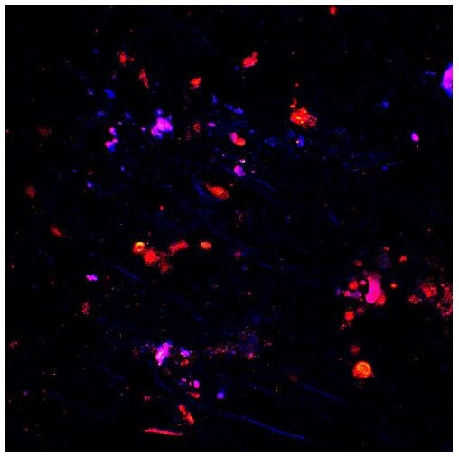
Immunocytochemistry/ Immunofluorescence: TNF-alpha Antibody - BSA Free [NBP1-19532] -
Immunocytochemistry/ Immunofluorescence: TNF-alpha Antibody - BSA Free [NBP1-19532] - Effects of femoral artery occlusion on TNF-alpha signal in sensory nerves. (A) Seventy-two hours of femoral artery occlusion increased the levels of TNF-alpha in the DRG tissues as compared with them in DRG of control limbs (n = 6 in each group); & PTX given into the hindlimb muscles attenuated amplification of TNF-alpha in the DRG tissues of limbs with femoral artery occlusion (n = 8). ∗P < 0.05, occlusion group vs. control & occlusion with prior PTX. (B) Immunofluorescence was used to examine double-labeling for TNF-alpha & peripherin/NF-200 (n = 3). Peripherin was used to label DRG neurons that project thin C-fibers. NF200 was used to identify A-fibers of DRG neurons. Representative photomicrographs show co-existence of TNF-alpha & peripherin staining in DRG neurons (top panel), whereas few TNF-alpha & NF-200 staining were observed in DRG neurons (bottom panel). Arrows indicate representative positive cells for both TNF-alpha & peripherin after they were merged. Scale bar = 50 μm. (C) Representative bands (left panel) & averaged data (right panel), demonstrating that femoral artery occlusion upregulated protein expression TNF-alpha receptor subtype TNFR1, but not TNFR2. A significant difference in TNFR1 was seen between control & occluded groups. ∗P < 0.05 vs. control. n = 6 in each group. Image collected & cropped by CiteAb from the following publication (https://pubmed.ncbi.nlm.nih.gov/30374312), licensed under a CC-BY license. Not internally tested by Novus Biologicals.
Immunocytochemistry/ Immunofluorescence: TNF-alpha Antibody - BSA Free [NBP1-19532] -
Immunocytochemistry/ Immunofluorescence: TNF-alpha Antibody - BSA Free [NBP1-19532] - Immunoflorescence staining results of TNF-alpha (96 h). A). Lamella of gill tissue (arrow). Control group. IF. 20 μm. B) Positive reaction in lamellar cells of gill tissue (arrows). Low dose toxic group. IF. 20 μm. C) Positivity in lamellar cells of gill tissue (arrow). High dose toxic group. IF. 20 μm. D) Liver tissue, vena centralis (arrow). Control group. IF. 20 μm. E) Positive reactions in hepatocytes (arrows). Low dose toxic group. IF. 20 μm. F) Positive reactions in hepatocytes (arrows). High dose toxic group. IF. 20 μm. G) Weak positive reactions in neurons (arrows). Control group. IF. 20 μm. H) Weak positivity in neurons (arrows). Low dose toxic group. IF. 20 μm. I) Positivity in neurons (arrows). High dose toxic group. IF. 20 μm. Image collected & cropped by CiteAb from the following publication (https://pubmed.ncbi.nlm.nih.gov/29321977), licensed under a CC-BY license. Not internally tested by Novus Biologicals.
Immunohistochemistry: TNF-alpha Antibody - BSA Free [NBP1-19532] -
Suppressive effects of ketogenic diet on the number of inflammatory cytokine-positive cells and inflammasome component protein-positive cells surrounding knee joint after repeated intra-articular blood injections. The immunohistochemical staining of knee joints on post intra-articular injection day 26 was compared, in the intra-articular saline injection + control diet (Saline + control diet) group (a, d, g, j, and m), the intra-articular blood injection + control diet (Blood + control diet) group (b, e, h, k, and n), and the intra-articular blood injection + ketogenic diet (Blood + ketogenic diet) group (c, f, i, l, and o) using anti-IL-1 beta antibody (a – c), anti-ASC antibody (d – f), anti-NLRP3 antibody (g – i), anti-TNF-alpha antibody (j – l), and anti-IL-6 antibody (m – o). The scale bar in (a) indicates 50 μm. The number of inflammatory cytokine-positive cells (IL-1 beta, TNF-alpha, and IL-6) and inflammasome component protein-positive cells (ASC and NLRP3) per 1 mm2 in the periarticular tissues was counted (p – t). White: Saline + control diet group, black: Blood + control diet group, and gray: Blood + ketogenic diet group (p – t). Data are shown as mean and SD. Intergroup comparisons were assessed by one-way ANOVA. Bonferroni multiple comparison test was used for comparisons among each group (*p < 0.05, **p < 0.01). Image collected and cropped by CiteAb from the following open publication (https://www.nature.com/articles/s41598-024-77074-6), licensed under a CC-BY license. Not internally tested by Novus Biologicals.
Immunohistochemistry: TNF-alpha Antibody - BSA Free [NBP1-19532] -
Histological and immunohistochemistry results of periapical lesions in each group. (A) Representative histopathological images in periapical areas after root canal filling; (B) The inflammation grade evaluation of periapical lesions (n = 10); (C) Representative immunohistochemistry (IHC) staining of MMP-9 in periapical areas after root canal filling. Green boxes indicate peripheral periodontal tissue; (D) Representative immunohistochemistry (IHC) staining of TNF-alpha in periapical areas after root canal filling. Green boxes indicate peripheral periodontal tissue. ns p > 0.05, not significant, * p < 0.05, ** p < 0.01. Image collected and cropped by CiteAb from the following open publication (https://pubmed.ncbi.nlm.nih.gov/36361925), licensed under a CC-BY license. Not internally tested by Novus Biologicals.Applications for TNF-alpha Antibody - BSA Free
Application
Recommended Usage
Immunocytochemistry/ Immunofluorescence
1:200 - 1:300
Immunohistochemistry
1:200
Immunohistochemistry-Paraffin
1:200
Western Blot
1:250 - 1:500
Application Notes
Western Blot data from customer review and citation (PMID: 24223475). Use in Immunohistochemistry-free floating reported in scientific literature (PMID: 26219646). Use in Immunohistochemistry-Frozen reported in scientific literature (PMID: 26725948). Use in FLOW reported in scientific literature (PMID: 26889045).
Reviewed Applications
Read 3 reviews rated 5 using NBP1-19532 in the following applications:
Flow Cytometry Panel Builder
Bio-Techne Knows Flow Cytometry
Save time and reduce costly mistakes by quickly finding compatible reagents using the Panel Builder Tool.
Advanced Features
- Spectra Viewer - Custom analysis of spectra from multiple fluorochromes
- Spillover Popups - Visualize the spectra of individual fluorochromes
- Antigen Density Selector - Match fluorochrome brightness with antigen density

Formulation, Preparation, and Storage
Purification
Immunogen affinity purified
Formulation
PBS
Format
BSA Free
Preservative
0.02% Sodium Azide
Concentration
1.0 mg/ml
Shipping
The product is shipped with polar packs. Upon receipt, store it immediately at the temperature recommended below.
Stability & Storage
Store at 4C short term. Aliquot and store at -20C long term. Avoid freeze-thaw cycles.
Background: TNF-alpha
TNF-alpha is critical for normal immune response; however, dysregulation of TNF-alpha production can result in various pathologies (2,4,5). Excessive production of pro-inflammatory cytokines including interleukin 1 (IL-1), IL-6, and TNF-alpha has been implicated in an array of autoimmune diseases like rheumatoid arthritis (RA), inflammatory bowel disease (IBD), and psoriasis (2,4,5). Anti-TNF monoclonal antibodies, including Infliximab, and soluble TNFR have been approved for the treatment of autoimmune and TNF-mediated diseases (5). Additionally, data suggests that TNF inhibitors can be beneficial for treating patients experiencing immune-related adverse events associated with immune checkpoint inhibitor cancer treatment (6).
References
1. Holbrook J, Lara-Reyna S, Jarosz-Griffiths H, McDermott M. Tumour necrosis factor signalling in health and disease. F1000Res. 2019;8:F1000 Faculty Rev-111. https://doi.org/10.12688/f1000research.17023.1
2. Jang DI, Lee AH, Shin HY, et al. The Role of Tumor Necrosis Factor Alpha (TNF-alpha) in Autoimmune Disease and Current TNF-alpha Inhibitors in Therapeutics. Int J Mol Sci. 2021;22(5):2719. https://doi.org/10.3390/ijms22052719
3. Horiuchi T, Mitoma H, Harashima S, Tsukamoto H, Shimoda T. Transmembrane TNF-alpha: structure, function and interaction with anti-TNF agents. Rheumatology (Oxford). 2010;49(7):1215-1228. https://doi.org/10.1093/rheumatology/keq031
4. Webster JD, Vucic D. The Balance of TNF Mediated Pathways Regulates Inflammatory Cell Death Signaling in Healthy and Diseased Tissues. Front Cell Dev Biol. 2020;8:365. https://doi.org/10.3389/fcell.2020.00365
5. Kalliolias GD, Ivashkiv LB. TNF biology, pathogenic mechanisms and emerging therapeutic strategies. Nat Rev Rheumatol. 2016; 12(1):49-62. https://doi.org/10.1038/nrrheum.2015.169
6. Chen AY, Wolchok JD, Bass AR. TNF in the era of immune checkpoint inhibitors: friend or foe?. Nat Rev Rheumatol. 2021;17(4):213-223. doi:10.1038/s41584-021-00584-4
Long Name
Tumor Necrosis Factor alpha
Alternate Names
Cachetin, DIF, TNF, TNF-A, TNFA, TNFalpha, TNFG1F, TNFSF1A, TNFSF2
Gene Symbol
TNF
UniProt
Additional TNF-alpha Products
Product Documents for TNF-alpha Antibody - BSA Free
Certificate of Analysis
To download a Certificate of Analysis, please enter a lot or batch number in the search box below.
Product Specific Notices for TNF-alpha Antibody - BSA Free
This product is for research use only and is not approved for use in humans or in clinical diagnosis. Primary Antibodies are guaranteed for 1 year from date of receipt.
Related Research Areas
Citations for TNF-alpha Antibody - BSA Free
 Powered by Bioz
Customer Reviews for TNF-alpha Antibody - BSA Free (3)
5 out of 5
3 Customer Ratings
Have you used TNF-alpha Antibody - BSA Free?
Submit a review and receive an Amazon gift card!
$25/€18/£15/$25CAN/¥2500 Yen for a review with an image
$10/€7/£6/$10CAN/¥1110 Yen for a review without an image
Submit a review
Customer Images
-(01-mg)_NBP1-19532_8991.jpg)
Showing
1
-
3 of
3 reviews
Showing All
Filter By:
-
Application: Immunocytochemistry/ImmunofluorescenceSample Tested: Primary neuronsSpecies: RatVerified Customer | Posted 10/13/2016
-
Application: ImmunofluorescenceSample Tested: trabecular meshwork (TM) region of pig eyesSpecies: OtherVerified Customer | Posted 01/07/2016Pig TM tissue stained with TNF alpha antibody

-
Application: Western BlotSample Tested: Recombinant his-tagged TNFa made in E. coli cellsSpecies: HumanVerified Customer | Posted 07/24/2014WB for His-tagged TNFa protein made in E. coli
There are no reviews that match your criteria.
Protocols
View specific protocols for TNF-alpha Antibody - BSA Free (NBP1-19532):
Immunocytochemistry Protocol
Culture cells to appropriate density in 35 mm culture dishes or 6-well plates.
1. Remove culture medium and add 10% formalin to the dish. Fix at room temperature for 30 minutes.
2. Remove the formalin and add ice cold methanol. Incubate for 5-10 minutes.
3. Remove methanol and add washing solution (i.e. PBS). Be sure to not let the specimen dry out. Wash three times for 10 minutes.
4. To block nonspecific antibody binding incubate in 10% normal goat serum from 1 hour to overnight at room temperature.
5. Add primary antibody at appropriate dilution and incubate at room temperature from 2 hours to overnight at room temperature.
6. Remove primary antibody and replace with washing solution. Wash three times for 10 minutes.
7. Add secondary antibody at appropriate dilution. Incubate for 1 hour at room temperature.
8. Remove antibody and replace with wash solution, then wash for 10 minutes. Add Hoechst 33258 to wash solution at 1:25,0000 and incubate for 10 minutes. Wash a third time for 10 minutes.
9. Cells can be viewed directly after washing. The plates can also be stored in PBS containing Azide covered in Parafilm (TM). Cells can also be cover-slipped using Fluoromount, with appropriate sealing.
*The above information is only intended as a guide. The researcher should determine what protocol best meets their needs. Please follow safe laboratory procedures.
Immunohistochemistry-Paraffin Embedded Sections
Antigen Unmasking:
Bring slides to a boil in 10 mM sodium citrate buffer (pH 6.0) then maintain at a sub-boiling temperature for 10 minutes. Cool slides on bench-top for 30 minutes.
Staining:
1. Wash sections in deionized water three times for 5 minutes each.
2. Wash sections in wash buffer for 5 minutes.
3. Block each section with 100-400 ul blocking solution for 1 hour at room temperature.
4. Remove blocking solution and add 100-400 ul diluted primary antibody. Incubate overnight at 4 C.
5. Remove antibody solution and wash sections in wash buffer three times for 5 minutes each.
6. Add 100-400 ul biotinylated diluted secondary antibody. Incubate 30 minutes at room temperature.
7. Remove secondary antibody solution and wash sections three times with wash buffer for 5 minutes each.
8. Add 100-400 ul Streptavidin-HRP reagent to each section and incubate for 30 minutes at room temperature.
9. Wash sections three times in wash buffer for 5 minutes each.
10. Add 100-400 ul DAB substrate to each section and monitor staining closely.
11. As soon as the sections develop, immerse slides in deionized water.
12. Counterstain sections in hematoxylin.
13. Wash sections in deionized water two times for 5 minutes each.
14. Dehydrate sections.
15. Mount coverslips.
Find general support by application which include: protocols, troubleshooting, illustrated assays, videos and webinars.
- 7-Amino Actinomycin D (7-AAD) Cell Viability Flow Cytometry Protocol
- Antigen Retrieval Protocol (PIER)
- Antigen Retrieval for Frozen Sections Protocol
- Appropriate Fixation of IHC/ICC Samples
- Cellular Response to Hypoxia Protocols
- Chromogenic IHC Staining of Formalin-Fixed Paraffin-Embedded (FFPE) Tissue Protocol
- Chromogenic Immunohistochemistry Staining of Frozen Tissue
- ClariTSA™ Fluorophore Kits
- Detection & Visualization of Antibody Binding
- Extracellular Membrane Flow Cytometry Protocol
- Flow Cytometry Protocol for Cell Surface Markers
- Flow Cytometry Protocol for Staining Membrane Associated Proteins
- Flow Cytometry Staining Protocols
- Flow Cytometry Troubleshooting Guide
- Fluorescent IHC Staining of Frozen Tissue Protocol
- Graphic Protocol for Heat-induced Epitope Retrieval
- Graphic Protocol for the Preparation and Fluorescent IHC Staining of Frozen Tissue Sections
- Graphic Protocol for the Preparation and Fluorescent IHC Staining of Paraffin-embedded Tissue Sections
- Graphic Protocol for the Preparation of Gelatin-coated Slides for Histological Tissue Sections
- ICC Cell Smear Protocol for Suspension Cells
- ICC Immunocytochemistry Protocol Videos
- ICC for Adherent Cells
- IHC Sample Preparation (Frozen sections vs Paraffin)
- Immunocytochemistry (ICC) Protocol
- Immunocytochemistry Troubleshooting
- Immunofluorescence of Organoids Embedded in Cultrex Basement Membrane Extract
- Immunofluorescent IHC Staining of Formalin-Fixed Paraffin-Embedded (FFPE) Tissue Protocol
- Immunohistochemistry (IHC) and Immunocytochemistry (ICC) Protocols
- Immunohistochemistry Frozen Troubleshooting
- Immunohistochemistry Paraffin Troubleshooting
- Intracellular Flow Cytometry Protocol Using Alcohol (Methanol)
- Intracellular Flow Cytometry Protocol Using Detergents
- Intracellular Nuclear Staining Flow Cytometry Protocol Using Detergents
- Intracellular Staining Flow Cytometry Protocol Using Alcohol Permeabilization
- Intracellular Staining Flow Cytometry Protocol Using Detergents to Permeabilize Cells
- Preparing Samples for IHC/ICC Experiments
- Preventing Non-Specific Staining (Non-Specific Binding)
- Primary Antibody Selection & Optimization
- Propidium Iodide Cell Viability Flow Cytometry Protocol
- Protocol for Heat-Induced Epitope Retrieval (HIER)
- Protocol for Liperfluo
- Protocol for Making a 4% Formaldehyde Solution in PBS
- Protocol for VisUCyte™ HRP Polymer Detection Reagent
- Protocol for the Characterization of Human Th22 Cells
- Protocol for the Characterization of Human Th9 Cells
- Protocol for the Fluorescent ICC Staining of Cell Smears - Graphic
- Protocol for the Fluorescent ICC Staining of Cultured Cells on Coverslips - Graphic
- Protocol for the Preparation & Fixation of Cells on Coverslips
- Protocol for the Preparation and Chromogenic IHC Staining of Frozen Tissue Sections
- Protocol for the Preparation and Chromogenic IHC Staining of Frozen Tissue Sections - Graphic
- Protocol for the Preparation and Chromogenic IHC Staining of Paraffin-embedded Tissue Sections
- Protocol for the Preparation and Chromogenic IHC Staining of Paraffin-embedded Tissue Sections - Graphic
- Protocol for the Preparation and Fluorescent ICC Staining of Cells on Coverslips
- Protocol for the Preparation and Fluorescent ICC Staining of Non-adherent Cells
- Protocol for the Preparation and Fluorescent ICC Staining of Stem Cells on Coverslips
- Protocol for the Preparation and Fluorescent IHC Staining of Frozen Tissue Sections
- Protocol for the Preparation and Fluorescent IHC Staining of Paraffin-embedded Tissue Sections
- Protocol for the Preparation of Gelatin-coated Slides for Histological Tissue Sections
- Protocol for the Preparation of a Cell Smear for Non-adherent Cell ICC - Graphic
- Protocol: Annexin V and PI Staining by Flow Cytometry
- Protocol: Annexin V and PI Staining for Apoptosis by Flow Cytometry
- R&D Systems Quality Control Western Blot Protocol
- TUNEL and Active Caspase-3 Detection by IHC/ICC Protocol
- The Importance of IHC/ICC Controls
- Troubleshooting Guide: Fluorokine Flow Cytometry Kits
- Troubleshooting Guide: Immunohistochemistry
- Troubleshooting Guide: Western Blot Figures
- Western Blot Conditions
- Western Blot Protocol
- Western Blot Protocol for Cell Lysates
- Western Blot Troubleshooting
- Western Blot Troubleshooting Guide
- View all Protocols, Troubleshooting, Illustrated assays and Webinars
FAQs for TNF-alpha Antibody - BSA Free
Showing
1
-
5 of
7 FAQs
Showing All
-
Q: Can your TNF-alpha products be used to treat TBI victims and therefore avoid the perispinal injections with Enbrel?
A: I am very sorry, but all of our products are for scientific research use only, and none are intended or approved for use in humans.
-
Q: I am interested in a TNF alpha antibody, cross reactive for human, rat and mouse (host species: rabbit). Could your product NBP1-19532 be used in western blotting applications? Or do you have a similar product in your catalog which could fit with my request?
A:
The antibody you mention, NBP1-19532, has not yet been validated in Western blot. I would instead recommend either NBP1-67821 or NB600-587. These are both rabbit polyclonal antibodies that cross-reacts with human, mouse and rat and have been used in Western blotting.
-
Q: I need to know about TNF-alpha antibody kits for mice.
A:
We currently have two TNFalpha ELISA kits specific for mouse: catalog numbers KA0257 and NBP1-92670.
-
Q: I would like to ask you for help. I need an antibody for Elisa to detect human TNF alpha in a supernatant from a cell culture (meaning supernatant after centrifugation of collected cell suspension from a plate well). Which of your antibodies against human tnf alpha would be suitable? I would like to buy only the primary antibody.
A:
I would recommend catalog number NBP1-67821 or NB600-587; however, a full list of our anti-human TNF alpha antibodies suitable for use in ELISA can be found using this link.
-
Q: I would like to learn about the TNF alpha antibody (NBP1-19532) immunohistochemistry protocol. Is there any specific protocol you have?
A:
Our lab did not use any customized protocol for the IHC-P validation of this antibody. Please see our general IHC-P protocol.
-
Q: Is there any positive control tissue besides human colon carcinoma tissue that can be used for for paraffin embedded IHC testing?
A: You can use skin, lung, liver or mammary tumor tissue etc as a positive control for your assay.
-
Q: We ordered a TNF alpha antibody from you back in May, and I am inquiring about the expiration date. The antibody we ordered was cat #NBP1-19532 and lot # 360986.. The data sheet says that short term storage at 4C and -20C for long term storage but that they are only guaranteed for 6 months. My question is are you considering the "short term storage" at 4C to be the 6 month expiration and -20C the antibody should still be OK? Typically we give our antibodies that are stored at -20C a two year expiration if one is not given.
A: We suggest to store the antibody at 4 degrees for no longer than a week. After this the product should be stored at -20 degrees and we are not sure if this is can be used after the 6 month period. It is likely that the antibody should be fine however we only guarantee our products for up to 6 months.
-
Q: Can your TNF-alpha products be used to treat TBI victims and therefore avoid the perispinal injections with Enbrel?
A: I am very sorry, but all of our products are for scientific research use only, and none are intended or approved for use in humans.
-
Q: I am interested in a TNF alpha antibody, cross reactive for human, rat and mouse (host species: rabbit). Could your product NBP1-19532 be used in western blotting applications? Or do you have a similar product in your catalog which could fit with my request?
A:
The antibody you mention, NBP1-19532, has not yet been validated in Western blot. I would instead recommend either NBP1-67821 or NB600-587. These are both rabbit polyclonal antibodies that cross-reacts with human, mouse and rat and have been used in Western blotting.
-
Q: I need to know about TNF-alpha antibody kits for mice.
A:
We currently have two TNFalpha ELISA kits specific for mouse: catalog numbers KA0257 and NBP1-92670.
-
Q: I would like to ask you for help. I need an antibody for Elisa to detect human TNF alpha in a supernatant from a cell culture (meaning supernatant after centrifugation of collected cell suspension from a plate well). Which of your antibodies against human tnf alpha would be suitable? I would like to buy only the primary antibody.
A:
I would recommend catalog number NBP1-67821 or NB600-587; however, a full list of our anti-human TNF alpha antibodies suitable for use in ELISA can be found using this link.
-
Q: I would like to learn about the TNF alpha antibody (NBP1-19532) immunohistochemistry protocol. Is there any specific protocol you have?
A:
Our lab did not use any customized protocol for the IHC-P validation of this antibody. Please see our general IHC-P protocol.
-
Q: Is there any positive control tissue besides human colon carcinoma tissue that can be used for for paraffin embedded IHC testing?
A: You can use skin, lung, liver or mammary tumor tissue etc as a positive control for your assay.
-
Q: We ordered a TNF alpha antibody from you back in May, and I am inquiring about the expiration date. The antibody we ordered was cat #NBP1-19532 and lot # 360986.. The data sheet says that short term storage at 4C and -20C for long term storage but that they are only guaranteed for 6 months. My question is are you considering the "short term storage" at 4C to be the 6 month expiration and -20C the antibody should still be OK? Typically we give our antibodies that are stored at -20C a two year expiration if one is not given.
A: We suggest to store the antibody at 4 degrees for no longer than a week. After this the product should be stored at -20 degrees and we are not sure if this is can be used after the 6 month period. It is likely that the antibody should be fine however we only guarantee our products for up to 6 months.
Loading...
Associated Pathways


IL-15 Signaling Pathways and their Primary Biological Effects in Different Immune Cell Types
 Innate Lymphoid Cell Differentiation Pathways
 mTOR Signaling Pathway
 NOD-like Receptor Signaling Pathways
 Th1 Differentiation Pathway
 TNF Superfamily Pathway: Human Ligand-Receptor Interactions & their Associated Functions
 Toll-Like Receptor Signaling Pathways
