AKT1 [p Ser473] Antibody (17F6.B11) - BSA Free
Novus Biologicals | Catalog # NBP1-69923
Key Product Details
Validated by
Biological Validation
Species Reactivity
Validated:
Human, Mouse, Rat, Monkey
Cited:
Human, Mouse
Applications
Validated:
Immunohistochemistry, Immunohistochemistry-Paraffin, Western Blot, ELISA, Flow Cytometry, Immunocytochemistry/ Immunofluorescence, Immunoprecipitation, Dot Blot
Cited:
Western Blot
Label
Unconjugated
Antibody Source
Monoclonal Mouse IgG1 kappa Clone # 17F6.B11
Format
BSA Free
Loading...
Product Specifications
Immunogen
AKT1 [p Ser473] Antibody (17F6.B11) was produced by repeated immunizations with a synthetic peptide corresponding to residues surrounding S473 of human AKT11 protein, followed by hybridoma development. (Uniprot: P31749)
Reactivity Notes
A BLAST analysis was used to suggest cross-reactivity with AKT1 pS473 from human, mouse, rat and chimpanzee sources based on 100% homology with the immunizing sequence. Cross-reactivity with AKT1 from other sources has not been determined. Cross-reactivity with AKT2 and AKT3 has not been determined.
Modification
p Ser473
Specificity
This phospho specific monoclonal antibody is specific for phosphorylated human and mouse AKT protein at S473. A BLAST analysis was used to suggest cross-reactivity with AKT pS473 from human, mouse, rat and chimpanzee sources based on 100% homology with the immunizing sequence. Cross-reactivity with AKT from other sources has not been determined. Cross-reactivity with AKT2 and AKT3 has not been determined.
Clonality
Monoclonal
Host
Mouse
Isotype
IgG1 kappa
Description
This antibody Monoclonal Antibody was purified from concentrated tissue culture supernate by Protein A chromatography
Store this antibody at -20C prior to opening. Aliquot contents and freeze at -20C or below for extended storage. Avoid cycles of freezing and thawing. Centrifuge product if not completely clear after standing at room temperature. This product is stable for several weeks at 4C as an undiluted liquid. Dilute only prior to immediate use.
Store this antibody at -20C prior to opening. Aliquot contents and freeze at -20C or below for extended storage. Avoid cycles of freezing and thawing. Centrifuge product if not completely clear after standing at room temperature. This product is stable for several weeks at 4C as an undiluted liquid. Dilute only prior to immediate use.
Scientific Data Images for AKT1 [p Ser473] Antibody (17F6.B11) - BSA Free
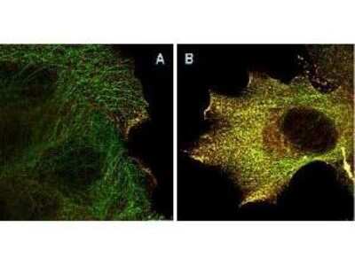
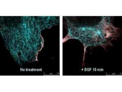
Immunocytochemistry/Immunofluorescence: AKT1 [p Ser473] Antibody (17F6.B11) [NBP1-69923] - A431 cells. Panel A: serum starved, unstimulated cells. Panel B: serum starved, EGF stimulated for 15 mins. A massive increase in AKT-pS473 activation, as measured by intensity signal, peaked at 15 minutes and was associated with depolymerized tubulin. Panel A shows STED data (AKT-pS473, red channel) collected simultaneously with confocal signal (alpha-tubulin, green channel). Upon stimulation of cells with EGF, a rapid activation of AKT is observed (Panel B) along with a coincident change in the tubulin organization (yellow signal), as well as an extensive cell shape-change (cell membrane folding) and accumulation of AKT pS473 at the cell periphery.
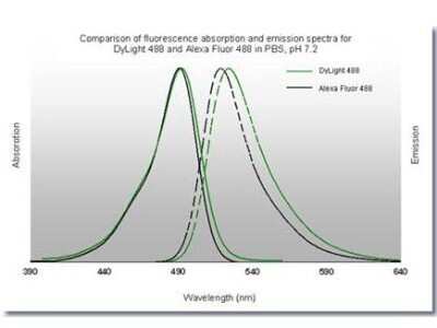
Flow Cytometry: AKT1 [p Ser473] Antibody (17F6.B11) [NBP1-69923] - Analysis using the DyLight 488 conjugate of AKT1 phospho Ser473 antibody. Image shows anti-histone detection using a DyLight 488 conjugate (green). Anti-Tubulin was detected using a DyLight 549 conjugate (red). Nuclei were counter-stained using DAPI (blue).
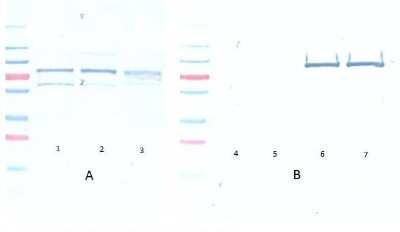
Western Blot: AKT1 [p Ser473] Antibody (17F6.B11) [NBP1-69923] - Western Blot of AKT1 [p Ser473] antibody (17F6.B11). A: Lane 1) PDGF stimulated NIH 3T3 cells [10ul]. Lane 2) NIH 3T3 cells [10ul]. Lane 3) Hela whole cell lysate [10ul] (weak signal). B: Lane 4) GST negative control protein [100ng]. Lane 5) GST negative control protein [25ng]. Lane 6) AKT 1 recombinant protein [100ng]. Lane 7) AKT 1 recombinant protein [25ng].Block: 5% BSA overnight at 4C.Primary antibody: Monoclonal anti-AKT antibody used at 1:1000 for overnight at 4C.Secondary antibody: HRP Conjugated goat anti-mouse 1:25K for 45 min at RT.Detection: TMB for 20 minutes, rinsed with deionized water, dried and scanned on conventional flatbed scanner.
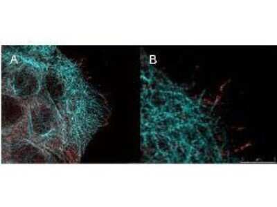
Immunocytochemistry/Immunofluorescence: AKT1 [p Ser473] Antibody (17F6.B11) [NBP1-69923] - High resolution STED immunofluorescence nanoscopy of AKT11 [p Ser473] antibody (17F6.B11). Tissue: A431 cells. The merge images (A) and at high magnification (B) show phosphorylated AKT1 colocalized with the distal microtubules. Fixation: 4% paraformaldehyde for 5 min and after washes blocked with 10% NGS/0.2% Triton X-100 for 30 min.Antigen retrieval: serum deprivation for 12 h.Primary antibody: AKT1 pS473 antibody at 10 ug/mL and alpha-tubulin (cyan) at 1.4 ug/mL for 1 h at RT.Secondary antibody: Atto 647N anti-Mouse IgG (ATTO TEC GmbH), and DyLight(TM)488 anti-Rabbit IgG were used at 1.0 ug/mL for 1h at RT for indirect detection.Localization: AKT1 pS473 is in the cytoplasm and also organized at the periphery of the cell.Staining: AKT1 pS473 as red signal with bis-benzimide (blue) nuclear counterstain.
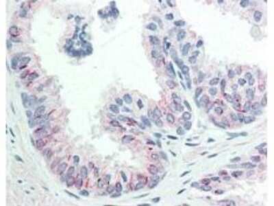
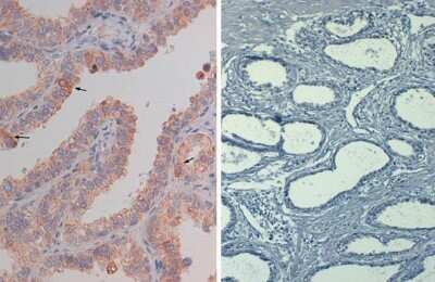
Immunohistochemistry: AKT1 [p Ser473] Antibody (17F6.B11) [NBP1-69923] - Immunohistochemistry of AKT11 [p Ser473] antibody (17F6.B11). Tissue: human prostate tissue. Fixation: formalin fixed paraffin embedded. Antigen retrieval: not required.Primary antibody: AKT1 pS473 antibody at 20 ug/mL for 1 h at RT.Secondary antibody: Dako's Techmate streptavidin-biotin reagents at 1:10,000 for 45 min at RT.Localization: AKT1 pS473 is nuclear and occasionally cytoplasmic.Staining: AKT1 pS473 as precipitated red signal with hematoxylin purple nuclear counterstain.
AKT1-p-Ser473-Antibody-17F6-B11-Western-Blot-NBP1-69923-img0037.jpg
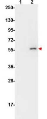
Western Blot: AKT1 [p Ser473] Antibody (17F6.B11) [NBP1-69923] - Western Blot of AKT11 [p Ser473] antibody (17F6.B11). Lane 1: untreated NIH/3T3 cell lysates. Lane 2: detects phosphorylated AKT1 (indicated by arrowhead at ~56 kDa) on PDGF stimulated NIH/3T3 cell lysates. Load: 10 ug per lane.Primary antibody: AKT1 pS473 antibody at 1:10,000 in TBS with 0.05% Tween-20 with 1% BSA, for 1 h at 4 C.Secondary antibody: HRP conjugated Gt-a-Mouse IgG was used at a 1:20,000 dilution for 1 h at 4 C with FemtoMax(TM) enhanced chemiluminescent reagent.
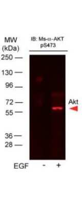
Western Blot: AKT1 [p Ser473] Antibody (17F6.B11) [NBP1-69923] - Western Blot of AKT11 [p Ser473] antibody (17F6.B11). Lane 1: A431 cells. Lane 2: A431 cells stimulated for 15 min with EGF. Load: 35 ug per lane.Primary antibody: AKT1pS473 antibody at 1:400 for overnight at 4C.Secondary antibody: DyLight(TM)649 Conjugated Anti-AKT1 pS473 Monoclonal Antibody at 1:10,000 for 45 min at RT.Block: Blocking Buffer for Fluorescent Western Blotting overnight at 4C.Predicted/Observed size: 56kDa. Other band(s): none.
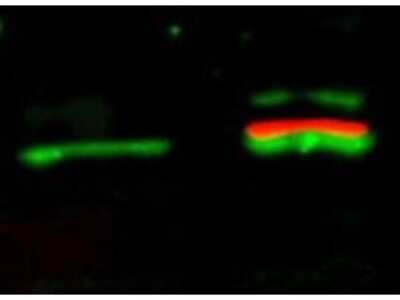
Western Blot: AKT1 [p Ser473] Antibody (17F6.B11) [NBP1-69923] - Western Blot of AKT1 [p Ser473] antibody (17F6.B11). Lane 1: unstimulated NIH/3T3 lysates contain inactive unphosphorylated AKT11, green band. Lane 2: PDGF stimulated NIH/3T3 lysate contains both inactive (green band) and activated phosphorylated AKT11 (red band). Load: 10 ug per lane.Primary antibody: rabbit anti-AKT1 (pan) and mouse anti-AKT1 pS473 specific antibodies at 1:400 for overnight at 4C.Secondary antibody: DyLight(TM) 549 conjugated anti-rabbit IgG (green) and DyLight(TM) 649 conjugated anti-mouse IgG (red) secondary antibodies at 1:10,000 for 45 min at RT.Block: 5% BLOTTO overnight at 4C.
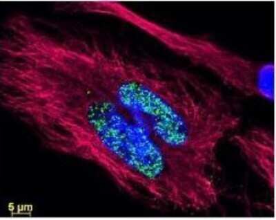
Immunocytochemistry/Immunofluorescence: AKT1 [p Ser473] Antibody (17F6.B11) [NBP1-69923] - Analysis of DyLight 488 conjugate of NBP1-69923. This image shows anti-histone detection using a DyLight 488 conjugate (green). Anti-Tubulin was detected using a DyLight 549 conjugate (red). Nuclei were counter-stained using DAPI (blue).
Immunocytochemistry/Immunofluorescence: AKT1 [p Ser473] Antibody (17F6.B11) [NBP1-69923]

Immunocytochemistry/Immunofluorescence: AKT1 [p Ser473] Antibody (17F6.B11) [NBP1-69923] - Immunofluorescence Microscopy of AKT11 [p Ser473] antibody (17F6.B11) using STED nanoscopy to evaluate AKT1 activation and migration. Tissue: A431 cells. Antigen retrieval: Panel A: serum starved, unstimulated cells. Panel B: serum starved, EGF stimulated for 15 mins.A massive increase in AKT1-pS473 activation, as measured by intensity signal, peaked at 15 minutes and was associated with depolymerized tubulin.Staining: Panel A shows STED data (AKT1-pS473, red channel) collected simultaneously with confocal signal (a-tubulin, green channel). Upon stimulation of cells with EGF, a rapid activation of AKT1 is observed (Panel B) along with a coincident change in the tubulin organization (yellow signal), as well as an extensive cell shape-change (cell membrane folding) and accumulation of AKT1pS473 at the cell periphery.
Immunohistochemistry-Paraffin: AKT1 [p Ser473] Antibody (17F6.B11) [NBP1-69923] - Analysis of Biotin conjugate of NBP1-69923. 20 ug/mL for 1 h at RT Secondary antibody: Streptavidin Conj. HRP 10 ug/ml Localization: nuclear and occasionally cytoplasmic Staining: antibody as precipitated red signal with a hematoxylin purple nuclear count
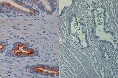
Immunohistochemistry-Paraffin: AKT1 [p Ser473] Antibody (17F6.B11) [NBP1-69923] - Analysis of FFPE prostate tissue using AKT1 phospho Ser473 antibody (left panel). Negative control (right panel). Antigen retrieval with heat and pressure in citrate buffer pH 6.2. AKT1 phospho Ser473 antibody at 20 ug/mL. Secondary antibody Streptavidin-HRP at 10 ug/mL. Hematoxylin nuclear counterstain (purple). Image using the Biotin format of this antibody.
![AKT1 [p Ser473] Antibody (17F6.B11)](https://resources.rndsystems.com/images/products/nbp1-69923_mouse-monoclonal-akt1-p-ser473-antibody-17f6-b11-235202318143116.jpg "AKT1 [p Ser473] Antibody (17F6.B11)")
AKT1 [p Ser473] Antibody (17F6.B11)
Immunohistochemistry of Mouse anti-AKT pS473 antibody. Tissue: human prostate tissue. Fixation: formalin fixed paraffin embedded. Antigen retrieval: not required. Primary antibody: AKT pS473 antibody at 20 ug/mL for 1 h at RT. Secondary antibody: Dako's Techmate streptavidin-biotin reagents at 1:10,000 for 45 min at RT. Localization: AKT pS473 is nuclear and occasionally cytoplasmic. Staining: AKT pS473 as precipitated red signal with hematoxylin purple nuclear counterstain.![AKT1 [p Ser473] Antibody (17F6.B11)](https://resources.rndsystems.com/images/products/nbp1-69923_mouse-monoclonal-akt1-p-ser473-antibody-17f6-b11-255202313136.jpg "AKT1 [p Ser473] Antibody (17F6.B11)")
AKT1 [p Ser473] Antibody (17F6.B11)
Western Blot of Mouse Anti-AKTpS473 antibody. Lane 1: A431 cells. Lane 2: A431 cells stimulated for 15 min with EGF. Load: 35 ug per lane. Primary antibody: AKTpS473 antibody at 1:400 for overnight at 4C. Secondary antibody: DyLight(TM)649 Conjugated Anti-AKT1 [p Ser473] Antibody (17F6.B11) at 1:10,000 for 45 min at RT. Block: Blocking Buffer for Fluorescent Western Blotting overnight at 4C. Predicted/Observed size: 56kDa. Other band(s): none.Applications for AKT1 [p Ser473] Antibody (17F6.B11) - BSA Free
Application
Recommended Usage
ELISA
1:20000
Flow Cytometry
1:10-1:1000
Immunocytochemistry/ Immunofluorescence
1:500-1:3000
Immunohistochemistry
20 ug/ml
Immunohistochemistry-Paraffin
1:10-1:500
Western Blot
1:500-1:3000
Application Notes
This product is tested in ELISA, immunofluorescence, immunohistochemistry, and western blotting. Expect a band approximately 56 kDa in size corresponding to phosphorylated AKT protein by western blotting in the appropriate cell lysate or extract. This phospho-specific monoclonal antibody reacts with human and mouse AKT pS473 and shows minimal reactivity by ELISA against the non-phosphorylated form of the immunizing peptide. Specific conditions for reactivity should be optimized by the end user. For immunohistochemistry use formalin-fixed paraffin-embedded sections. No pre-treatment of sample is required.
Flow Cytometry Panel Builder
Bio-Techne Knows Flow Cytometry
Save time and reduce costly mistakes by quickly finding compatible reagents using the Panel Builder Tool.
Advanced Features
- Spectra Viewer - Custom analysis of spectra from multiple fluorochromes
- Spillover Popups - Visualize the spectra of individual fluorochromes
- Antigen Density Selector - Match fluorochrome brightness with antigen density

Formulation, Preparation, and Storage
Purification
Protein A purified
Formulation
0.02 M Potassium Phosphate, 0.15 M Sodium Chloride, pH 7.2
Format
BSA Free
Preservative
0.01% Sodium Azide
Concentration
Please see the vial label for concentration. If unlisted please contact technical services.
Shipping
The product is shipped with polar packs. Upon receipt, store it immediately at the temperature recommended below.
Stability & Storage
Store at -20C. Avoid freeze-thaw cycles.
Background: Akt1
The main function of AKT is to control inhibition of apoptosis and promote cell proliferation. Survival factors can activate AKT Ser473 and Thr308 phosphorylation sites in a transcription-independent manner, resulting in the inactivation of apoptotic signaling transduction through the tumor suppressor PTEN, an antagonist to PI3-K (5). PTEN exerts enzymatic activity as a phosphatidylinositol-3,4,5-trisphosphate (PIP3) phosphatase, opposing PI3K activity by decreasing availability of PIP3 to proliferating cells, leading to overexpression and inappropriate activation of AKT noted in many types of cancer.
AKT1 function has been linked to overall physiological growth and function (2). AKT1 has been correlated with proteus syndrome, a rare disorder characterized by overgrowth of various tissues caused by a mosaic variant in the AKT1 gene in humans.
AKT2 is strongly correlated with Type II diabetes, including phenotypes of insulin resistance, hyperglycemia and atherosclerosis (2, 6).
The function of AKT3 is specifically associated to brain development, where disruptions to AKT3 are correlated with microcephaly, hemimegalencephaly, megalencephaly and intellectual disabilities (2).
References
1. Ersahin, T., Tuncbag, N., & Cetin-Atalay, R. (2015). The PI3K/AKT/mTOR interactive pathway. Mol Biosyst, 11(7), 1946-1954. doi:10.1039/c5mb00101c
2. Cohen, M. M., Jr. (2013). The AKT genes and their roles in various disorders. Am J Med Genet A, 161a(12), 2931-2937. doi:10.1002/ajmg.a.36101
3. Georgescu, M. M. (2010). PTEN Tumor Suppressor Network in PI3K-Akt Pathway Control. Genes Cancer, 1(12), 1170-1177. doi:10.1177/1947601911407325
4. Mishra, P., Paital, B., Jena, S., Swain, S. S., Kumar, S., Yadav, M. K.,... Samanta, L. (2019). Possible activation of NRF2 by Vitamin E/Curcumin against altered thyroid hormone induced oxidative stress via NFkB/AKT/mTOR/KEAP1 signalling in rat heart. Sci Rep, 9(1), 7408. doi:10.1038/s41598-019-43320-5
5. Wedel, S., Hudak, L., Seibel, J. M., Juengel, E., Oppermann, E., Haferkamp, A., & Blaheta, R. A. (2011). Critical analysis of simultaneous blockage of histone deacetylase and multiple receptor tyrosine kinase in the treatment of prostate cancer. Prostate, 71(7), 722-735. doi:10.1002/pros.21288
6. Rotllan, N., Chamorro-Jorganes, A., Araldi, E., Wanschel, A. C., Aryal, B., Aranda, J. F.,... Fernandez-Hernando, C. (2015). Hematopoietic Akt2 deficiency attenuates the progression of atherosclerosis. Faseb j, 29(2), 597-610. doi:10.1096/fj.14-262097
Long Name
v-Akt Murine Thymoma Viral Oncogene Homolog 1
Alternate Names
PKB alpha, PRKBA, RAC-alpha
Gene Symbol
AKT1
UniProt
Additional Akt1 Products
Product Documents for AKT1 [p Ser473] Antibody (17F6.B11) - BSA Free
Certificate of Analysis
To download a Certificate of Analysis, please enter a lot or batch number in the search box below.
Product Specific Notices for AKT1 [p Ser473] Antibody (17F6.B11) - BSA Free
This product is for research use only and is not approved for use in humans or in clinical diagnosis. Primary Antibodies are guaranteed for 1 year from date of receipt.
Citations for AKT1 [p Ser473] Antibody (17F6.B11) - BSA Free
 Powered by Bioz
Customer Reviews for AKT1 [p Ser473] Antibody (17F6.B11) - BSA Free
There are currently no reviews for this product. Be the first to review AKT1 [p Ser473] Antibody (17F6.B11) - BSA Free and earn rewards!
Have you used AKT1 [p Ser473] Antibody (17F6.B11) - BSA Free?
Submit a review and receive an Amazon gift card!
$25/€18/£15/$25CAN/¥2500 Yen for a review with an image
$10/€7/£6/$10CAN/¥1110 Yen for a review without an image
Submit a review
Protocols
Find general support by application which include: protocols, troubleshooting, illustrated assays, videos and webinars.
- 7-Amino Actinomycin D (7-AAD) Cell Viability Flow Cytometry Protocol
- Antigen Retrieval Protocol (PIER)
- Antigen Retrieval for Frozen Sections Protocol
- Appropriate Fixation of IHC/ICC Samples
- Cellular Response to Hypoxia Protocols
- Chromogenic IHC Staining of Formalin-Fixed Paraffin-Embedded (FFPE) Tissue Protocol
- Chromogenic Immunohistochemistry Staining of Frozen Tissue
- ClariTSA™ Fluorophore Kits
- Detection & Visualization of Antibody Binding
- ELISA Sample Preparation & Collection Guide
- ELISA Troubleshooting Guide
- Extracellular Membrane Flow Cytometry Protocol
- Flow Cytometry Protocol for Cell Surface Markers
- Flow Cytometry Protocol for Staining Membrane Associated Proteins
- Flow Cytometry Staining Protocols
- Flow Cytometry Troubleshooting Guide
- Fluorescent IHC Staining of Frozen Tissue Protocol
- Graphic Protocol for Heat-induced Epitope Retrieval
- Graphic Protocol for the Preparation and Fluorescent IHC Staining of Frozen Tissue Sections
- Graphic Protocol for the Preparation and Fluorescent IHC Staining of Paraffin-embedded Tissue Sections
- Graphic Protocol for the Preparation of Gelatin-coated Slides for Histological Tissue Sections
- How to Run an R&D Systems DuoSet ELISA
- How to Run an R&D Systems Quantikine ELISA
- How to Run an R&D Systems Quantikine™ QuicKit™ ELISA
- ICC Cell Smear Protocol for Suspension Cells
- ICC Immunocytochemistry Protocol Videos
- ICC for Adherent Cells
- IHC Sample Preparation (Frozen sections vs Paraffin)
- Immunocytochemistry (ICC) Protocol
- Immunocytochemistry Troubleshooting
- Immunofluorescence of Organoids Embedded in Cultrex Basement Membrane Extract
- Immunofluorescent IHC Staining of Formalin-Fixed Paraffin-Embedded (FFPE) Tissue Protocol
- Immunohistochemistry (IHC) and Immunocytochemistry (ICC) Protocols
- Immunohistochemistry Frozen Troubleshooting
- Immunohistochemistry Paraffin Troubleshooting
- Immunoprecipitation Protocol
- Intracellular Flow Cytometry Protocol Using Alcohol (Methanol)
- Intracellular Flow Cytometry Protocol Using Detergents
- Intracellular Nuclear Staining Flow Cytometry Protocol Using Detergents
- Intracellular Staining Flow Cytometry Protocol Using Alcohol Permeabilization
- Intracellular Staining Flow Cytometry Protocol Using Detergents to Permeabilize Cells
- Preparing Samples for IHC/ICC Experiments
- Preventing Non-Specific Staining (Non-Specific Binding)
- Primary Antibody Selection & Optimization
- Propidium Iodide Cell Viability Flow Cytometry Protocol
- Protocol for Heat-Induced Epitope Retrieval (HIER)
- Protocol for Liperfluo
- Protocol for Making a 4% Formaldehyde Solution in PBS
- Protocol for VisUCyte™ HRP Polymer Detection Reagent
- Protocol for the Characterization of Human Th22 Cells
- Protocol for the Characterization of Human Th9 Cells
- Protocol for the Fluorescent ICC Staining of Cell Smears - Graphic
- Protocol for the Fluorescent ICC Staining of Cultured Cells on Coverslips - Graphic
- Protocol for the Preparation & Fixation of Cells on Coverslips
- Protocol for the Preparation and Chromogenic IHC Staining of Frozen Tissue Sections
- Protocol for the Preparation and Chromogenic IHC Staining of Frozen Tissue Sections - Graphic
- Protocol for the Preparation and Chromogenic IHC Staining of Paraffin-embedded Tissue Sections
- Protocol for the Preparation and Chromogenic IHC Staining of Paraffin-embedded Tissue Sections - Graphic
- Protocol for the Preparation and Fluorescent ICC Staining of Cells on Coverslips
- Protocol for the Preparation and Fluorescent ICC Staining of Non-adherent Cells
- Protocol for the Preparation and Fluorescent ICC Staining of Stem Cells on Coverslips
- Protocol for the Preparation and Fluorescent IHC Staining of Frozen Tissue Sections
- Protocol for the Preparation and Fluorescent IHC Staining of Paraffin-embedded Tissue Sections
- Protocol for the Preparation of Gelatin-coated Slides for Histological Tissue Sections
- Protocol for the Preparation of a Cell Smear for Non-adherent Cell ICC - Graphic
- Protocol: Annexin V and PI Staining by Flow Cytometry
- Protocol: Annexin V and PI Staining for Apoptosis by Flow Cytometry
- Quantikine HS ELISA Kit Assay Principle, Alkaline Phosphatase
- Quantikine HS ELISA Kit Principle, Streptavidin-HRP Polymer
- R&D Systems Quality Control Western Blot Protocol
- Sandwich ELISA (Colorimetric) – Biotin/Streptavidin Detection Protocol
- Sandwich ELISA (Colorimetric) – Direct Detection Protocol
- TUNEL and Active Caspase-3 Detection by IHC/ICC Protocol
- The Importance of IHC/ICC Controls
- Troubleshooting Guide: ELISA
- Troubleshooting Guide: Fluorokine Flow Cytometry Kits
- Troubleshooting Guide: Immunohistochemistry
- Troubleshooting Guide: Western Blot Figures
- Western Blot Conditions
- Western Blot Protocol
- Western Blot Protocol for Cell Lysates
- Western Blot Troubleshooting
- Western Blot Troubleshooting Guide
- View all Protocols, Troubleshooting, Illustrated assays and Webinars
FAQs for AKT1 [p Ser473] Antibody (17F6.B11) - BSA Free
Showing
1
-
5 of
5 FAQs
Showing All
-
Q: Do your HRP-conjugated antibodies contain sodium azide?
A: No. None of our HRP-conjugated antibodies contain sodium azide as this agent inhibits the activity of HRP.
-
Q: How do I choose secondary antibodies to label the same cells when I have two primary antibodies from the same host?
A: Use isotype-specific secondary antibodies if the primary antibodies are of different isotypes. You can also make direct conjugates of the primary antibodies by use of antibody labeling kits, dyes, or custom conjugations (please contact Technical Support for custom orders).
-
Q: I am looking for a antibody that recognizes human Akt1 but NOT Akt2 or 3, for Western blot analyses. I also want that antibody to recognize Akt1 regardless of its phosphorylated form.
A: At the moment we do not have an AKT1 antibody that definitively does not react with either AKT2 or AKT3.
-
Q: What is the molecular weight of your antibodies?
A: All IgG antibodies are approximately 150 kDa (each heavy chain is about 50 kDa and each light chain is about 25 kDa).
-
Q: Why are many of your antibodies formulated with sodium azide and BSA?
A: Sodium azide is a preservative which is added to prevent bacterial growth. BSA is added as a protein stabilizer.
-
Q: Do your HRP-conjugated antibodies contain sodium azide?
A: No. None of our HRP-conjugated antibodies contain sodium azide as this agent inhibits the activity of HRP.
-
Q: How do I choose secondary antibodies to label the same cells when I have two primary antibodies from the same host?
A: Use isotype-specific secondary antibodies if the primary antibodies are of different isotypes. You can also make direct conjugates of the primary antibodies by use of antibody labeling kits, dyes, or custom conjugations (please contact Technical Support for custom orders).
-
Q: I am looking for a antibody that recognizes human Akt1 but NOT Akt2 or 3, for Western blot analyses. I also want that antibody to recognize Akt1 regardless of its phosphorylated form.
A: At the moment we do not have an AKT1 antibody that definitively does not react with either AKT2 or AKT3.
-
Q: What is the molecular weight of your antibodies?
A: All IgG antibodies are approximately 150 kDa (each heavy chain is about 50 kDa and each light chain is about 25 kDa).
-
Q: Why are many of your antibodies formulated with sodium azide and BSA?
A: Sodium azide is a preservative which is added to prevent bacterial growth. BSA is added as a protein stabilizer.
Loading...
Associated Pathways


IL-2 Signaling Pathways
 IL-4 Signaling Pathways
 IL-7 Signaling Pathways
 IL-9 Signaling Pathways
 IL-15 Signaling Pathways
 IL-21 Signaling Pathways
 mTOR Signaling Pathway
 Notch Signaling Pathways
 TGF-beta Signaling Pathways
 VEGF - VEGF R2 Signaling Pathways
