HA Tag Antibody (1049F)
R&D Systems | Catalog # MAB0601
Recombinant Monoclonal Antibody.

Key Product Details
Species Reactivity
Validated:
Multi-Species
Cited:
Human
Applications
Validated:
Western Blot, Immunocytochemistry, Simple Western, Immunoprecipitation
Cited:
Western Blot
Label
Unconjugated
Antibody Source
Monoclonal Rabbit IgG Clone # 1049F
Loading...
Product Specifications
Immunogen
HA Tag peptide
Accession # ABB51961
Accession # ABB51961
Specificity
Detects N-terminal and C-terminal HA-tagged proteins in Western blot.
Clonality
Monoclonal
Host
Rabbit
Isotype
IgG
Scientific Data Images for HA Tag Antibody (1049F)
Detection of HA-tagged proteins by Western Blot.
Western blot shows lysates of HEK293 human embryonic kidney cell line transfected with N-terminal HA-tagged MICA, C-terminal HA-tagged TRANCE, and C-terminal HA-tagged LRRN5. PVDF membrane was probed with 0.1 µg/mL of Rabbit Anti-HA Monoclonal Antibody (Catalog # MAB0601) followed by HRP-conjugated Anti-Rabbit IgG Secondary Antibody (Catalog # HAF008). Specific bands were detected for HA-tagged proteins at approximately 70 kDa, 40 kDa, and 77 kDa (as indicated). This experiment was conducted under reducing conditions and using Immunoblot Buffer Group 1.
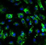
HA Tag in NS0 Mouse Cell Line.
HA was detected in immersion fixed NS0 mouse myeloma cell line transfected with HA-tagged WISP-2 using Rabbit Anti-HA Monoclonal Antibody (Catalog # MAB0601) at 3 µg/mL for 3 hours at room temperature. Cells were stained using the NorthernLights™ 557-conjugated Anti-Rabbit IgG Secondary Antibody (red; Catalog # NL004) and counterstained with DAPI (blue). Specific staining was localized to cytoplasm. View our protocol for Fluorescent ICC Staining of Non-adherent Cells.
Detection of HA-tagged proteins by Simple WesternTM.
Simple Western lane view shows lysates of HEK293 human embryonic kidney cell line transfected with C-terminal HA-tagged LRRN5 and N-terminal HA-tagged MICA, loaded at 0.1 mg/mL. Specific bands were detected for HA-tagged proteins at approximately 87 and 106 kDa (as indicated) using 1 µg/mL of Rabbit Anti-HA Monoclonal Antibody (Catalog # MAB0601). This experiment was conducted under reducing conditions and using the 12-230 kDa separation system.Applications for HA Tag Antibody (1049F)
Application
Recommended Usage
Immunocytochemistry
8-25 µg/mL
Sample: Immersion fixed NS0 mouse myeloma cell line transfected with Hemagglutinin/HA-tagged WISP-2
Sample: Immersion fixed NS0 mouse myeloma cell line transfected with Hemagglutinin/HA-tagged WISP-2
Immunoprecipitation
1-4 µg/100 µg cell lysate
Sample: HEK293 human embryonic kidney cell line transfected with C-terminal HA-tagged LRRN5 and N-terminal HA-tagged MICA, see our available Western blot detection antibodies.
Sample: HEK293 human embryonic kidney cell line transfected with C-terminal HA-tagged LRRN5 and N-terminal HA-tagged MICA, see our available Western blot detection antibodies.
Simple Western
1 µg/mL
Sample: HEK293 human embryonic kidney cell line transfected with C-terminal HA-tagged LRRN5 and N-terminal HA-tagged MICA
Sample: HEK293 human embryonic kidney cell line transfected with C-terminal HA-tagged LRRN5 and N-terminal HA-tagged MICA
Western Blot
0.1 µg/mL
Sample: HEK293 human embryonic kidney cell line transfected with N-terminal HA-tagged MICA, C-terminal HA-tagged TRANCE, and C-terminal HA-tagged LRRN5
Sample: HEK293 human embryonic kidney cell line transfected with N-terminal HA-tagged MICA, C-terminal HA-tagged TRANCE, and C-terminal HA-tagged LRRN5
Reviewed Applications
Read 1 review rated 4 using MAB0601 in the following applications:
Formulation, Preparation, and Storage
Purification
Protein A or G purified from cell culture supernatant
Reconstitution
Reconstitute at 0.5 mg/mL in sterile PBS.
Loading...
Formulation
Lyophilized from a 0.2 μm filtered solution in PBS with Trehalose.
Shipping
The product is shipped at ambient temperature. Upon receipt, store it immediately at the temperature recommended below.
Stability & Storage
Use a manual defrost freezer and avoid repeated freeze-thaw cycles.
- 12 months from date of receipt, -20 to -70 °C as supplied.
- 1 month, 2 to 8 °C under sterile conditions after reconstitution.
- 6 months, -20 to -70 °C under sterile conditions after reconstitution.
Calculators
Background: HA Tag
References
1. Wilks, S., Graaf, M. D., Smith, D. J., & Burke, D. F. (2012). A review of influenza haemagglutinin receptor binding as it relates to pandemic properties. Vaccine, 30(29), 4369-4376. doi:10.1016/j.vaccine.2012.02.076
2. Wu, N. C., & Wilson, I. A. (2019). Influenza hemagglutinin structures and antibody recognition. Cold Spring Harbor Perspectives in Medicine, 10(8). doi:10.1101/cshperspect.a038778
3. Zhao, X., Li, G., & Liang, S. (2013). Several affinity tags commonly used in chromatographic purification. Journal of Analytical Methods in Chemistry, 2013, 1-8. doi:10.1155/2013/581093
4. Kimple, M. E., Brill, A. L., & Pasker, R. L. (2013). Overview of affinity tags for protein purification. Current Protocols in Protein Science, 73(1). doi:10.1002/0471140864.ps0909s73
5. Schembri, L., Dalibart, R., Tomasello, F., Legembre, P., Ichas, F., & Giorgi, F. D. (2007). The HA tag is cleaved and loses immunoreactivity during apoptosis. Nature Methods, 4(2), 107-108. doi:10.1038/nmeth0207-107
Alternate Names
HA Epitope Tag
Gene Symbol
HA
UniProt
Additional HA Tag Products
Product Documents for HA Tag Antibody (1049F)
Certificate of Analysis
To download a Certificate of Analysis, please enter a lot or batch number in the search box below.
Note: Certificate of Analysis not available for kit components.
Product Specific Notices for HA Tag Antibody (1049F)
For research use only
Related Research Areas
Citations for HA Tag Antibody (1049F)
 Powered by Bioz
Customer Reviews for HA Tag Antibody (1049F) (1)
4 out of 5
1 Customer Rating
Have you used HA Tag Antibody (1049F)?
Submit a review and receive an Amazon gift card!
$25/€18/£15/$25CAN/¥2500 Yen for a review with an image
$10/€7/£6/$10CAN/¥1110 Yen for a review without an image
Submit a review
Customer Images
Showing
1
-
1 of
1 review
Showing All
Filter By:
-
Application: ImmunohistochemistrySample Tested: HUVEC human umbilical vein endothelial cellsSpecies: HumanVerified Customer | Posted 12/08/2018

There are no reviews that match your criteria.
Protocols
Find general support by application which include: protocols, troubleshooting, illustrated assays, videos and webinars.
- Appropriate Fixation of IHC/ICC Samples
- Cellular Response to Hypoxia Protocols
- ClariTSA™ Fluorophore Kits
- Detection & Visualization of Antibody Binding
- ICC Cell Smear Protocol for Suspension Cells
- ICC Immunocytochemistry Protocol Videos
- ICC for Adherent Cells
- Immunocytochemistry (ICC) Protocol
- Immunocytochemistry Troubleshooting
- Immunofluorescence of Organoids Embedded in Cultrex Basement Membrane Extract
- Immunohistochemistry (IHC) and Immunocytochemistry (ICC) Protocols
- Immunoprecipitation Protocol
- Preparing Samples for IHC/ICC Experiments
- Preventing Non-Specific Staining (Non-Specific Binding)
- Primary Antibody Selection & Optimization
- Protocol for VisUCyte™ HRP Polymer Detection Reagent
- Protocol for the Fluorescent ICC Staining of Cell Smears - Graphic
- Protocol for the Fluorescent ICC Staining of Cultured Cells on Coverslips - Graphic
- Protocol for the Preparation and Fluorescent ICC Staining of Cells on Coverslips
- Protocol for the Preparation and Fluorescent ICC Staining of Non-adherent Cells
- Protocol for the Preparation and Fluorescent ICC Staining of Stem Cells on Coverslips
- Protocol for the Preparation of a Cell Smear for Non-adherent Cell ICC - Graphic
- R&D Systems Quality Control Western Blot Protocol
- TUNEL and Active Caspase-3 Detection by IHC/ICC Protocol
- The Importance of IHC/ICC Controls
- Troubleshooting Guide: Western Blot Figures
- Western Blot Conditions
- Western Blot Protocol
- Western Blot Protocol for Cell Lysates
- Western Blot Troubleshooting
- Western Blot Troubleshooting Guide
- View all Protocols, Troubleshooting, Illustrated assays and Webinars
Loading...