Activin isoforms and other members of the TGF-beta superfamily exert their biological effects by binding to heteromeric complexes of a type I and a type II serine‑threonine kinase receptor, both of which are essential for signal transduction. To date, seven type I and five type II receptors, including the two type I and the two type II activin receptors, designated ActR-I(A), ActR-IB, ActR-II(A), and ActR-IIB, have been cloned from mammals. Through alternative mRNA splicing, multiple ActR-IIB isoforms can also be generated, adding to the complexity of the activin receptor system. Different activin isoforms bind with different high-affinities to the various type II isoforms. Type I activin receptors do not bind directly to activin but will associate with the type II receptor-activin complex and initiate signal transduction. Besides the activin isoforms, ActR-II will also bind inhibin, BMP-2 and BMP-7 with lower affinities. ActR-I can also bind and form signaling complexes with the BMP-2/7-bound BMPR-II. Activin type I receptors are highly conserved. Human, mouse and bovine type IA activin receptors share greater than 98% amino acid sequence homology. Recombinant soluble activin type I receptor does not bind activin.
Human Activin RIA/ALK-2 Antibody (71412)
R&D Systems | Catalog # MAB637

Key Product Details
Species Reactivity
Validated:
Human
Cited:
Human, Mouse
Applications
Validated:
Western Blot, Immunocytochemistry
Cited:
Immunohistochemistry-Paraffin, Immunohistochemistry-Frozen, Western Blot, Neutralization, Flow Cytometry
Label
Unconjugated
Antibody Source
Monoclonal Mouse IgG1 Clone # 71412
Loading...
Product Specifications
Immunogen
S. frugiperda insect ovarian cell line Sf 21-derived recombinant human Activin RIA/ALK‑2
Asp23-Val124
Accession # Q04771
Asp23-Val124
Accession # Q04771
Specificity
Detects human Activin RIA/ALK‑2 in direct ELISAs and Western blots.
Clonality
Monoclonal
Host
Mouse
Isotype
IgG1
Scientific Data Images for Human Activin RIA/ALK-2 Antibody (71412)
Activin RIA/ALK‑2 in HUVEC Human Cells.
Activin RIA/ALK-2 was detected in immersion fixed HUVEC human umbilical vein endothelial cells using Mouse Anti-Human Activin RIA/ALK-2 Monoclonal Antibody (Catalog # MAB637) at 10 µg/mL for 3 hours at room temperature. Cells were stained using the NorthernLights™ 557-conjugated Anti-Mouse IgG Secondary Antibody (orange; Catalog # NL007) and counterstained with DAPI (blue). Specific staining was localized to cytoplasm. View our protocol for Fluorescent ICC Staining of Cells on Coverslips.Applications for Human Activin RIA/ALK-2 Antibody (71412)
Application
Recommended Usage
Immunocytochemistry
8-25 µg/mL
Sample: Immersion fixed HUVEC human umbilical vein endothelial cells
Sample: Immersion fixed HUVEC human umbilical vein endothelial cells
Western Blot
1 µg/mL
Sample: Recombinant Human Activin RIA/ALK-2 Fc Chimera (Catalog # 637-AR)
under non-reducing conditions only
Sample: Recombinant Human Activin RIA/ALK-2 Fc Chimera (Catalog # 637-AR)
under non-reducing conditions only
Reviewed Applications
Read 1 review rated 3 using MAB637 in the following applications:
Formulation, Preparation, and Storage
Purification
Protein A or G purified from ascites
Reconstitution
Reconstitute at 0.5 mg/mL in sterile PBS. For liquid material, refer to CoA for concentration.
Loading...
Formulation
Lyophilized from a 0.2 μm filtered solution in PBS with Trehalose. *Small pack size (SP) is supplied either lyophilized or as a 0.2 µm filtered solution in PBS.
Shipping
Lyophilized product is shipped at ambient temperature. Liquid small pack size (-SP) is shipped with polar packs. Upon receipt, store immediately at the temperature recommended below.
Stability & Storage
Use a manual defrost freezer and avoid repeated freeze-thaw cycles.
- 12 months from date of receipt, -20 to -70 °C as supplied.
- 1 month, 2 to 8 °C under sterile conditions after reconstitution.
- 6 months, -20 to -70 °C under sterile conditions after reconstitution.
Calculators
Background: Activin RIA/ALK-2
References
- Attisano, L. et al. (1996) Mol. and Cell. Biol. 16:1066.
- Woodruff, T.K. (1998) Biochem. Pharmacology 55:953.
Long Name
Activin Receptor IA
Alternate Names
ActivinRIA, ACVR1, ALK-2, ALK2
Gene Symbol
ACVR1
UniProt
Additional Activin RIA/ALK-2 Products
Product Documents for Human Activin RIA/ALK-2 Antibody (71412)
Certificate of Analysis
To download a Certificate of Analysis, please enter a lot or batch number in the search box below.
Note: Certificate of Analysis not available for kit components.
Product Specific Notices for Human Activin RIA/ALK-2 Antibody (71412)
For research use only
Related Research Areas
Citations for Human Activin RIA/ALK-2 Antibody (71412)
 Powered by Bioz
Customer Reviews for Human Activin RIA/ALK-2 Antibody (71412) (1)
3 out of 5
1 Customer Rating
Have you used Human Activin RIA/ALK-2 Antibody (71412)?
Submit a review and receive an Amazon gift card!
$25/€18/£15/$25CAN/¥2500 Yen for a review with an image
$10/€7/£6/$10CAN/¥1110 Yen for a review without an image
Submit a review
Customer Images
Showing
1
-
1 of
1 review
Showing All
Filter By:
-
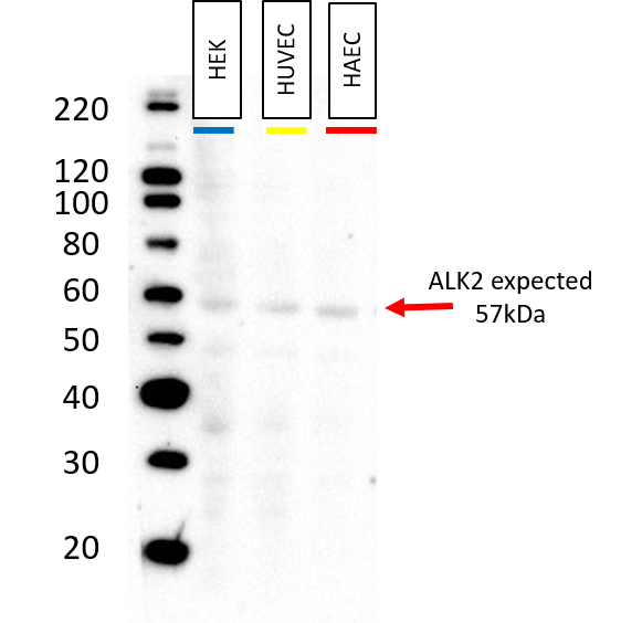
Application: Western BlotSample Tested: HUVEC human umbilical vein endothelial cells, HAEC human aortic endothelial cells and HEK293T human embryonic kidney cell lineSpecies: HumanVerified Customer | Posted 08/27/201810ug of protein collected from untreated HEK, HUVEC, and HAEC cells was loaded into each well. Blocked in Prometheus OneBlock Western-CL Blocking Buffer for 1hr @ room temp Primary antibody: Anti-ALK2 MAB637, mouse, RnD 1:500 overnight @ 4C Secondary antibody: goat-alpha -mouse HRP (Thermo) 1:10,000 1 hr @ room temp Substrate: Luminata Forte Western HRP Substrate Millipore 2 min Imaging: 300 sec exposure

There are no reviews that match your criteria.
Protocols
Find general support by application which include: protocols, troubleshooting, illustrated assays, videos and webinars.
- Appropriate Fixation of IHC/ICC Samples
- Cellular Response to Hypoxia Protocols
- ClariTSA™ Fluorophore Kits
- Detection & Visualization of Antibody Binding
- ICC Cell Smear Protocol for Suspension Cells
- ICC Immunocytochemistry Protocol Videos
- ICC for Adherent Cells
- Immunocytochemistry (ICC) Protocol
- Immunocytochemistry Troubleshooting
- Immunofluorescence of Organoids Embedded in Cultrex Basement Membrane Extract
- Immunohistochemistry (IHC) and Immunocytochemistry (ICC) Protocols
- Preparing Samples for IHC/ICC Experiments
- Preventing Non-Specific Staining (Non-Specific Binding)
- Primary Antibody Selection & Optimization
- Protocol for VisUCyte™ HRP Polymer Detection Reagent
- Protocol for the Fluorescent ICC Staining of Cell Smears - Graphic
- Protocol for the Fluorescent ICC Staining of Cultured Cells on Coverslips - Graphic
- Protocol for the Preparation and Fluorescent ICC Staining of Cells on Coverslips
- Protocol for the Preparation and Fluorescent ICC Staining of Non-adherent Cells
- Protocol for the Preparation and Fluorescent ICC Staining of Stem Cells on Coverslips
- Protocol for the Preparation of a Cell Smear for Non-adherent Cell ICC - Graphic
- R&D Systems Quality Control Western Blot Protocol
- TUNEL and Active Caspase-3 Detection by IHC/ICC Protocol
- The Importance of IHC/ICC Controls
- Troubleshooting Guide: Western Blot Figures
- Western Blot Conditions
- Western Blot Protocol
- Western Blot Protocol for Cell Lysates
- Western Blot Troubleshooting
- Western Blot Troubleshooting Guide
- View all Protocols, Troubleshooting, Illustrated assays and Webinars
Loading...