Asialoglycoprotein receptor 2 (ASGR2) is a subunit of the Asialoglycoprotein receptor. The asialoglycoprotein receptor is a hetero-oligomeric protein composed of major and minor subunits, which are encoded by different genes. The protein encoded by this gene is the less abundant minor subunit. The asialoglycoprotein receptor may facilitate hepatic infection by multiple viruses including hepatitis B, and is also a target for liver-specific drug delivery. Expressed on hepatocytes, the Asialoglycoprotein receptor mediates endocytosis and lysosomal degradation of glycoproteins to mediate serum glycoprotein homeostasis. The ASGR receptor binds plasma glycoproteins which have had the terminal sialic acid residue removed. An alternatively spliced variant, H2, which can be shed, has been proposed as a marker for liver fibrosis.
Human ASGR2 Antibody (2327C)
R&D Systems | Catalog # MAB9970
Recombinant Monoclonal Antibody.

Key Product Details
Species Reactivity
Human
Applications
Immunohistochemistry, Western Blot, Flow Cytometry
Label
Unconjugated
Antibody Source
Recombinant Monoclonal Rabbit IgG Clone # 2327C
Loading...
Product Specifications
Immunogen
Chinese hamster ovary cell line CHO-derived recombinant human ASGR2
Gln80-Ala311
Accession # P07307
Gln80-Ala311
Accession # P07307
Specificity
Detects human ASGR2 in direct ELISAs.
Clonality
Monoclonal
Host
Rabbit
Isotype
IgG
Scientific Data Images for Human ASGR2 Antibody (2327C)
Detection of Human ASGR2 by Western Blot.
Western blot shows lysates of human liver tissue. PVDF membrane was probed with 2 µg/mL of Rabbit Anti-Human ASGR2 Monoclonal Antibody (Catalog # MAB9970) followed by HRP-conjugated Anti-Rabbit IgG Secondary Antibody (Catalog # HAF008). A specific band was detected for ASGR2 at approximately 45 kDa (as indicated). This experiment was conducted under reducing conditions and using Immunoblot Buffer Group 1.
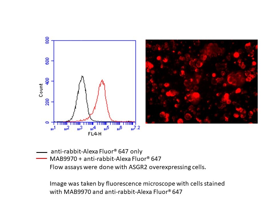
Detection of ASGR2 in HEK293 Human Cell Line Transfected with Human ASGR2 and eGFP by Flow Cytometry.
HEK293 human embryonic kidney cell line transfected with (A) human ASGR2 or (B) irrelevant transfectants and eGFP was stained with Mouse Anti-Human ASGR2 Monoclonal Antibody (Catalog # MAB9970) followed by APC-conjugated Anti-Rabbit IgG Secondary Antibody (Catalog # F0111). Quadrant markers were set based on control antibody staining (Catalog # MAB1050). View our protocol for Staining Membrane-associated Proteins.
ASGR2 in Human Liver.
ASGR2 was detected in immersion fixed paraffin-embedded sections of human liver using Rabbit Anti-Human ASGR2 Monoclonal Antibody (Catalog # MAB9970) at 1 µg/mL for 1 hour at room temperature followed by incubation with the Anti-Rabbit IgG VisUCyte™ HRP Polymer Antibody (Catalog # VC003). Tissue was stained using DAB (brown) and counterstained with hematoxylin (blue). Specific staining was localized to cytoplasm in hepatocytes. View our protocol for IHC Staining with VisUCyte HRP Polymer Detection Reagents.Applications for Human ASGR2 Antibody (2327C)
Application
Recommended Usage
Flow Cytometry
0.25 µg/106 cells
Sample: HEK293 Human Cell Line Transfected with Human ASGR2 and eGFP
Sample: HEK293 Human Cell Line Transfected with Human ASGR2 and eGFP
Immunohistochemistry
1-25 µg/mL
Sample: Immersion fixed paraffin-embedded sections of human liver
Sample: Immersion fixed paraffin-embedded sections of human liver
Western Blot
2 µg/mL
Sample: Human liver tissue
Sample: Human liver tissue
Reviewed Applications
Read 1 review rated 5 using MAB9970 in the following applications:
Flow Cytometry Panel Builder
Bio-Techne Knows Flow Cytometry
Save time and reduce costly mistakes by quickly finding compatible reagents using the Panel Builder Tool.
Advanced Features
- Spectra Viewer - Custom analysis of spectra from multiple fluorochromes
- Spillover Popups - Visualize the spectra of individual fluorochromes
- Antigen Density Selector - Match fluorochrome brightness with antigen density

Formulation, Preparation, and Storage
Purification
Protein A or G purified from hybridoma culture supernatant
Reconstitution
Reconstitute at 0.5 mg/mL in sterile PBS. For liquid material, refer to CoA for concentration.
Loading...
Formulation
Lyophilized from a 0.2 μm filtered solution in PBS with Trehalose. *Small pack size (SP) is supplied either lyophilized or as a 0.2 µm filtered solution in PBS.
Shipping
Lyophilized product is shipped at ambient temperature. Liquid small pack size (-SP) is shipped with polar packs. Upon receipt, store immediately at the temperature recommended below.
Stability & Storage
Use a manual defrost freezer and avoid repeated freeze-thaw cycles.
- 12 months from date of receipt, -20 to -70 °C, as supplied.
- 1 month, 2 to 8 °C under sterile conditions after opening.
- 6 months, -20 to -70 °C under sterile conditions after opening.
Calculators
Background: ASGR2
Long Name
Asialoglycoprotein Receptor 2
Alternate Names
ASGPR2, CLEC4H2, HBXBP
Gene Symbol
ASGR2
UniProt
Additional ASGR2 Products
Product Documents for Human ASGR2 Antibody (2327C)
Certificate of Analysis
To download a Certificate of Analysis, please enter a lot or batch number in the search box below.
Note: Certificate of Analysis not available for kit components.
Product Specific Notices for Human ASGR2 Antibody (2327C)
For research use only
Related Research Areas
Customer Reviews for Human ASGR2 Antibody (2327C) (1)
5 out of 5
1 Customer Rating
Have you used Human ASGR2 Antibody (2327C)?
Submit a review and receive an Amazon gift card!
$25/€18/£15/$25CAN/¥2500 Yen for a review with an image
$10/€7/£6/$10CAN/¥1110 Yen for a review without an image
Submit a review
Customer Images
Showing
1
-
1 of
1 review
Showing All
Filter By:
-
Application: Flow CytometrySample Tested: ASGR over-expressing cellsSpecies: HumanVerified Customer | Posted 10/10/2019Compared three ASGR2 antibodies, MAB9970 stains better than the other 2 antibodies with less background and less non-specific bindings in flow assay and ICC.

There are no reviews that match your criteria.
Protocols
Find general support by application which include: protocols, troubleshooting, illustrated assays, videos and webinars.
- 7-Amino Actinomycin D (7-AAD) Cell Viability Flow Cytometry Protocol
- Antigen Retrieval Protocol (PIER)
- Antigen Retrieval for Frozen Sections Protocol
- Appropriate Fixation of IHC/ICC Samples
- Cellular Response to Hypoxia Protocols
- Chromogenic IHC Staining of Formalin-Fixed Paraffin-Embedded (FFPE) Tissue Protocol
- Chromogenic Immunohistochemistry Staining of Frozen Tissue
- ClariTSA™ Fluorophore Kits
- Detection & Visualization of Antibody Binding
- Extracellular Membrane Flow Cytometry Protocol
- Flow Cytometry Protocol for Cell Surface Markers
- Flow Cytometry Protocol for Staining Membrane Associated Proteins
- Flow Cytometry Staining Protocols
- Flow Cytometry Troubleshooting Guide
- Fluorescent IHC Staining of Frozen Tissue Protocol
- Graphic Protocol for Heat-induced Epitope Retrieval
- Graphic Protocol for the Preparation and Fluorescent IHC Staining of Frozen Tissue Sections
- Graphic Protocol for the Preparation and Fluorescent IHC Staining of Paraffin-embedded Tissue Sections
- Graphic Protocol for the Preparation of Gelatin-coated Slides for Histological Tissue Sections
- IHC Sample Preparation (Frozen sections vs Paraffin)
- Immunofluorescent IHC Staining of Formalin-Fixed Paraffin-Embedded (FFPE) Tissue Protocol
- Immunohistochemistry (IHC) and Immunocytochemistry (ICC) Protocols
- Immunohistochemistry Frozen Troubleshooting
- Immunohistochemistry Paraffin Troubleshooting
- Intracellular Flow Cytometry Protocol Using Alcohol (Methanol)
- Intracellular Flow Cytometry Protocol Using Detergents
- Intracellular Nuclear Staining Flow Cytometry Protocol Using Detergents
- Intracellular Staining Flow Cytometry Protocol Using Alcohol Permeabilization
- Intracellular Staining Flow Cytometry Protocol Using Detergents to Permeabilize Cells
- Preparing Samples for IHC/ICC Experiments
- Preventing Non-Specific Staining (Non-Specific Binding)
- Primary Antibody Selection & Optimization
- Propidium Iodide Cell Viability Flow Cytometry Protocol
- Protocol for Heat-Induced Epitope Retrieval (HIER)
- Protocol for Liperfluo
- Protocol for Making a 4% Formaldehyde Solution in PBS
- Protocol for VisUCyte™ HRP Polymer Detection Reagent
- Protocol for the Characterization of Human Th22 Cells
- Protocol for the Characterization of Human Th9 Cells
- Protocol for the Preparation & Fixation of Cells on Coverslips
- Protocol for the Preparation and Chromogenic IHC Staining of Frozen Tissue Sections
- Protocol for the Preparation and Chromogenic IHC Staining of Frozen Tissue Sections - Graphic
- Protocol for the Preparation and Chromogenic IHC Staining of Paraffin-embedded Tissue Sections
- Protocol for the Preparation and Chromogenic IHC Staining of Paraffin-embedded Tissue Sections - Graphic
- Protocol for the Preparation and Fluorescent IHC Staining of Frozen Tissue Sections
- Protocol for the Preparation and Fluorescent IHC Staining of Paraffin-embedded Tissue Sections
- Protocol for the Preparation of Gelatin-coated Slides for Histological Tissue Sections
- Protocol: Annexin V and PI Staining by Flow Cytometry
- Protocol: Annexin V and PI Staining for Apoptosis by Flow Cytometry
- R&D Systems Quality Control Western Blot Protocol
- TUNEL and Active Caspase-3 Detection by IHC/ICC Protocol
- The Importance of IHC/ICC Controls
- Troubleshooting Guide: Fluorokine Flow Cytometry Kits
- Troubleshooting Guide: Immunohistochemistry
- Troubleshooting Guide: Western Blot Figures
- Western Blot Conditions
- Western Blot Protocol
- Western Blot Protocol for Cell Lysates
- Western Blot Troubleshooting
- Western Blot Troubleshooting Guide
- View all Protocols, Troubleshooting, Illustrated assays and Webinars
Loading...