Human B7-H2, also called B7RP-1, B7h, LICOS, and GL50, is a 60 kDa member of the B7 family of immune costimulatory proteins, which includes B7-1, B7-2, B7-H1 (PD-L1), PD-L2, and B7-H3. B7 proteins are members of the immunoglobulin (Ig) superfamily, the extracellular domains contain 2 Ig-like domains and all members have short cytoplasmic domains. Family members share about 20‑25% amino acid identity. Within the extracellular domain, human B7-H2 shares 49% and 54% amino acid sequence identity with human and rat B7-H2, respectively. B7-H2 has been identified as the ligand for ICOS, a member of the CD28 family of costimulatory receptors. Human B7-H2 is a 309 amino acid (aa) protein with a putative 18 aa signal peptide, a 239 aa extracellular domain, an 18 aa transmembrane region, and a 33 aa cytoplasmic domain. Human B7-H2 is expressed constitutively on resting B cells, dendritic cells, and at low levels on monocytes. The B7-H2/ICOS interaction appears to play roles in T cell dependent B cell activation and Th differentiation.

Key Product Details
Species Reactivity
Validated:
Human
Cited:
Human
Applications
Validated:
Western Blot, Flow Cytometry, CyTOF-ready
Cited:
Immunohistochemistry-Paraffin, Western Blot, Flow Cytometry
Label
Unconjugated
Antibody Source
Polyclonal Goat IgG
Loading...
Product Specifications
Immunogen
Mouse myeloma cell line NS0-derived recombinant human B7-H2
Asp19-Ser258
Accession # O75144
Asp19-Ser258
Accession # O75144
Specificity
Detects human B7-H2 in direct ELISAs and Western blots. In direct ELISAs and Western blots, less than 1% cross‑reactivity with recombinant mouse B7-H2 and recombinant human B7-H1 is observed.
Clonality
Polyclonal
Host
Goat
Isotype
IgG
Scientific Data Images for Human B7-H2 Antibody
Detection of B7‑H2 by Western Blot.
Western blot shows lysate of U937 human histiocytic lymphoma cell line. PVDF membrane was probed with 2 µg/mL of Goat Anti-Human B7-H2 Antigen Affinity-purified Polyclonal Antibody (Catalog # AF165) followed by HRP-conjugated Anti-Goat IgG Secondary Antibody (Catalog # HAF017). A specific band was detected for B7-H2 at approximately 75 kDa (as indicated). This experiment was conducted under reducing conditions and using Immunoblot Buffer Group 1.
Detection of B7‑H2 in U937 Human Cell Line by Flow Cytometry.
U937 human histiocytic lymphoma cell line was stained with Goat Anti-Human B7-H2 Antigen Affinity-purified Polyclonal Anti-body (Catalog # AF165, filled histogram) or isotype control antibody (Catalog # AB-108-C, open histogram), followed by Phycoerythrin-conjugated Anti-Goat IgG Secondary Antibody (Catalog # F0107).Applications for Human B7-H2 Antibody
Application
Recommended Usage
CyTOF-ready
Ready to be labeled using established conjugation methods. No BSA or other carrier proteins that could interfere with conjugation.
Flow Cytometry
0.25 µg/106 cells
Sample: U937 human histiocytic lymphoma cell line
Sample: U937 human histiocytic lymphoma cell line
Western Blot
2 µg/mL
Sample: U937 human histiocytic lymphoma cell line
Sample: U937 human histiocytic lymphoma cell line
Reviewed Applications
Read 1 review rated 5 using AF165 in the following applications:
Flow Cytometry Panel Builder
Bio-Techne Knows Flow Cytometry
Save time and reduce costly mistakes by quickly finding compatible reagents using the Panel Builder Tool.
Advanced Features
- Spectra Viewer - Custom analysis of spectra from multiple fluorochromes
- Spillover Popups - Visualize the spectra of individual fluorochromes
- Antigen Density Selector - Match fluorochrome brightness with antigen density

Formulation, Preparation, and Storage
Purification
Antigen Affinity-purified
Reconstitution
Reconstitute at 0.2 mg/mL in sterile PBS. For liquid material, refer to CoA for concentration.
Loading...
Formulation
Lyophilized from a 0.2 μm filtered solution in PBS with Trehalose. *Small pack size (SP) is supplied either lyophilized or as a 0.2 µm filtered solution in PBS.
Shipping
Lyophilized product is shipped at ambient temperature. Liquid small pack size (-SP) is shipped with polar packs. Upon receipt, store immediately at the temperature recommended below.
Stability & Storage
Use a manual defrost freezer and avoid repeated freeze-thaw cycles.
- 12 months from date of receipt, -20 to -70 °C as supplied.
- 1 month, 2 to 8 °C under sterile conditions after reconstitution.
- 6 months, -20 to -70 °C under sterile conditions after reconstitution.
Calculators
Background: B7-H2
References
- Coyle, A.J. and J.C. Gutierrez-Ramos (2001) Nat. Immunol. 2:203.
- Ling, V. et al. (2000) J. Immunol. 164:1653.
- Wang, S. et al. (2000) Blood 96:2808.
- Brodie, D. et al. (2000) Curr. Biol. 10:333.
- Mages, H.W. et al. (2000) Eur. J. Immunol. 30:1040.
- Swallow, M.M. et al. (1999) Immunity 11:423.
- Yoshinaga, S.K. et al. (1999) Nature 402:827.
Long Name
B7 Homolog 2
Alternate Names
B7H2, B7RP-1, CD275, GL50, ICOSL, ICOSLG
Gene Symbol
ICOSLG
UniProt
Additional B7-H2 Products
Product Documents for Human B7-H2 Antibody
Certificate of Analysis
To download a Certificate of Analysis, please enter a lot or batch number in the search box below.
Note: Certificate of Analysis not available for kit components.
Product Specific Notices for Human B7-H2 Antibody
For research use only
Citations for Human B7-H2 Antibody
 Powered by Bioz
Customer Reviews for Human B7-H2 Antibody (1)
5 out of 5
1 Customer Rating
Have you used Human B7-H2 Antibody?
Submit a review and receive an Amazon gift card!
$25/€18/£15/$25CAN/¥2500 Yen for a review with an image
$10/€7/£6/$10CAN/¥1110 Yen for a review without an image
Submit a review
Customer Images
Showing
1
-
1 of
1 review
Showing All
Filter By:
-
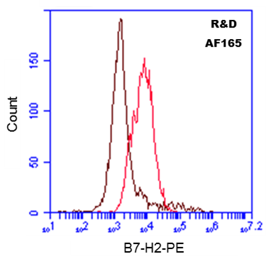
Application: Flow CytometrySample Tested: MCF-7 human breast cancer cell lineSpecies: HumanVerified Customer | Posted 03/22/2017Detection of B7-H2 in MCF-7. 106 cells were incubated with 0.50 ug of Goat anti-Human B7-H2 antibody (red), or Goat Isotype Control IgG (brown), followed by PE-conjugated anti-Goat secondary antibody (1:100). Unfixed cells.

There are no reviews that match your criteria.
Protocols
Find general support by application which include: protocols, troubleshooting, illustrated assays, videos and webinars.
- 7-Amino Actinomycin D (7-AAD) Cell Viability Flow Cytometry Protocol
- Cellular Response to Hypoxia Protocols
- Extracellular Membrane Flow Cytometry Protocol
- Flow Cytometry Protocol for Cell Surface Markers
- Flow Cytometry Protocol for Staining Membrane Associated Proteins
- Flow Cytometry Staining Protocols
- Flow Cytometry Troubleshooting Guide
- Intracellular Flow Cytometry Protocol Using Alcohol (Methanol)
- Intracellular Flow Cytometry Protocol Using Detergents
- Intracellular Nuclear Staining Flow Cytometry Protocol Using Detergents
- Intracellular Staining Flow Cytometry Protocol Using Alcohol Permeabilization
- Intracellular Staining Flow Cytometry Protocol Using Detergents to Permeabilize Cells
- Propidium Iodide Cell Viability Flow Cytometry Protocol
- Protocol for Liperfluo
- Protocol for the Characterization of Human Th22 Cells
- Protocol for the Characterization of Human Th9 Cells
- Protocol: Annexin V and PI Staining by Flow Cytometry
- Protocol: Annexin V and PI Staining for Apoptosis by Flow Cytometry
- R&D Systems Quality Control Western Blot Protocol
- Troubleshooting Guide: Fluorokine Flow Cytometry Kits
- Troubleshooting Guide: Western Blot Figures
- Western Blot Conditions
- Western Blot Protocol
- Western Blot Protocol for Cell Lysates
- Western Blot Troubleshooting
- Western Blot Troubleshooting Guide
- View all Protocols, Troubleshooting, Illustrated assays and Webinars
Loading...