Human BAFF/BLyS/TNFSF13B Antibody (137314)
R&D Systems | Catalog # MAB1241
Clone 137314 was used by HLDA to establish CD designation

Key Product Details
Species Reactivity
Validated:
Human
Cited:
Human
Applications
Validated:
Western Blot, Flow Cytometry, CyTOF-ready
Cited:
Western Blot, Flow Cytometry, ELISA Development
Label
Unconjugated
Antibody Source
Monoclonal Mouse IgG1 Clone # 137314
Loading...
Product Specifications
Immunogen
E. coli-derived recombinant human BAFF/BLyS/TNFSF13B
Ala81-Leu285
Accession # Q9Y275
Ala81-Leu285
Accession # Q9Y275
Specificity
Detects human BAFF/BLyS/TNFSF13B in direct ELISAs and Western blots. In Western blots, approximately 5% cross‑reactivity with recombinant human (rh) TL1A/TNSF15 is observed and no cross-reactivity with rhAPRIL, rhTNF-alpha, rhFas Ligand, rhGITR Ligand, rhLIGHT, rhTRAIL, rhTRANCE, or rhTWEAK is observed.
Clonality
Monoclonal
Host
Mouse
Isotype
IgG1
Scientific Data Images for Human BAFF/BLyS/TNFSF13B Antibody (137314)
Detection of BAFF/BLyS/TNFSF13B in THP-1 cells by Flow Cytometry
THP-1 cells were stained with Mouse Anti-Human BAFF/BLyS/TNFSF13B Monoclonal Antibody (Catalog # MAB1241, filled histogram) or isotype control antibody (Catalog # MAB002, open histogram) followed by Phycoerythrin-conjugated Anti-Mouse IgG Secondary Antibody (Catalog # F0102B). View our protocol for Staining Membrane-associated Proteins.Applications for Human BAFF/BLyS/TNFSF13B Antibody (137314)
Application
Recommended Usage
CyTOF-ready
Ready to be labeled using established conjugation methods. No BSA or other carrier proteins that could interfere with conjugation.
Flow Cytometry
0.25 µg/106 cells
Sample: THP-1 cells
Sample: THP-1 cells
Western Blot
1 µg/mL
Sample: Recombinant Human BAFF/BLyS/TNFSF13B (Catalog # 2149-BF)
Sample: Recombinant Human BAFF/BLyS/TNFSF13B (Catalog # 2149-BF)
Reviewed Applications
Read 2 reviews rated 5 using MAB1241 in the following applications:
Flow Cytometry Panel Builder
Bio-Techne Knows Flow Cytometry
Save time and reduce costly mistakes by quickly finding compatible reagents using the Panel Builder Tool.
Advanced Features
- Spectra Viewer - Custom analysis of spectra from multiple fluorochromes
- Spillover Popups - Visualize the spectra of individual fluorochromes
- Antigen Density Selector - Match fluorochrome brightness with antigen density

Formulation, Preparation, and Storage
Purification
Protein A or G purified from hybridoma culture supernatant
Reconstitution
Reconstitute at 0.5 mg/mL in sterile PBS. For liquid material, refer to CoA for concentration.
Loading...
Formulation
Lyophilized from a 0.2 μm filtered solution in PBS with Trehalose. See Certificate of Analysis for details.
*Small pack size (-SP) is supplied either lyophilized or as a 0.2 µm filtered solution in PBS.
*Small pack size (-SP) is supplied either lyophilized or as a 0.2 µm filtered solution in PBS.
Shipping
Lyophilized product is shipped at ambient temperature. Liquid small pack size (-SP) is shipped with polar packs. Upon receipt, store immediately at the temperature recommended below.
Stability & Storage
Use a manual defrost freezer and avoid repeated freeze-thaw cycles.
- 12 months from date of receipt, -20 to -70 °C as supplied.
- 1 month, 2 to 8 °C under sterile conditions after reconstitution.
- 6 months, -20 to -70 °C under sterile conditions after reconstitution.
Calculators
Background: BAFF/BLyS/TNFSF13B
References
- Schneider, P. et al. (1999) J. Exp. Med. 189:1747.
- Mukhopadhyay, A. et al. (1999) J. Biol. Chem. 274:15978.
- Karpusas, M. et al. (2002) J. Mol. Biol. 315:1145.
- Liu, Y. et al. (2002) Cell 108:383.
- Cheema, G.S. et al. (2001) Arthr. Rheum. 44:1313.
- Marsters, S.A. et al. (2000) Curr. Biol. 10:785.
- Thompson, J.S. et al. (2001) Science 293:2108.
- Ng, L.G. et al. (2004) J. Immunol. 173:807.
- Roschke, V. et al. (2002) J. Immunol. 169:4314.
- Batten, M. et al. (2000) J. Exp. Med. 192:1453.
- Avery, D.T. et al. (2003) J. Clin. Invest. 112:286.
Long Name
B cell Activating Factor
Alternate Names
BLyS, CD257, TALL1, THANK, TNFSF13B, ZTNF4
Gene Symbol
TNFSF13B
UniProt
Additional BAFF/BLyS/TNFSF13B Products
Product Documents for Human BAFF/BLyS/TNFSF13B Antibody (137314)
Certificate of Analysis
To download a Certificate of Analysis, please enter a lot or batch number in the search box below.
Note: Certificate of Analysis not available for kit components.
Product Specific Notices for Human BAFF/BLyS/TNFSF13B Antibody (137314)
For research use only
Citations for Human BAFF/BLyS/TNFSF13B Antibody (137314)
 Powered by Bioz
Customer Reviews for Human BAFF/BLyS/TNFSF13B Antibody (137314) (2)
5 out of 5
2 Customer Ratings
Have you used Human BAFF/BLyS/TNFSF13B Antibody (137314)?
Submit a review and receive an Amazon gift card!
$25/€18/£15/$25CAN/¥2500 Yen for a review with an image
$10/€7/£6/$10CAN/¥1110 Yen for a review without an image
Submit a review
Customer Images

Showing
1
-
2 of
2 reviews
Showing All
Filter By:
-
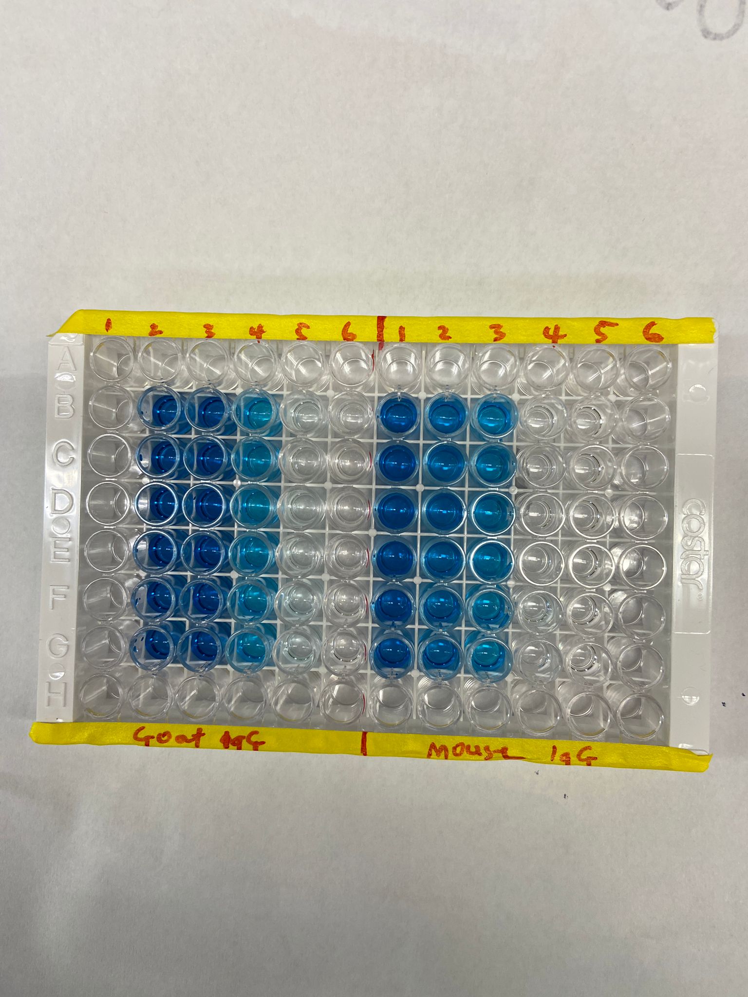
Application: ELISASample Tested: Recombinant proteinSpecies: HumanVerified Customer | Posted 09/09/2024Binding signal (blue color) vs no binding (clear)Just like AF124, I used this MAB1241 to detect rhBAFF after incubation with BCMA in ELISA. I got an expected signal showing binding compared to control (no signal).

-
Application: Western BlotSample Tested: Jurkat whole cell extracts and Jurkat whole cell extractSpecies: HumanVerified Customer | Posted 02/08/2022
There are no reviews that match your criteria.
Protocols
Find general support by application which include: protocols, troubleshooting, illustrated assays, videos and webinars.
- 7-Amino Actinomycin D (7-AAD) Cell Viability Flow Cytometry Protocol
- Cellular Response to Hypoxia Protocols
- Extracellular Membrane Flow Cytometry Protocol
- Flow Cytometry Protocol for Cell Surface Markers
- Flow Cytometry Protocol for Staining Membrane Associated Proteins
- Flow Cytometry Staining Protocols
- Flow Cytometry Troubleshooting Guide
- Intracellular Flow Cytometry Protocol Using Alcohol (Methanol)
- Intracellular Flow Cytometry Protocol Using Detergents
- Intracellular Nuclear Staining Flow Cytometry Protocol Using Detergents
- Intracellular Staining Flow Cytometry Protocol Using Alcohol Permeabilization
- Intracellular Staining Flow Cytometry Protocol Using Detergents to Permeabilize Cells
- Propidium Iodide Cell Viability Flow Cytometry Protocol
- Protocol for Liperfluo
- Protocol for the Characterization of Human Th22 Cells
- Protocol for the Characterization of Human Th9 Cells
- Protocol: Annexin V and PI Staining by Flow Cytometry
- Protocol: Annexin V and PI Staining for Apoptosis by Flow Cytometry
- R&D Systems Quality Control Western Blot Protocol
- Troubleshooting Guide: Fluorokine Flow Cytometry Kits
- Troubleshooting Guide: Western Blot Figures
- Western Blot Conditions
- Western Blot Protocol
- Western Blot Protocol for Cell Lysates
- Western Blot Troubleshooting
- Western Blot Troubleshooting Guide
- View all Protocols, Troubleshooting, Illustrated assays and Webinars
Loading...
Associated Pathways
