B and T Lymphocyte Attenuator (BTLA) also known as CD272, is a 70kDa type I transmembrane glycoprotein of the CD28 family of T cell costimulatory molecules. BTLA functions as a coinhibitor on activated T cells and on B cells and dendritic cells. The extracellular domain (ECD) of this Ig superfamily protein contains one
V‑type Ig-like domain, which interacts with the herpesvirus entry mediator (HVEM/TNFSF14). The amino acid sequence of the ECD of human BTLA shares only 34% identity to that of mouse BTLA. One human BTLA variant lacking the transmembrane sequence has been reported.

Key Product Details
Species Reactivity
Human
Applications
Western Blot, Flow Cytometry, CyTOF-ready
Label
Unconjugated
Antibody Source
Polyclonal Goat IgG
Loading...
Product Specifications
Immunogen
Mouse myeloma cell line NS0-derived recombinant human BTLA
Ile25-Ser150
Accession # AAP44003
Ile25-Ser150
Accession # AAP44003
Specificity
Detects human BTLA in direct ELISAs and Western blots.
Clonality
Polyclonal
Host
Goat
Isotype
IgG
Scientific Data Images for Human BTLA Antibody
Detection of Human BTLA by Western Blot.
Western blot shows lysates of HEK293T human embryonic kidney cell line either mock transfected or transfected with human BTLA. PVDF membrane was probed with 0.5 µg/mL of Goat Anti-Human BTLA Antigen Affinity-purified Polyclonal Antibody (Catalog # AF3354) followed by HRP-conjugated Anti-Goat IgG Secondary Antibody (Catalog # HAF017). Specific bands were detected for BTLA at approximately 70-80 kDa (as indicated). This experiment was conducted under reducing conditions and using Immunoblot Buffer Group 1.Applications for Human BTLA Antibody
Application
Recommended Usage
CyTOF-ready
Ready to be labeled using established conjugation methods. No BSA or other carrier proteins that could interfere with conjugation.
Flow Cytometry
0.25 µg/106 cells
Sample: Human whole blood lymphocytes
Sample: Human whole blood lymphocytes
Western Blot
0.5 µg/mL
Sample: HEK293T human embryonic kidney cell line transfected with human BTLA
Sample: HEK293T human embryonic kidney cell line transfected with human BTLA
Reviewed Applications
Read 1 review rated 5 using AF3354 in the following applications:
Flow Cytometry Panel Builder
Bio-Techne Knows Flow Cytometry
Save time and reduce costly mistakes by quickly finding compatible reagents using the Panel Builder Tool.
Advanced Features
- Spectra Viewer - Custom analysis of spectra from multiple fluorochromes
- Spillover Popups - Visualize the spectra of individual fluorochromes
- Antigen Density Selector - Match fluorochrome brightness with antigen density

Formulation, Preparation, and Storage
Purification
Antigen Affinity-purified
Reconstitution
Reconstitute at 0.2 mg/mL in sterile PBS. For liquid material, refer to CoA for concentration.
Loading...
Formulation
Lyophilized from a 0.2 μm filtered solution in PBS with Trehalose. *Small pack size (SP) is supplied either lyophilized or as a 0.2 µm filtered solution in PBS.
Shipping
Lyophilized product is shipped at ambient temperature. Liquid small pack size (-SP) is shipped with polar packs. Upon receipt, store immediately at the temperature recommended below.
Stability & Storage
Use a manual defrost freezer and avoid repeated freeze-thaw cycles.
- 12 months from date of receipt, -20 to -70 °C as supplied.
- 1 month, 2 to 8 °C under sterile conditions after reconstitution.
- 6 months, -20 to -70 °C under sterile conditions after reconstitution.
Calculators
Background: BTLA
Long Name
B- And T-Lymphocyte Attenuator
Alternate Names
CD272
Gene Symbol
BTLA
UniProt
Additional BTLA Products
Product Documents for Human BTLA Antibody
Certificate of Analysis
To download a Certificate of Analysis, please enter a lot or batch number in the search box below.
Note: Certificate of Analysis not available for kit components.
Product Specific Notices for Human BTLA Antibody
For research use only
Citations for Human BTLA Antibody
 Powered by Bioz
Customer Reviews for Human BTLA Antibody (1)
5 out of 5
1 Customer Rating
Have you used Human BTLA Antibody?
Submit a review and receive an Amazon gift card!
$25/€18/£15/$25CAN/¥2500 Yen for a review with an image
$10/€7/£6/$10CAN/¥1110 Yen for a review without an image
Submit a review
Customer Images
Showing
1
-
1 of
1 review
Showing All
Filter By:
-
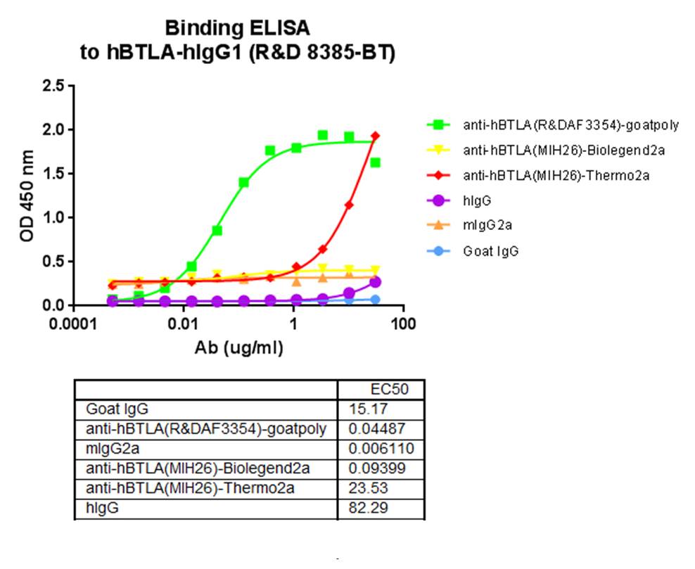
Application: ELISASample Tested: NS0 mouse myeloma cell lineSpecies: HumanVerified Customer | Posted 12/20/2017Observed binding via ELISA to R&D huBTLA-hIgG1 (8385-BT). More efficient binding observed when compared to Biolegend and Thermo anti-huBTLA antibodies.

There are no reviews that match your criteria.
Protocols
Find general support by application which include: protocols, troubleshooting, illustrated assays, videos and webinars.
- 7-Amino Actinomycin D (7-AAD) Cell Viability Flow Cytometry Protocol
- Cellular Response to Hypoxia Protocols
- Extracellular Membrane Flow Cytometry Protocol
- Flow Cytometry Protocol for Cell Surface Markers
- Flow Cytometry Protocol for Staining Membrane Associated Proteins
- Flow Cytometry Staining Protocols
- Flow Cytometry Troubleshooting Guide
- Intracellular Flow Cytometry Protocol Using Alcohol (Methanol)
- Intracellular Flow Cytometry Protocol Using Detergents
- Intracellular Nuclear Staining Flow Cytometry Protocol Using Detergents
- Intracellular Staining Flow Cytometry Protocol Using Alcohol Permeabilization
- Intracellular Staining Flow Cytometry Protocol Using Detergents to Permeabilize Cells
- Propidium Iodide Cell Viability Flow Cytometry Protocol
- Protocol for Liperfluo
- Protocol for the Characterization of Human Th22 Cells
- Protocol for the Characterization of Human Th9 Cells
- Protocol: Annexin V and PI Staining by Flow Cytometry
- Protocol: Annexin V and PI Staining for Apoptosis by Flow Cytometry
- R&D Systems Quality Control Western Blot Protocol
- Troubleshooting Guide: Fluorokine Flow Cytometry Kits
- Troubleshooting Guide: Western Blot Figures
- Western Blot Conditions
- Western Blot Protocol
- Western Blot Protocol for Cell Lysates
- Western Blot Troubleshooting
- Western Blot Troubleshooting Guide
- View all Protocols, Troubleshooting, Illustrated assays and Webinars
Loading...