The cadherin superfamily is a large family of membrane-associated glycoproteins that engage in homotypic, calcium‑dependent, cell-to-cell adhesion events. The superfamily can be divided into at least four subfamilies based on the extracellular (EC) regions and cytoplasmic domains (1, 2). These include classical cadherins, desmosomal cadherins, protocadherins, and cadherin-like molecules that contain a variable number of EC and transmembrane (TM) domains (1). Cadherin‑6, also known as KCAD or K-cadherin, is a classical cadherin of 110‑120 kD that has at least one full length and two alternate splice forms ranging in size from 105‑120 kDa (3). Human Cadherin-6 is synthesized as a 790 amino acid (aa) type I transmembrane glycoprotein that contains a 18 aa signal peptide, a 35 aa prosequence, a 562 aa extracellular region, a 21 aa transmembrane segment, and a 154 aa cytoplasmic domain (4, 5). There are five EC cadherin domains that are approximately 110 aa in length. This pattern is consistent with classical cadherin family molecules that are modular in their extracellular region and mediate calcium‑dependent cell‑to‑cell adhesion through their Ca++-binding repeats (2). Due to the absence of a His-Ala-Val motif in its most N-terminal cadherin repeat, Cadherin-6 can be further classified as a type II classical cadherin (4). One Cadherin‑6 splice variant (termed 6/2) shows a 9 aa substitution for the 94 aa that span residues 283 to 376 of the full-length extracellular region (3). A second splice variant shows a 36 aa substitution for the C-terminal 163 aa of the transmembrane and cytoplasmic region (6). Human Cadherin‑6 shares 98% aa sequence identity with rat Cadherin‑6, plus 60% and 58% aa identity with human cadherin 8 and 11, respectively, within the extracellular regions. Cadherin-6 has high expression in kidney, brain, and cerebellum, and low expression in lung, pancreas, gastric mucosa, and cytotrophoblasts (4, 5, 7, 8, 9). Cadherin-6 is also found in renal, lung, and ovariancarcinoma (7, 10). As a classic cadherin, Cadherin-6 will form homodimers and promote intercellular adhesion with itself and possibly cadherin-9 and -14 (4, 11).

Key Product Details
Species Reactivity
Human
Applications
Immunohistochemistry
Label
Unconjugated
Antibody Source
Monoclonal Mouse IgG3 Clone # 427903
Loading...
Product Specifications
Immunogen
Mouse myeloma cell line NS0-derived recombinant human Cadherin‑6/KCAD
Ser54-Ala615
Accession # P55285
Ser54-Ala615
Accession # P55285
Specificity
Detects human Cadherin‑6/KCAD in direct ELISAs.
Clonality
Monoclonal
Host
Mouse
Isotype
IgG3
Scientific Data Images for Human Cadherin-6/KCAD Antibody (427903)
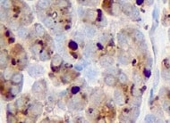
Cadherin‑6/KCAD in Human Liver Cancer Tissue.
Cadherin‑6/KCAD was detected in immersion fixed paraffin-embedded sections of human liver cancer tissue using 8 µg/mL Mouse Anti-Human Cadherin‑6/KCAD Monoclonal Antibody (Catalog # MAB27151) overnight at 4 °C. Tissue was stained with the Anti-Mouse HRP-DAB Cell & Tissue Staining Kit (brown; Catalog # CTS002) and counterstained with hematoxylin (blue). View our protocol for Chromogenic IHC Staining of Paraffin-embedded Tissue Sections.
Cadherin‑6/KCAD in Human Kidney Cancer Tissue.
Cadherin-6/KCAD was detected in immersion fixed paraffin-embedded sections of human kidney cancer tissue using 25 µg/mL Mouse Anti-Human Cadherin-6/KCAD Monoclonal Antibody (Catalog # MAB27151) overnight at 4 °C. Tissue was stained with the Anti-Mouse HRP-DAB Cell & Tissue Staining Kit (brown; Catalog # CTS002) and counterstained with hematoxylin (blue). View our protocol for Chromogenic IHC Staining of Paraffin-embedded Tissue Sections.Applications for Human Cadherin-6/KCAD Antibody (427903)
Application
Recommended Usage
Immunohistochemistry
8-25 µg/mL
Sample: Immersion fixed paraffin-embedded sections of human kidney cancer tissue and immersion fixed paraffin-embedded sections of human liver cancer tissue
Sample: Immersion fixed paraffin-embedded sections of human kidney cancer tissue and immersion fixed paraffin-embedded sections of human liver cancer tissue
Reviewed Applications
Read 1 review rated 5 using MAB27151 in the following applications:
Formulation, Preparation, and Storage
Purification
Protein A or G purified from hybridoma culture supernatant
Reconstitution
Reconstitute at 0.5 mg/mL in sterile PBS. For liquid material, refer to CoA for concentration.
Loading...
Formulation
Lyophilized from a 0.2 μm filtered solution in Tris and NaCl with Trehalose. *Small pack size (SP) is supplied either lyophilized or as a 0.2 µm filtered solution in PBS.
Shipping
Lyophilized product is shipped at ambient temperature. Liquid small pack size (-SP) is shipped with polar packs. Upon receipt, store immediately at the temperature recommended below.
Stability & Storage
Use a manual defrost freezer and avoid repeated freeze-thaw cycles.
- 12 months from date of receipt, -20 to -70 °C as supplied.
- 1 month, 2 to 8 °C under sterile conditions after reconstitution.
- 6 months, -20 to -70 °C under sterile conditions after reconstitution.
Calculators
Background: Cadherin-6/KCAD
References
- Koch, A.W. et al. (2004) Cell. Mol. Life Sci. 61:1884.
- Angst, B.D. et al. (2001) J. Cell Sci. 114:629.
- Mbalaviele, G. et al. (1998) J. Cell Biol. 141:1467.
- Shimoyama, Y. et al. (2000) Biochem. J. 349:159.
- Shimoyama, Y. et al. (1995) Cancer Res. 55:2206.
- GenBank Accession # P55285.
- Xiang Y.Y. et al. (1994) Cancer Res. 54:3034.
- Marthiens V. et al. (2002) Mol. Cell Neurosci. 20:458.
- MacCalman C.D. et al. (1998) Am J Reprod. Immunol. 39:96.
- Sella, G.C. et al. (2001) Cancer Res. 61:6977.
- Shimoyama, Y. et al. (1999) J. Biol. Chem. 274:11987.
Alternate Names
Cadherin6, CDH6, K-Cadherin
Entrez Gene IDs
1004 (Human)
Gene Symbol
CDH6
UniProt
Additional Cadherin-6/KCAD Products
Product Documents for Human Cadherin-6/KCAD Antibody (427903)
Certificate of Analysis
To download a Certificate of Analysis, please enter a lot or batch number in the search box below.
Note: Certificate of Analysis not available for kit components.
Product Specific Notices for Human Cadherin-6/KCAD Antibody (427903)
For research use only
Related Research Areas
Customer Reviews for Human Cadherin-6/KCAD Antibody (427903) (1)
5 out of 5
1 Customer Rating
Have you used Human Cadherin-6/KCAD Antibody (427903)?
Submit a review and receive an Amazon gift card!
$25/€18/£15/$25CAN/¥2500 Yen for a review with an image
$10/€7/£6/$10CAN/¥1110 Yen for a review without an image
Submit a review
Customer Images
Showing
1
-
1 of
1 review
Showing All
Filter By:
-
Application: ImmunohistochemistrySample Tested: Thyroid cancer tissueSpecies: HumanVerified Customer | Posted 02/02/2022

There are no reviews that match your criteria.
Protocols
Find general support by application which include: protocols, troubleshooting, illustrated assays, videos and webinars.
- Antigen Retrieval Protocol (PIER)
- Antigen Retrieval for Frozen Sections Protocol
- Appropriate Fixation of IHC/ICC Samples
- Cellular Response to Hypoxia Protocols
- Chromogenic IHC Staining of Formalin-Fixed Paraffin-Embedded (FFPE) Tissue Protocol
- Chromogenic Immunohistochemistry Staining of Frozen Tissue
- ClariTSA™ Fluorophore Kits
- Detection & Visualization of Antibody Binding
- Fluorescent IHC Staining of Frozen Tissue Protocol
- Graphic Protocol for Heat-induced Epitope Retrieval
- Graphic Protocol for the Preparation and Fluorescent IHC Staining of Frozen Tissue Sections
- Graphic Protocol for the Preparation and Fluorescent IHC Staining of Paraffin-embedded Tissue Sections
- Graphic Protocol for the Preparation of Gelatin-coated Slides for Histological Tissue Sections
- IHC Sample Preparation (Frozen sections vs Paraffin)
- Immunofluorescent IHC Staining of Formalin-Fixed Paraffin-Embedded (FFPE) Tissue Protocol
- Immunohistochemistry (IHC) and Immunocytochemistry (ICC) Protocols
- Immunohistochemistry Frozen Troubleshooting
- Immunohistochemistry Paraffin Troubleshooting
- Preparing Samples for IHC/ICC Experiments
- Preventing Non-Specific Staining (Non-Specific Binding)
- Primary Antibody Selection & Optimization
- Protocol for Heat-Induced Epitope Retrieval (HIER)
- Protocol for Making a 4% Formaldehyde Solution in PBS
- Protocol for VisUCyte™ HRP Polymer Detection Reagent
- Protocol for the Preparation & Fixation of Cells on Coverslips
- Protocol for the Preparation and Chromogenic IHC Staining of Frozen Tissue Sections
- Protocol for the Preparation and Chromogenic IHC Staining of Frozen Tissue Sections - Graphic
- Protocol for the Preparation and Chromogenic IHC Staining of Paraffin-embedded Tissue Sections
- Protocol for the Preparation and Chromogenic IHC Staining of Paraffin-embedded Tissue Sections - Graphic
- Protocol for the Preparation and Fluorescent IHC Staining of Frozen Tissue Sections
- Protocol for the Preparation and Fluorescent IHC Staining of Paraffin-embedded Tissue Sections
- Protocol for the Preparation of Gelatin-coated Slides for Histological Tissue Sections
- TUNEL and Active Caspase-3 Detection by IHC/ICC Protocol
- The Importance of IHC/ICC Controls
- Troubleshooting Guide: Immunohistochemistry
- View all Protocols, Troubleshooting, Illustrated assays and Webinars
Loading...