The CD31 adhesion molecule, also known as PECAM-1, is expressed in large amounts on endothelial cells at intercellular junctions and on T cell subsets, and to a lesser extent on platelets and most other leukocytes such as monocytes and neutrophils. CD31 binds to itself homotypically, and also to the leukocyte integrin alpha v beta 3 heterotypically. CD31 is required for the transendothelial migration of leukocytes through intercellular junctions of vascular endothelial cells. CD31 has been found in human plasma, and the presence of this circulating isoform is suggested to modulate the transendothelial migration of leukocytes.

Key Product Details
Validated by
Knockout/Knockdown
Species Reactivity
Validated:
Human
Cited:
Human, Mouse, Bovine, Primate, Primate - Macaca fuscata (Japanese Macaque), Primate - Macaca mulatta (Rhesus Macaque), Rabbit, Transgenic Mouse, Xenograft
Applications
Validated:
Knockout Validated, Immunohistochemistry, Western Blot, Immunocytochemistry, Simple Western
Cited:
Immunohistochemistry, Immunohistochemistry-Paraffin, Immunohistochemistry-Frozen, Western Blot, Flow Cytometry, Immunocytochemistry, ELISA Capture, FACS, Flourescense Microscopy
Label
Unconjugated
Antibody Source
Polyclonal Sheep IgG
Loading...
Product Specifications
Immunogen
Chinese hamster ovary cell line CHO-derived recombinant human CD31
Extracellular domain
Extracellular domain
Specificity
Detects human CD31 in direct ELISAs and Western blots. In direct ELISAs, less than 5% cross-reactivity with recombinant mouse CD31 is observed.
Clonality
Polyclonal
Host
Sheep
Isotype
IgG
Scientific Data Images for Human CD31/PECAM-1 Antibody
Detection of Human CD31/PECAM‑1 by Western Blot.
Western blot shows lysates of HepG2 human hepatocellular carcinoma cell line. PVDF membrane was probed with 1 µg/mL of Sheep Anti-Human CD31/PECAM-1 Antigen Affinity-purified Polyclonal Antibody (Catalog # AF806) followed by HRP-conjugated Anti-Sheep IgG Secondary Antibody (HAF016). A specific band was detected for CD31/PECAM-1 at approximately 130 kDa (as indicated). This experiment was conducted under reducing conditions and using Immunoblot Buffer Group 1.
CD31/PECAM‑1 in HUVECs.
CD31/PECAM-1 was detected in immersion fixed HUVEC human umbilical vein endothelial cells using 10 µg/mL Sheep Anti-Human CD31/PECAM-1 Antigen Affinity-purified Polyclonal Antibody (Catalog # AF806) for 3 hours at room temperature. Cells were stained with the NorthernLights™ 557-conjugated Anti-Sheep IgG Secondary Antibody (red; Catalog # NL010) and counterstained with DAPI (blue). View our protocol for Fluorescent ICC Staining of Cells on Coverslips.
Detection of Human CD31/PECAM‑1 by Simple WesternTM.
Left: Simple Western lane view shows lysates of HUVEC human umbilical vein endothelial cells, loaded at 0.1 mg/ml. A specific band was detected for CD31/PECAM‑1 at approximately 155 kDa (as indicated) using both 10 µg/ml and 50 µg/ml of Sheep Anti-Human CD31/PECAM‑1 Antigen Affinity-purified Polyclonal Antibody (Catalog # AF806) followed by 1:50 dilution of HRP-conjugated Anti-Sheep IgG Secondary Antibody (Catalog # HAF016) in Milk-free Antibody Diluent (Catalog # 043-524). This experiment was conducted under reducing conditions and using the 12-230kDa separation system. Right: Simple Western electropherogram showing the same Sheep Anti-Human CD31/PECAM‑1 Antigen Affinity-purified Polyclonal Antibody (Catalog # AF806) tested at 10 µg/ml (blue line) and 50 µg/ml (green line) in the HUVEC human umbilical vein endothelial cells.
CD31/PECAM‑1 Specificity is Shown by Immunocytochemistry in Knockout Cell Line.
CD31/PECAM‑1 was detected in immersion fixed THP‑1 human acute monocytic leukemia cell line but is not detected in CD31/PECAM‑1 knockout (KO) THP‑1 human cell line using Sheep Anti-Human CD31/PECAM‑1 Antigen Affinity-purified Polyclonal Antibody (Catalog # AF806) at 5 µg/mL for 3 hours at room temperature. Cells were stained using the NorthernLights™ 557-conjugated Anti-Sheep IgG Secondary Antibody (red; NL010) and counterstained with DAPI (blue). Specific staining was localized to cell membrane. Staining was performed using our protocol for Fluorescent ICC Staining of Non-adherent Cells.
Detection of CD31/PECAM‑1 in human liver.
CD31/PECAM‑1 was detected in immersion fixed paraffin-embedded sections of human liver using Sheep Anti-Human CD31/PECAM‑1 Antigen Affinity-purified Polyclonal Antibody (Catalog # AF806) at 5 µg/mL for 1 hour at room temperature followed by incubation with the Anti-Goat IgG VisUCyte™ HRP Polymer Antibody (Catalog # VC004). Before incubation with the primary antibody, tissue was subjected to heat-induced epitope retrieval using VisUCyte Antigen Retrieval Reagent-Basic (Catalog # VCTS021). Tissue was stained using DAB (brown) and counterstained with hematoxylin (blue). Specific staining was localized to endothelial cells. View our protocol for IHC Staining with VisUCyte HRP Polymer Detection Reagents.
Detection of CD31/PECAM‑1 in human liver.
CD31/PECAM‑1 was detected in immersion fixed paraffin-embedded sections of human liver using Sheep Anti-Human CD31/PECAM‑1 Antigen Affinity-purified Polyclonal Antibody (Catalog # AF806) at 5 µg/mL for 1 hour at room temperature followed by incubation with the Anti-Goat IgG VisUCyte™ HRP Polymer Antibody (Catalog # VC004). Before incubation with the primary antibody, tissue was subjected to heat-induced epitope retrieval using VisUCyte Antigen Retrieval Reagent-Basic (Catalog # VCTS021). Tissue was stained using DAB (brown) and counterstained with hematoxylin (blue). Specific staining was localized to endothelial cells and sinusoids. View our protocol for IHC Staining with VisUCyte HRP Polymer Detection Reagents.
Detection of Human CD31/PECAM-1 by Immunocytochemistry/Immunofluorescence
Expression pattern of integrin receptors and type IV collagen in normal pancreatic tissue. A. H&E staining of normal exocrine pancreas with acini structures and a duct in the insert. The area shown is representative for all panels in B. B. Merged immunofluorescence staining of integrin alpha 1, alpha 2, and beta 1 (in red), and type IV collagen (green in upper row), or the endothelial marker CD31 (green in lower row). Type IV collagen is present in the vascular-, ductal-, and acini-BMs. Integrin alpha 1 is only expressed in endothelial cells and integrin alpha 2 only in the ductal epithelium. Integrin beta 1 is found in the ductal-, endothelial-, and at the base of the acini-cells. Arrows indicate examples of colocalization. Magnification x40 in all panels. Cell nuclei are stained by DAPI (in blue). Image collected and cropped by CiteAb from the following publication (https://bmccancer.biomedcentral.com/articles/10.1186/1471-2407-13-154), licensed under a CC-BY license. Not internally tested by R&D Systems.Applications for Human CD31/PECAM-1 Antibody
Application
Recommended Usage
Immunocytochemistry
5-15 µg/mL
Sample: Immersion fixed HUVEC human umbilical vein endothelial cells
Sample: Immersion fixed HUVEC human umbilical vein endothelial cells
Immunohistochemistry
5-25 µg/mL
Sample: Immersion fixed paraffin-embedded sections of human liver
Sample: Immersion fixed paraffin-embedded sections of human liver
Knockout Validated
CD31/PECAM‑1 was detected in immersion fixed THP‑1 human acute monocytic leukemia cell line but is not detected in CD31/PECAM‑1 knockout (KO) THP‑1 human cell line.
Simple Western
10-50 µg/mL
Sample: HUVEC human umbilical vein endothelial cells
Sample: HUVEC human umbilical vein endothelial cells
Western Blot
1 µg/mL
Sample: HepG2 human hepatocellular carcinoma cell line
Sample: HepG2 human hepatocellular carcinoma cell line
Reviewed Applications
Read 3 reviews rated 4.7 using AF806 in the following applications:
Formulation, Preparation, and Storage
Purification
Antigen Affinity-purified
Reconstitution
Reconstitute at 0.2 mg/mL in sterile PBS. For liquid material, refer to CoA for concentration.
Loading...
Formulation
Lyophilized from a 0.2 μm filtered solution in PBS with Trehalose. See Certificate of Analysis for details.
*Small pack size (-SP) is supplied either lyophilized or as a 0.2 µm filtered solution in PBS.
*Small pack size (-SP) is supplied either lyophilized or as a 0.2 µm filtered solution in PBS.
Shipping
Lyophilized product is shipped at ambient temperature. Liquid small pack size (-SP) is shipped with polar packs. Upon receipt, store immediately at the temperature recommended below.
Stability & Storage
Use a manual defrost freezer and avoid repeated freeze-thaw cycles.
- 12 months from date of receipt, -20 to -70 °C as supplied.
- 1 month, 2 to 8 °C under sterile conditions after reconstitution.
- 6 months, -20 to -70 °C under sterile conditions after reconstitution.
Calculators
Background: CD31/PECAM-1
Long Name
Platelet Endothelial Cell Adhesion Molecule 1
Alternate Names
CD31, EndoCAM, PECA1, PECAM-1, PECAM1
Gene Symbol
PECAM1
Additional CD31/PECAM-1 Products
Product Documents for Human CD31/PECAM-1 Antibody
Certificate of Analysis
To download a Certificate of Analysis, please enter a lot or batch number in the search box below.
Note: Certificate of Analysis not available for kit components.
Product Specific Notices for Human CD31/PECAM-1 Antibody
For research use only
Related Research Areas
Citations for Human CD31/PECAM-1 Antibody
 Powered by Bioz
Customer Reviews for Human CD31/PECAM-1 Antibody (3)
4.7 out of 5
3 Customer Ratings
Have you used Human CD31/PECAM-1 Antibody?
Submit a review and receive an Amazon gift card!
$25/€18/£15/$25CAN/¥2500 Yen for a review with an image
$10/€7/£6/$10CAN/¥1110 Yen for a review without an image
Submit a review
Customer Images
Showing
1
-
3 of
3 reviews
Showing All
Filter By:
-
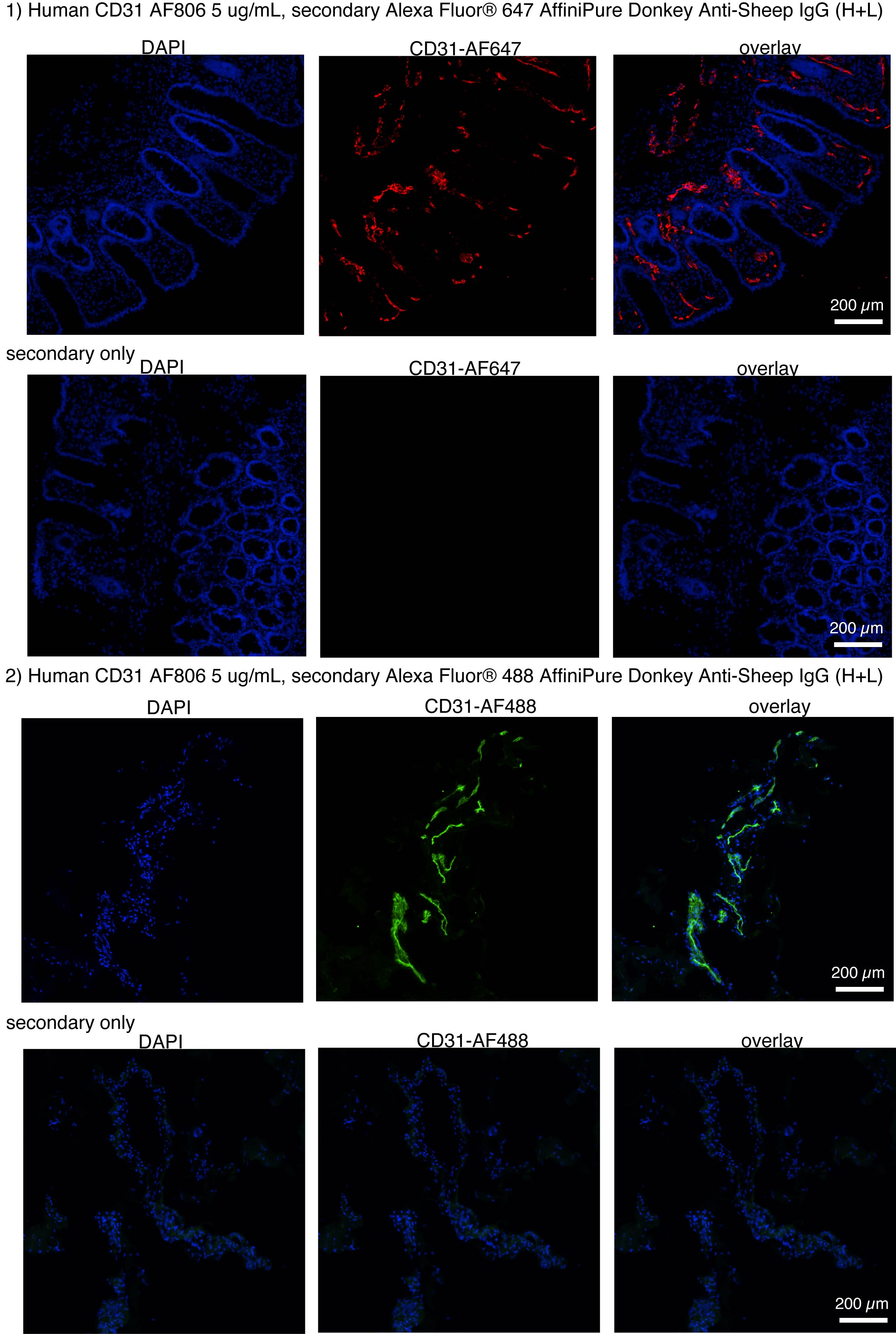
Application: Immunohistochemistry-FrozenSample Tested: Human Colon and colonSpecies: HumanVerified Customer | Posted 08/21/2023IF of healthy human colon 8 micrometer cryosections fixed with 4% PFA. 2 different secondary antibodies were used. Please see Primary antibody concentrations and fluorescence channels designation on the image.Immunofluorescence on freshly-frozen PFA-fixed cryosections

-
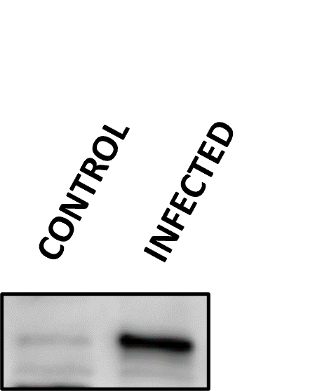
Application: Western BlotSample Tested: Cell LysatesSpecies: HumanVerified Customer | Posted 05/27/2022
-
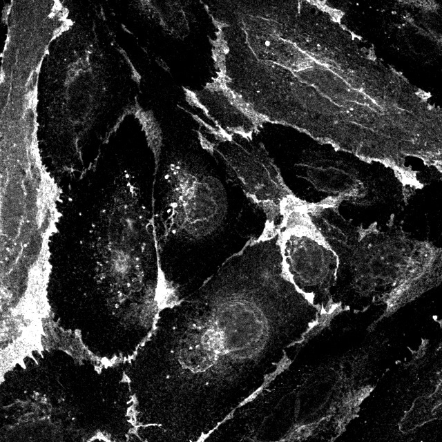
Application: Immunocytochemistry/ImmunofluorescenceSample Tested: HUVEC human umbilical vein endothelial cellsSpecies: HumanVerified Customer | Posted 08/04/2021
There are no reviews that match your criteria.
Protocols
Find general support by application which include: protocols, troubleshooting, illustrated assays, videos and webinars.
- Antigen Retrieval Protocol (PIER)
- Antigen Retrieval for Frozen Sections Protocol
- Appropriate Fixation of IHC/ICC Samples
- Cellular Response to Hypoxia Protocols
- Chromogenic IHC Staining of Formalin-Fixed Paraffin-Embedded (FFPE) Tissue Protocol
- Chromogenic Immunohistochemistry Staining of Frozen Tissue
- ClariTSA™ Fluorophore Kits
- Detection & Visualization of Antibody Binding
- Fluorescent IHC Staining of Frozen Tissue Protocol
- Graphic Protocol for Heat-induced Epitope Retrieval
- Graphic Protocol for the Preparation and Fluorescent IHC Staining of Frozen Tissue Sections
- Graphic Protocol for the Preparation and Fluorescent IHC Staining of Paraffin-embedded Tissue Sections
- Graphic Protocol for the Preparation of Gelatin-coated Slides for Histological Tissue Sections
- ICC Cell Smear Protocol for Suspension Cells
- ICC Immunocytochemistry Protocol Videos
- ICC for Adherent Cells
- IHC Sample Preparation (Frozen sections vs Paraffin)
- Immunocytochemistry (ICC) Protocol
- Immunocytochemistry Troubleshooting
- Immunofluorescence of Organoids Embedded in Cultrex Basement Membrane Extract
- Immunofluorescent IHC Staining of Formalin-Fixed Paraffin-Embedded (FFPE) Tissue Protocol
- Immunohistochemistry (IHC) and Immunocytochemistry (ICC) Protocols
- Immunohistochemistry Frozen Troubleshooting
- Immunohistochemistry Paraffin Troubleshooting
- Preparing Samples for IHC/ICC Experiments
- Preventing Non-Specific Staining (Non-Specific Binding)
- Primary Antibody Selection & Optimization
- Protocol for Heat-Induced Epitope Retrieval (HIER)
- Protocol for Making a 4% Formaldehyde Solution in PBS
- Protocol for VisUCyte™ HRP Polymer Detection Reagent
- Protocol for the Fluorescent ICC Staining of Cell Smears - Graphic
- Protocol for the Fluorescent ICC Staining of Cultured Cells on Coverslips - Graphic
- Protocol for the Preparation & Fixation of Cells on Coverslips
- Protocol for the Preparation and Chromogenic IHC Staining of Frozen Tissue Sections
- Protocol for the Preparation and Chromogenic IHC Staining of Frozen Tissue Sections - Graphic
- Protocol for the Preparation and Chromogenic IHC Staining of Paraffin-embedded Tissue Sections
- Protocol for the Preparation and Chromogenic IHC Staining of Paraffin-embedded Tissue Sections - Graphic
- Protocol for the Preparation and Fluorescent ICC Staining of Cells on Coverslips
- Protocol for the Preparation and Fluorescent ICC Staining of Non-adherent Cells
- Protocol for the Preparation and Fluorescent ICC Staining of Stem Cells on Coverslips
- Protocol for the Preparation and Fluorescent IHC Staining of Frozen Tissue Sections
- Protocol for the Preparation and Fluorescent IHC Staining of Paraffin-embedded Tissue Sections
- Protocol for the Preparation of Gelatin-coated Slides for Histological Tissue Sections
- Protocol for the Preparation of a Cell Smear for Non-adherent Cell ICC - Graphic
- R&D Systems Quality Control Western Blot Protocol
- TUNEL and Active Caspase-3 Detection by IHC/ICC Protocol
- The Importance of IHC/ICC Controls
- Troubleshooting Guide: Immunohistochemistry
- Troubleshooting Guide: Western Blot Figures
- Western Blot Conditions
- Western Blot Protocol
- Western Blot Protocol for Cell Lysates
- Western Blot Troubleshooting
- Western Blot Troubleshooting Guide
- View all Protocols, Troubleshooting, Illustrated assays and Webinars
Loading...