The G protein-coupled receptor, RDC1, belongs to a subgroup of chemokine receptors and has been designated CXCR7. CXCR7 can bind with high-affinity to CXCL12/SDF-1 and CXCL11/I‑TAC. It is also a co-receptor for several HIV and SIV strains. In their N-termini and extracellular loops 1, 2, and 3, human and mouse CXCR7 share 84%, 100%, 96%, and 86% amino acid sequence identity, respectively. Reports of mRNA levels and/or protein expression (as assessed using anti-CXCR7, clone 9C4) (J. Biol. Chem. 2005, 280(42):35760, J. Immunol. 2006, 176(4):2197) indicate that CXCR7 occurs on a wide variety of tissues and cells including monocytes, B cells, T cells and mature dendritic cells. In contrast, based on ligand binding analysis and receptor level (as assessed using anti-CXCR7, clone 11G8), surface expression of CXCR7 was reported to be restricted to tumor cells, activated endothelial cells, fetal liver cells, and few other cell types (J. Exp. Med. 2006, 203(9):2201). The basis of these inconsistent observations is not known but may be attributed to cell context and the use of different antibodies that may recognize different epitopes.
Human CXCR7/RDC-1 Antibody (11G8)
R&D Systems | Catalog # MAB42273

Key Product Details
Validated by
Species Reactivity
Validated:
Cited:
Applications
Validated:
Cited:
Label
Antibody Source
Product Specifications
Immunogen
Accession # P25106
Specificity
Clonality
Host
Isotype
Scientific Data Images for Human CXCR7/RDC-1 Antibody (11G8)
Detection of CXCR7/RDC‑1 in MCF‑7 Human Cell Line by Flow Cytometry.
MCF-7 human breast cancer cell line was stained with Mouse Anti-Human CXCR7/RDC-1 Monoclonal Antibody (Catalog # MAB42273, filled histogram) or isotype control antibody (Catalog # MAB002, open histogram), followed by Allophycocyanin-conjugated Anti-Mouse IgG F(ab')2Secondary Antibody (Catalog # F0101B).
CXCR7/RDC‑1 in Human Squamous Cell Carcinoma.
CXCR7/RDC-1 was detected in immersion fixed paraffin-embedded sections of human squamous cell carcinoma using Mouse Anti-Human CXCR7/RDC-1 Monoclonal Antibody (Catalog # MAB42273) at 5 µg/mL for 1 hour at room temperature followed by incubation with the Anti-Mouse IgG VisUCyte™ HRP Polymer Antibody (Catalog # VC001). Tissue was stained using DAB (brown) and counterstained with hematoxylin (blue). Specific staining was localized to cytoplasm and plasma membrane in cancer cells. View our protocol for IHC Staining with VisUCyte HRP Polymer Detection Reagents.
Detection of Human CXCR7/RDC-1 by Functional
CXCL12 induces CXCR4/CXCR7 and cell polarization in HUVECs. HUVECs were cultured on plastic Petri dishes (pretreated with PDL) with BSA stripes (the control group) and BSA plus CXCL12 stripes (CXCL12 group) for 5 min. HUVECs were fixed and stained with antibodies against CXCR4 (purple), CXCR7 (blue) and F-Actin (red). The micro stripes of fluorescein-conjugated BSA (A) or CXCL12 (H) are shown. CXCR4 (B), CXCR7 (C) and F-Actin (D) in HUVECs cultured on BSA stripes stayed in the resting state, while the polarization of CXCR4 (arrow: I), CXCR7 (arrow: J) and F-Actin (arrow: K) was observed in HUVECs cultured on CXCL12 stripes for 5 min. Moreover, the HUVECs in the BSA control group showed no morphological changes (E–G). The CXCL12 group HUVECs shows polarization towards the CXCL12 stripe (L–N). Scale bar: 10 μm. Image collected and cropped by CiteAb from the following publication (https://pubmed.ncbi.nlm.nih.gov/28811579), licensed under a CC-BY license. Not internally tested by R&D Systems.
Detection of CXCR7/RDC-1 by Western Blot
Impact of CXCR7 over-expression on the E2-dependent and -independent growth of MCF-7 cells.MCF-7 cells were transiently transfected with either a control expression vector or one containing the human CXCR7 open reading frame. (A) Total protein extracts were prepared 48 h after transfection, and a Western blot analysis was performed to confirm CXCR7 over-expression. (B) Transfected cells were cultured in the presence of EtOH (−) or 10−8 M E2 (+) for seven days. E2-dependent and E2-independent cell growth rates were then evaluated by cell count of three independent experiments (n = 3). The results are expressed as a percentage of the relative cell number obtained from control cells treated with E2 (considered as 100%). Significant differences (p<0.05) between transfected cells in the absence of E2 are indicated by an asterisk and between transfected cells in the presence of E2 by a sharp symbol. (C) A proposed model for the involvement of the CXCL12 signaling axis in E2-dependent and -independent cell growth is shown. The binding of CXCL12 to CXCR4 and CXCR7 leads to the stimulation of cell growth through diverse pathways [28]. CXCR7 can also modulate CXCL12 availability by removing the chemokine from the extracellular space (left panel). Estrogens could stimulate cell growth by favoring the activation of CXCL12 through CXCR4 and reducing the expression of CXCR7 (right panel). Image collected and cropped by CiteAb from the following open publication (https://pubmed.ncbi.nlm.nih.gov/21695171), licensed under a CC-BY license. Not internally tested by R&D Systems.Applications for Human CXCR7/RDC-1 Antibody (11G8)
CyTOF-ready
Flow Cytometry
Sample: MCF‑7 human breast cancer cell line
Immunohistochemistry
Sample: Perfusion fixed paraffin-embedded sections of nude mice injected with human breast cancer tissue cells and immersion fixed paraffin-embedded sections of human squamous cell carcinoma
Reviewed Applications
Read 2 reviews rated 4 using MAB42273 in the following applications:
Flow Cytometry Panel Builder
Bio-Techne Knows Flow Cytometry
Save time and reduce costly mistakes by quickly finding compatible reagents using the Panel Builder Tool.
Advanced Features
- Spectra Viewer - Custom analysis of spectra from multiple fluorochromes
- Spillover Popups - Visualize the spectra of individual fluorochromes
- Antigen Density Selector - Match fluorochrome brightness with antigen density

Formulation, Preparation, and Storage
Purification
Reconstitution
Reconstitute at 0.5 mg/mL in sterile PBS. For liquid material, refer to CoA for concentration.
Formulation
Shipping
Stability & Storage
- 12 months from date of receipt, -20 to -70 °C as supplied.
- 1 month, 2 to 8 °C under sterile conditions after reconstitution.
- 6 months, -20 to -70 °C under sterile conditions after reconstitution.
Calculators
Background: CXCR7/RDC-1
Alternate Names
Gene Symbol
UniProt
Additional CXCR7/RDC-1 Products
Product Documents for Human CXCR7/RDC-1 Antibody (11G8)
Certificate of Analysis
To download a Certificate of Analysis, please enter a lot or batch number in the search box below.
Note: Certificate of Analysis not available for kit components.
Product Specific Notices for Human CXCR7/RDC-1 Antibody (11G8)
For research use only
Citations for Human CXCR7/RDC-1 Antibody (11G8)
 Powered by Bioz
Customer Reviews for Human CXCR7/RDC-1 Antibody (11G8) (2)
Have you used Human CXCR7/RDC-1 Antibody (11G8)?
Submit a review and receive an Amazon gift card!
$25/€18/£15/$25CAN/¥2500 Yen for a review with an image
$10/€7/£6/$10CAN/¥1110 Yen for a review without an image
Submit a review
Customer Images
-
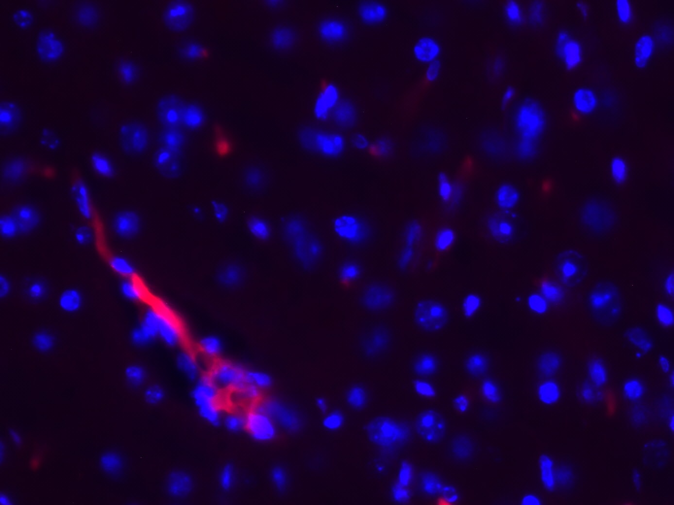
Application: Immunohistochemistry-FrozenSample Tested: Mouse brainSpecies: MouseVerified Customer | Posted 03/11/201910 µm Mouse brain cryosections stained with CXCR7 antibody (1:100) and Alexa Fluor 555 donkey anti mouse IgG. Antibody works quite well with mouse tissue, blood vessels are positive in the brain as they should be, for example.

-
Application: ELISASample Tested: See PMID 22457824Species: HumanVerified Customer | Posted 02/20/2015
There are no reviews that match your criteria.
Protocols
Find general support by application which include: protocols, troubleshooting, illustrated assays, videos and webinars.
- 7-Amino Actinomycin D (7-AAD) Cell Viability Flow Cytometry Protocol
- Antigen Retrieval Protocol (PIER)
- Antigen Retrieval for Frozen Sections Protocol
- Appropriate Fixation of IHC/ICC Samples
- Cellular Response to Hypoxia Protocols
- Chromogenic IHC Staining of Formalin-Fixed Paraffin-Embedded (FFPE) Tissue Protocol
- Chromogenic Immunohistochemistry Staining of Frozen Tissue
- ClariTSA™ Fluorophore Kits
- Detection & Visualization of Antibody Binding
- Extracellular Membrane Flow Cytometry Protocol
- Flow Cytometry Protocol for Cell Surface Markers
- Flow Cytometry Protocol for Staining Membrane Associated Proteins
- Flow Cytometry Staining Protocols
- Flow Cytometry Troubleshooting Guide
- Fluorescent IHC Staining of Frozen Tissue Protocol
- Graphic Protocol for Heat-induced Epitope Retrieval
- Graphic Protocol for the Preparation and Fluorescent IHC Staining of Frozen Tissue Sections
- Graphic Protocol for the Preparation and Fluorescent IHC Staining of Paraffin-embedded Tissue Sections
- Graphic Protocol for the Preparation of Gelatin-coated Slides for Histological Tissue Sections
- IHC Sample Preparation (Frozen sections vs Paraffin)
- Immunofluorescent IHC Staining of Formalin-Fixed Paraffin-Embedded (FFPE) Tissue Protocol
- Immunohistochemistry (IHC) and Immunocytochemistry (ICC) Protocols
- Immunohistochemistry Frozen Troubleshooting
- Immunohistochemistry Paraffin Troubleshooting
- Intracellular Flow Cytometry Protocol Using Alcohol (Methanol)
- Intracellular Flow Cytometry Protocol Using Detergents
- Intracellular Nuclear Staining Flow Cytometry Protocol Using Detergents
- Intracellular Staining Flow Cytometry Protocol Using Alcohol Permeabilization
- Intracellular Staining Flow Cytometry Protocol Using Detergents to Permeabilize Cells
- Preparing Samples for IHC/ICC Experiments
- Preventing Non-Specific Staining (Non-Specific Binding)
- Primary Antibody Selection & Optimization
- Propidium Iodide Cell Viability Flow Cytometry Protocol
- Protocol for Heat-Induced Epitope Retrieval (HIER)
- Protocol for Liperfluo
- Protocol for Making a 4% Formaldehyde Solution in PBS
- Protocol for VisUCyte™ HRP Polymer Detection Reagent
- Protocol for the Characterization of Human Th22 Cells
- Protocol for the Characterization of Human Th9 Cells
- Protocol for the Preparation & Fixation of Cells on Coverslips
- Protocol for the Preparation and Chromogenic IHC Staining of Frozen Tissue Sections
- Protocol for the Preparation and Chromogenic IHC Staining of Frozen Tissue Sections - Graphic
- Protocol for the Preparation and Chromogenic IHC Staining of Paraffin-embedded Tissue Sections
- Protocol for the Preparation and Chromogenic IHC Staining of Paraffin-embedded Tissue Sections - Graphic
- Protocol for the Preparation and Fluorescent IHC Staining of Frozen Tissue Sections
- Protocol for the Preparation and Fluorescent IHC Staining of Paraffin-embedded Tissue Sections
- Protocol for the Preparation of Gelatin-coated Slides for Histological Tissue Sections
- Protocol: Annexin V and PI Staining by Flow Cytometry
- Protocol: Annexin V and PI Staining for Apoptosis by Flow Cytometry
- TUNEL and Active Caspase-3 Detection by IHC/ICC Protocol
- The Importance of IHC/ICC Controls
- Troubleshooting Guide: Fluorokine Flow Cytometry Kits
- Troubleshooting Guide: Immunohistochemistry
- View all Protocols, Troubleshooting, Illustrated assays and Webinars
FAQs for Human CXCR7/RDC-1 Antibody (11G8)
-
Q: What light chain subclass is MAB42273 and MAB42273R?
A: MAB42273 and MAB42273R have a kappa light chain.
Associated Pathways
