Decorin is a small secreted chondroitin/dermatan sulfate proteoglycan in the family of small leucine-rich proteoglycans (SLRPs). SLRP family members are characterized by N-terminal and C-terminal cysteine-rich regions which flank the central region containing 10-12 tandem leucine-rich repeats (LRR) (1, 2). The human Decorin cDNA encodes a 359 amino acid (aa) precursor that includes a 16 aa signal sequence and a 14 aa propeptide. The 329 aa mature protein contains twelve LRR. Alternate splicing generates five isoforms with variable length deletions (3). Mature human and mouse Decorin share 80% aa sequence identity. In Decorin, serine 34 in the N-terminal domain is O-glycosylated. Naturally occurring Decorin proteoglycan has a molecular mass of approximately 100 kDa, and the deglycosylated Decorin core protein has a mass of approximately 40 kDa. Decorin binds to fibronectin, TGF-beta, and type I and type II collagens. The binding of Decorin to various molecules was reported to be mediated via the core protein. Decorin has been implicated in matrix assembly and has also been reported to suppress the growth of various tumor cell lines by activating the epidermal growth factor receptor.

Key Product Details
Validated by
Biological Validation
Species Reactivity
Validated:
Human
Cited:
Human, Mouse
Applications
Validated:
Western Blot, Simple Western
Cited:
Immunohistochemistry, Immunohistochemistry-Paraffin, Immunohistochemistry-Frozen, Western Blot, Immunocytochemistry, Immunoprecipitation
Label
Unconjugated
Antibody Source
Monoclonal Mouse IgG1 Clone # 115402
Loading...
Product Specifications
Immunogen
S. frugiperda insect ovarian cell line Sf 21-derived recombinant human Decorin
Gly17-Lys172
Accession # NP_598013.1
Gly17-Lys172
Accession # NP_598013.1
Specificity
Detects human Decorin in direct ELISAs and Western blots. In direct ELISAs, this antibody does not cross-react with recombinant mouse Decorin.
Clonality
Monoclonal
Host
Mouse
Isotype
IgG1
Scientific Data Images for Human Decorin Antibody (115402)
Detection of Human Decorin by Western Blot.
Western blot shows lysates of human uterus tissue and human heart tissue. PVDF membrane was probed with 1 µg/mL of Mouse Anti-Human Decorin Monoclonal Antibody (Catalog # MAB143) followed by HRP-conjugated Anti-Mouse IgG Secondary Antibody (Catalog # HAF018). A specific band was detected for Decorin at approximately 100 kDa (as indicated). This experiment was conducted under reducing conditions and using Immunoblot Buffer Group 1.
Detection of Human Decorin by Simple WesternTM.
Simple Western lane view shows lysates of human uterus tissue, loaded at 0.2 mg/mL. A specific band was detected for Decorin at approximately 145 kDa (as indicated) using 10 µg/mL of Mouse Anti-Human Decorin Monoclonal Antibody (Catalog # MAB143). This experiment was conducted under reducing conditions and using the 12-230 kDa separation system.
Detection of Human Decorin by Immunohistochemistry
Immunohistochemical staining of stromal protein expression in DCIS with sclerotic or myxoid stromaMicrophotographs displaying HE staining (A-B), and IHC staining for biglycan (C-D), decorin (E-F) and versican (G-H). Panels A-C-E-G display photographs of one DCIS lesion with sclerotic stroma; panels B-D-F-H display one DCIS lesion with myxoid stroma. This figure illustrates that myxoid DCIS present reduced periductal decorin staining and tend to have increased periductal versican and biglycan expression, whereas sclerotic DCIS generally present strong stromal decorin immunoreactivity, and tend to lack stromal versican and biglycan. Original magnification 100x. Image collected and cropped by CiteAb from the following publication (https://www.oncoscience.us/lookup/doi/10.18632/oncoscience.87), licensed under a CC-BY license. Not internally tested by R&D Systems.
Detection of Human Decorin by Western Blot
Treatment of CAFs with combined cytokines or cancer cell-derived secretomes affects ECM protein expression(A) Western blot showing the effects of bFGF, EGF and TGF-beta 1 treatment (10 ng/ml) on the expression of biglycan, decorin, alpha -SMA and versican in CAFs. (B) Western blot illustrating the effects of treatment with cancer cell-derived conditioned medium (CM) on the expression of biglycan, decorin, alpha -SMA and versican in CAFs. TGF-beta 1 treatment (1 and 10 ng/ml) was included as a positive control. Tubulin was used as loading control (A, B). Image collected and cropped by CiteAb from the following publication (https://www.oncoscience.us/lookup/doi/10.18632/oncoscience.87), licensed under a CC-BY license. Not internally tested by R&D Systems.
Detection of Human Decorin by Western Blot
Secretome analysis of WT, COG4p.G516R, and COG4-KO chondrosarcoma cells. (A) Coomassie blue staining of proteins in cell lysates and conditioned medium from COG4p.G516R clones #1, #2, WT controls and COG4 KO cells. (B) Volcano plot of differentially secreted proteins between COG4p.G516R and WT. (C) Volcano plot of differentially secreted proteins between COG4p.G516R and COG4-KO cells. A few top candidates confirmed by pathway analysis were labeled respectively. (D) Dot plot showing enriched pathways from Gene Set Enrichment Analysis with the Normalized Enrichment Score (NES) shown on the X-axis. The size of the dots represents the number of genes in the significant Data Set list associated with the GO term and the color of the dots represent the adjusted p- values. (E) Table showing a list of top 10 candidates which were decreased in secretions of COG4p.G516R with log 2 (Fold Change) and adjusted p-value shown for COG4p.G516R vs. WT and COG4p.G516R vs. COG4-KO. The number of peptides is equal to or more than two for all the candidates shown in table. (F) Western blotting of top candidates in medium decreased in both COG4p.G516R and COG4-KO cells compared to WT. GAPDH was from cell lysates with comparable amount as loading control. (G) Western blotting of top candidates only changed in COG4p.G516R compared to WT and COG4-KO cells. All experiments were performed in duplicate with similar results. Image collected and cropped by CiteAb from the following open publication (https://pubmed.ncbi.nlm.nih.gov/36393834), licensed under a CC-BY license. Not internally tested by R&D Systems.Applications for Human Decorin Antibody (115402)
Application
Recommended Usage
Simple Western
10 µg/mL
Sample: Human uterus tissue
Sample: Human uterus tissue
Western Blot
1 µg/mL
Sample: Human uterus tissue and human heart tissue
Sample: Human uterus tissue and human heart tissue
Reviewed Applications
Read 3 reviews rated 4.7 using MAB143 in the following applications:
Formulation, Preparation, and Storage
Purification
Protein A or G purified from hybridoma culture supernatant
Reconstitution
Reconstitute at 0.5 mg/mL in sterile PBS. For liquid material, refer to CoA for concentration.
Loading...
Formulation
Lyophilized from a 0.2 μm filtered solution in PBS with Trehalose. *Small pack size (SP) is supplied either lyophilized or as a 0.2 µm filtered solution in PBS.
Shipping
Lyophilized product is shipped at ambient temperature. Liquid small pack size (-SP) is shipped with polar packs. Upon receipt, store immediately at the temperature recommended below.
Stability & Storage
Use a manual defrost freezer and avoid repeated freeze-thaw cycles.
- 12 months from date of receipt, -20 to -70 °C as supplied.
- 1 month, 2 to 8 °C under sterile conditions after reconstitution.
- 6 months, -20 to -70 °C under sterile conditions after reconstitution.
Calculators
Background: Decorin
References
- Naito, Z. (2005) J. Nippon Med. Sch. 72:137.
- Matsushima, N. et al. (2005) Cell. Mol. Life Sci. 62:2771.
- Danielson, K. et al. (1993) Genomics 15:146.
Alternate Names
DCN, DSPG2, PG-II, PGS2, SLRR1B
Gene Symbol
DCN
UniProt
Additional Decorin Products
Product Documents for Human Decorin Antibody (115402)
Certificate of Analysis
To download a Certificate of Analysis, please enter a lot or batch number in the search box below.
Note: Certificate of Analysis not available for kit components.
Product Specific Notices for Human Decorin Antibody (115402)
For research use only
Related Research Areas
Citations for Human Decorin Antibody (115402)
 Powered by Bioz
Customer Reviews for Human Decorin Antibody (115402) (3)
4.7 out of 5
3 Customer Ratings
Have you used Human Decorin Antibody (115402)?
Submit a review and receive an Amazon gift card!
$25/€18/£15/$25CAN/¥2500 Yen for a review with an image
$10/€7/£6/$10CAN/¥1110 Yen for a review without an image
Submit a review
Customer Images
Showing
1
-
3 of
3 reviews
Showing All
Filter By:
-
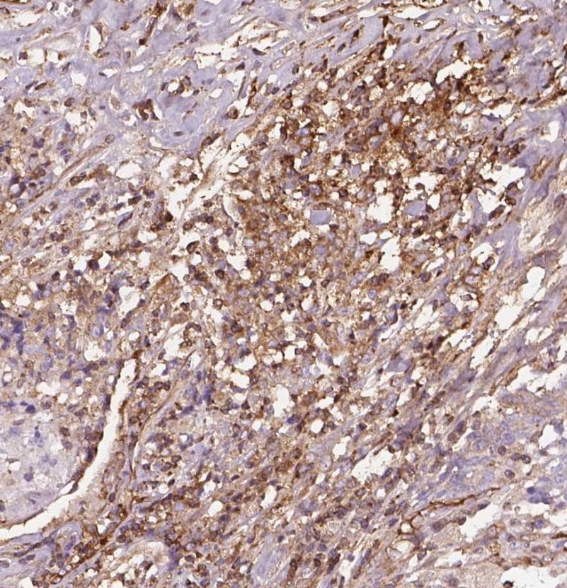
Application: ImmunohistochemistrySample Tested: Breast cancer tissueSpecies: HumanVerified Customer | Posted 01/18/2022

-
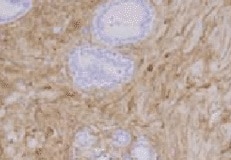
Application: Immunohistochemistry-ParaffinSample Tested: Mammary gland tissueSpecies: HumanVerified Customer | Posted 10/02/2021Mammary gland tissue. Decorin is present in inflammatory cells and fibroblasts.
-
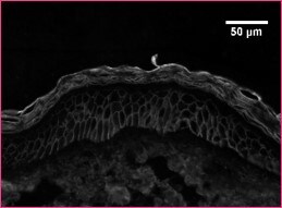
Application: Immunohistochemistry-ParaffinSample Tested: skinSpecies: HumanVerified Customer | Posted 04/11/2019
There are no reviews that match your criteria.
Protocols
Find general support by application which include: protocols, troubleshooting, illustrated assays, videos and webinars.
- Cellular Response to Hypoxia Protocols
- R&D Systems Quality Control Western Blot Protocol
- Troubleshooting Guide: Western Blot Figures
- Western Blot Conditions
- Western Blot Protocol
- Western Blot Protocol for Cell Lysates
- Western Blot Troubleshooting
- Western Blot Troubleshooting Guide
- View all Protocols, Troubleshooting, Illustrated assays and Webinars
Loading...
Associated Pathways

