
Key Product Details
Species Reactivity
Human
Applications
Flow Cytometry, CyTOF-ready
Label
Unconjugated
Antibody Source
Monoclonal Mouse IgG2A Clone # 889022
Loading...
Product Specifications
Immunogen
NS0 mouse myeloma cell line transfected with human Dopamine D5 R/DRD5
Met1-His477
Accession # P21918
Met1-His477
Accession # P21918
Specificity
Detects HEK293 human embryonic kidney cell line transfected with human Dopamine D5 R/DRD5 by Flow Cytometry. Does not detect untransfected or irrelevant transfected HEK293 cells.
Clonality
Monoclonal
Host
Mouse
Isotype
IgG2A
Scientific Data Images for Human Dopamine D5R/DRD5 Antibody (889022)
Detection of Dopamine D5 R/DRD5 in HEK293 Human Cell Line Transfected with Human Dopamine D5 R/DRD5 and eGFP by Flow Cytometry.
HEK293 human embryonic kidney cell line transfected with either (A) human Dopamine D5 R/DRD5 or (B) irrelevant transfectants and eGFP was stained with Mouse Anti-Human Dopamine D5 R/DRD5 Monoclonal Antibody (Catalog # MAB82861) followed by Allophycocyanin-conjugated Anti-Mouse IgG Secondary Antibody (Catalog # F0101B). Quadrant markers were set based on control antibody staining (Catalog # MAB003).Applications for Human Dopamine D5R/DRD5 Antibody (889022)
Application
Recommended Usage
CyTOF-ready
Ready to be labeled using established conjugation methods. No BSA or other carrier proteins that could interfere with conjugation.
Flow Cytometry
0.25 µg/106 cells
Sample: HEK293 human embryonic kidney cell line transfected with human Dopamine D5 R/DRD5 and eGFP
Sample: HEK293 human embryonic kidney cell line transfected with human Dopamine D5 R/DRD5 and eGFP
Reviewed Applications
Read 2 reviews rated 4 using MAB82861 in the following applications:
Flow Cytometry Panel Builder
Bio-Techne Knows Flow Cytometry
Save time and reduce costly mistakes by quickly finding compatible reagents using the Panel Builder Tool.
Advanced Features
- Spectra Viewer - Custom analysis of spectra from multiple fluorochromes
- Spillover Popups - Visualize the spectra of individual fluorochromes
- Antigen Density Selector - Match fluorochrome brightness with antigen density

Formulation, Preparation, and Storage
Purification
Protein A or G purified from hybridoma culture supernatant
Reconstitution
Reconstitute at 0.5 mg/mL in sterile PBS. For liquid material, refer to CoA for concentration.
Loading...
Formulation
Lyophilized from a 0.2 μm filtered solution in PBS with Trehalose. *Small pack size (SP) is supplied either lyophilized or as a 0.2 µm filtered solution in PBS.
Shipping
Lyophilized product is shipped at ambient temperature. Liquid small pack size (-SP) is shipped with polar packs. Upon receipt, store immediately at the temperature recommended below.
Stability & Storage
Use a manual defrost freezer and avoid repeated freeze-thaw cycles.
- 12 months from date of receipt, -20 to -70 °C as supplied.
- 1 month, 2 to 8 °C under sterile conditions after reconstitution.
- 6 months, -20 to -70 °C under sterile conditions after reconstitution.
Calculators
Background: Dopamine D5R/DRD5
References
- Sunahra, R. et al. (1991) Nature. 350:614.
- Weinshank, R. et al. (1991) J Biol Chem. 266:22427.
- Squassina, A. et al. (2008) Neurosci Lett. Feb 13.
- Golimbet, V. et al. (2008) Bull Exp Biol Med. Jan.
- Wei, J. et al. (2012) Addict Behav. 37:622.
- Thompson, D. et al. (2011) Traffic. 12:644.
Long Name
Dopamine D5 Receptor
Alternate Names
DBDR, Dopamine D5 R, DRD1B, DRD1L2, DRD5
Gene Symbol
DRD5
UniProt
Additional Dopamine D5R/DRD5 Products
Product Documents for Human Dopamine D5R/DRD5 Antibody (889022)
Certificate of Analysis
To download a Certificate of Analysis, please enter a lot or batch number in the search box below.
Note: Certificate of Analysis not available for kit components.
Product Specific Notices for Human Dopamine D5R/DRD5 Antibody (889022)
For research use only
Related Research Areas
Customer Reviews for Human Dopamine D5R/DRD5 Antibody (889022) (2)
4 out of 5
2 Customer Ratings
Have you used Human Dopamine D5R/DRD5 Antibody (889022)?
Submit a review and receive an Amazon gift card!
$25/€18/£15/$25CAN/¥2500 Yen for a review with an image
$10/€7/£6/$10CAN/¥1110 Yen for a review without an image
Submit a review
Customer Images
Showing
1
-
2 of
2 reviews
Showing All
Filter By:
-
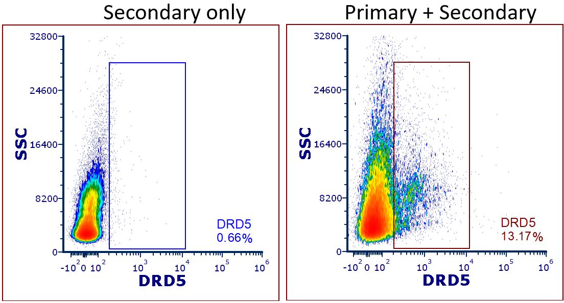
Application: Flow CytometrySample Tested: Whole bloodSpecies: HumanVerified Customer | Posted 12/17/2020Whole blood was diluted with PBS, overlaid atop Ficoll, and centrifuged to separate the PBMC layer. The PBMC layer was washed twice with PBS and cells were aliquoted into centrifuge tubes for staining. Cells were counted and concentration was adjusted to 10,000 cells per uL. 100 uL of cells was aliquoted into each centrifuge tube for surface staining. 1uL of anti-DRD5 antibody was added to the 'primary + secondary' condition. Cells were incubated on ice for 30 mins protected from light. Following incubation, cells were washed twice with PBS and then fixed at room temperature for 30 mins protected from light. Cells were washed twice with permeabilization buffer and resuspended in 100 uL perm buffer for secondary staining. 1uL of secondary antibody (R&D systems, Cat#F0102B) was added to both the 'secondary only' and 'primary + secondary' condition. Cells were incubated at room temperature for 30 mins protected from light. Following 2x perm buffer washes, cells were resuspended in 500uL PBS for flow cytometry acquisition. 'Secondary only' condition (left) was used to determine the gate placement.'Primary + secondary' condition (right) shows successful flow cytometry staining for DRD5 in total PBMC fraction.

-
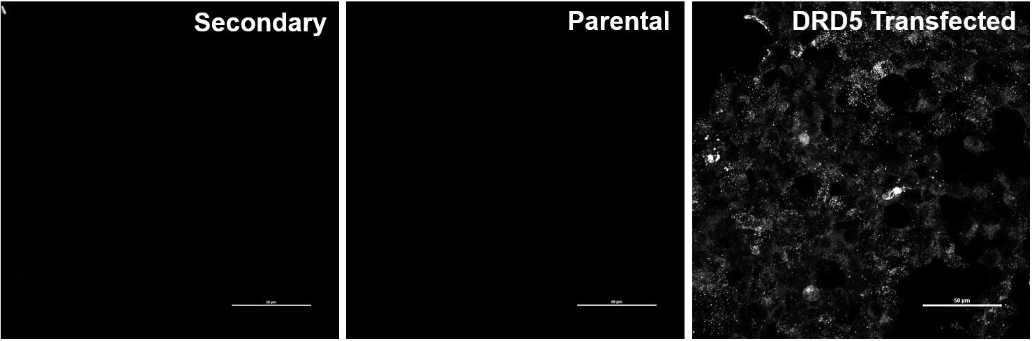
Application: Immunocytochemistry/ImmunofluorescenceSample Tested: 293T human embryonic kidney cell line and HEK293 human embryonic kidney cell lineSpecies: HumanVerified Customer | Posted 12/01/2020We transfected HEK cells with a DRD5 plasmid and stained them for DRD5 at a 1:1000 concentration. Secondary only and parental HEK cells were used as controls to confirm specific staining.
There are no reviews that match your criteria.
Protocols
Find general support by application which include: protocols, troubleshooting, illustrated assays, videos and webinars.
- 7-Amino Actinomycin D (7-AAD) Cell Viability Flow Cytometry Protocol
- Extracellular Membrane Flow Cytometry Protocol
- Flow Cytometry Protocol for Cell Surface Markers
- Flow Cytometry Protocol for Staining Membrane Associated Proteins
- Flow Cytometry Staining Protocols
- Flow Cytometry Troubleshooting Guide
- Intracellular Flow Cytometry Protocol Using Alcohol (Methanol)
- Intracellular Flow Cytometry Protocol Using Detergents
- Intracellular Nuclear Staining Flow Cytometry Protocol Using Detergents
- Intracellular Staining Flow Cytometry Protocol Using Alcohol Permeabilization
- Intracellular Staining Flow Cytometry Protocol Using Detergents to Permeabilize Cells
- Propidium Iodide Cell Viability Flow Cytometry Protocol
- Protocol for Liperfluo
- Protocol for the Characterization of Human Th22 Cells
- Protocol for the Characterization of Human Th9 Cells
- Protocol: Annexin V and PI Staining by Flow Cytometry
- Protocol: Annexin V and PI Staining for Apoptosis by Flow Cytometry
- Troubleshooting Guide: Fluorokine Flow Cytometry Kits
- View all Protocols, Troubleshooting, Illustrated assays and Webinars
Loading...