EphB4, also known as Htk, Myk1, Tyro11, and Mdk2, is a member of the Eph receptor tyrosine kinase family and binds Ephrin-B2. The A and B class Eph proteins have a common structural organization (1-4). The human EphB4 cDNA encodes a 987 amino acid (aa) precursor that includes a 15 aa signal sequence, a 524 aa extracellular domain (ECD), a 21 aa transmembrane segment, and a 427 aa cytoplasmic domain (5). The ECD contains an N-terminal globular domain, a cysteine-rich domain, and two fibronectin type III domains. The cytoplasmic domain contains a juxtamembrane motif with two tyrosine residues which are the major autophosphorylation sites, a kinase domain, and a conserved sterile alpha motif (SAM) (5). Activation of kinase activity occurs after membrane-bound or clustered ligand recognition and binding. The ECD of human EphB4 shares 89% aa sequence identity with mouse EphB4 and 42-45% aa sequence identity with human EphB1, 2, and 3. EphB4 is expressed preferentially on venous endothelial cells (EC) and inhibits cell-cell adhesion, chemotaxis, and angiogenesis. Opposing effects are induced by signaling through Ephrin-B2 expressed on arterial EC: adhesion, endothelial cell migration, and vessel sprouting (6). EphB4 singaling contributes to new vascularization by guiding venous EC away from Ephrin-B2 expressing EC. Ephrin-B2 signaling induces arterial EC to migrate towards nascent EphB4 expressing vessels (6). The combination of forward signaling through EphB4 and reverse signaling through Ephrin-B2 promotes in vivo mammary tumor growth and
tumor-associated angiogenesis (7). EphB4 promotes the differentiation of megakaryocytic and erythroid progenitors but not granulocytic or monocytic progenitors (8, 9).

Key Product Details
Validated by
Knockout/Knockdown
Species Reactivity
Validated:
Human
Cited:
Human, Mouse
Applications
Validated:
Knockout Validated, Immunohistochemistry, Western Blot, Flow Cytometry, Simple Western, CyTOF-ready
Cited:
Immunohistochemistry, Immunohistochemistry-Frozen, Western Blot, Immunocytochemistry, Immunoprecipitation
Label
Unconjugated
Antibody Source
Polyclonal Goat IgG
Loading...
Product Specifications
Immunogen
Mouse myeloma cell line NS0-derived recombinant human EphB4
Leu16-Ala539
Accession # P54760
Leu16-Ala539
Accession # P54760
Specificity
Detects human EphB4 in direct ELISAs and Western blots. In direct ELISAs, less than 10% cross-reactivity with recombinant mouse (rm) EphB4 is observed and less than 1% cross-reactivity with recombinant human (rh) EphA4, rhEphA3, rhEphB2, and rhEphB3 is observed.
Clonality
Polyclonal
Host
Goat
Isotype
IgG
Scientific Data Images for Human EphB4 Antibody
Detection of Human EphB4 by Western Blot.
Western blot shows lysates of K562 human chronic myelogenous leukemia cell line, COLO 205 human colorectal adenocarcinoma cell line, ZR-75 human breast cancer cell line, and HUVEC human umbilical vein endothelial cells. PVDF membrane was probed with 2 µg/mL of Goat Anti-Human EphB4 Antigen Affinity-purified Polyclonal Antibody (Catalog # AF3038) followed by HRP-conjugated Anti-Goat IgG Secondary Antibody (Catalog # HAF017). A specific band was detected for EphB4 at approximately 120 kDa (as indicated). This experiment was conducted under reducing conditions and using Immunoblot Buffer Group 1.
Detection of EphB4 in MCF‑7 Human Cell Line by Flow Cytometry.
MCF-7 human breast cancer cell line was stained with Goat Anti-Human EphB4 Antigen Affinity-purified Polyclonal Antibody (Catalog # AF3038, filled histogram) or isotype control antibody (Catalog # AB-108-C, open histogram), followed by Phycoerythrin-conjugated Anti-Goat IgG Secondary Antibody (Catalog # F0107). View our protocol for Staining Membrane-associated Proteins.
EphB4 in Human Kidney.
EphB4 was detected in immersion fixed paraffin-embedded sections of human kidney using 15 µg/mL Goat Anti-Human EphB4 Antigen Affinity-purified Polyclonal Antibody (Catalog # AF3038) overnight at 4 °C. Tissue was stained with the Anti-Goat HRP-DAB Cell & Tissue Staining Kit (brown; Catalog # CTS008) and counterstained with hematoxylin (blue). View our protocol for Chromogenic IHC Staining of Paraffin-embedded Tissue Sections.
Detection of Human EphB4 by Simple WesternTM.
Simple Western lane view shows lysates of HUVEC human umbilical vein endothelial cells, loaded at 0.2 mg/mL. A specific band was detected for EphB4 at approximately 127 kDa (as indicated) using 50 µg/mL of Goat Anti-Human EphB4 Antigen Affinity-purified Polyclonal Antibody (Catalog # AF3038) followed by 1:50 dilution of HRP-conjugated Anti-Goat IgG Secondary Antibody (Catalog # HAF109). This experiment was conducted under reducing conditions and using the 12-230 kDa separation system.
Western Blot Shows Human EphB4 Specificity by Using Knockout Cell Line.
Western blot shows lysates of HEK293T human embryonic kidney parental cell line and EphB4 knockout HEK293T cell line (KO). PVDF membrane was probed with 2 µg/mL of Goat Anti-Human EphB4 Antigen Affinity-purified Polyclonal Antibody (Catalog # AF3038) followed by HRP-conjugated Anti-Goat IgG Secondary Antibody (Catalog # HAF017). A specific band was detected for EphB4 at approximately 140 kDa (as indicated) in the parental HEK293T cell line, but is not detectable in knockout HEK293T cell line. GAPDH (Catalog # AF5718) is shown as a loading control. This experiment was conducted under reducing conditions and using Immunoblot Buffer Group 1.
Detection of Mouse EphB4 by Western Blot
EphB4 expression in transfected A375 melanoma cells and corresponding tumor xenografts. Western blot analysis of EphB4 and its ligand EphrinB2 in A375, A375-pIRES, and A375-EphB4 whole cell lysates (A) and tumor lysates (E). Anti-beta -actin served as loading control. (B) Relative mRNA expression of EphB4 and EphrinB2 in A375, A375-pIRES, and A375-EphB4 cells, analyzed by quantitative real-time RT-PCR was normalized to the constitutive expression level of beta -actin and to expression level in wild-type A375 melanoma cells using the delta delta CT method (2−∆∆ct) resulting in a value of 1 for A375 cells. Values represent mean ± SEM of at least three independent experiments each performed in triplicate (* p < 0.05, *** p < 0.001). (C) EphB4 phosphorylation was analyzed by pEphB4-ELISA in whole cell lysates of A375 EphB4 cells incubated with different concentrations of sEphrinB2-Fc (0; 0,5; 1 µg/mL) for 15, 30, and 60 min. Values represent mean ± SD from one of at least three independent experiments, each performed in duplicate. To rule out influence of sEphrinB2 on total EphB4 protein amount, EphB4 was analyzed in the same cell lysates by western blot analysis (figure shows one representative blot out of three independent experiments performed in duplicate) ranging from 92% to 112% of control as calculated after densitometric analysis. Anti beta -actin served as loading control. (D) Immunohistochemical detection of EphB4 in acetone-fixed cryosections of A375 pIRES and A375 EphB4 tumor xenografts using goat anti EphB4 antibody. Sections stained without the primary antibody served as negative control. Scale bar 50 µm. Image collected and cropped by CiteAb from the following publication (https://pubmed.ncbi.nlm.nih.gov/29462967), licensed under a CC-BY license. Not internally tested by R&D Systems.
Detection of Mouse EphB4 by Western Blot
EphB4 expression in transfected A375 melanoma cells and corresponding tumor xenografts. Western blot analysis of EphB4 and its ligand EphrinB2 in A375, A375-pIRES, and A375-EphB4 whole cell lysates (A) and tumor lysates (E). Anti-beta -actin served as loading control. (B) Relative mRNA expression of EphB4 and EphrinB2 in A375, A375-pIRES, and A375-EphB4 cells, analyzed by quantitative real-time RT-PCR was normalized to the constitutive expression level of beta -actin and to expression level in wild-type A375 melanoma cells using the delta delta CT method (2−∆∆ct) resulting in a value of 1 for A375 cells. Values represent mean ± SEM of at least three independent experiments each performed in triplicate (* p < 0.05, *** p < 0.001). (C) EphB4 phosphorylation was analyzed by pEphB4-ELISA in whole cell lysates of A375 EphB4 cells incubated with different concentrations of sEphrinB2-Fc (0; 0,5; 1 µg/mL) for 15, 30, and 60 min. Values represent mean ± SD from one of at least three independent experiments, each performed in duplicate. To rule out influence of sEphrinB2 on total EphB4 protein amount, EphB4 was analyzed in the same cell lysates by western blot analysis (figure shows one representative blot out of three independent experiments performed in duplicate) ranging from 92% to 112% of control as calculated after densitometric analysis. Anti beta -actin served as loading control. (D) Immunohistochemical detection of EphB4 in acetone-fixed cryosections of A375 pIRES and A375 EphB4 tumor xenografts using goat anti EphB4 antibody. Sections stained without the primary antibody served as negative control. Scale bar 50 µm. Image collected and cropped by CiteAb from the following publication (https://pubmed.ncbi.nlm.nih.gov/29462967), licensed under a CC-BY license. Not internally tested by R&D Systems.Applications for Human EphB4 Antibody
Application
Recommended Usage
CyTOF-ready
Ready to be labeled using established conjugation methods. No BSA or other carrier proteins that could interfere with conjugation.
Flow Cytometry
0.25 µg/106 cells
Sample: MCF‑7 human breast cancer cell line
Sample: MCF‑7 human breast cancer cell line
Immunohistochemistry
5-15 µg/mL
Sample: Immersion fixed paraffin-embedded sections of human kidney
Sample: Immersion fixed paraffin-embedded sections of human kidney
Knockout Validated
EphB4
is specifically detected in HEK293T human embryonic kidney parental cell line but is not detectable in
EphB4 knockout HEK293T cell line.
Simple Western
50 µg/mL
Sample: HUVEC human umbilical vein endothelial cells
Sample: HUVEC human umbilical vein endothelial cells
Western Blot
2 µg/mL
Sample: K562 human chronic myelogenous leukemia cell line, COLO 205 human colorectal adenocarcinoma cell line, ZR‑75 human breast cancer cell line, and HUVEC human umbilical vein endothelial cells
Sample: K562 human chronic myelogenous leukemia cell line, COLO 205 human colorectal adenocarcinoma cell line, ZR‑75 human breast cancer cell line, and HUVEC human umbilical vein endothelial cells
Reviewed Applications
Read 1 review rated 5 using AF3038 in the following applications:
Flow Cytometry Panel Builder
Bio-Techne Knows Flow Cytometry
Save time and reduce costly mistakes by quickly finding compatible reagents using the Panel Builder Tool.
Advanced Features
- Spectra Viewer - Custom analysis of spectra from multiple fluorochromes
- Spillover Popups - Visualize the spectra of individual fluorochromes
- Antigen Density Selector - Match fluorochrome brightness with antigen density

Formulation, Preparation, and Storage
Purification
Antigen Affinity-purified
Reconstitution
Reconstitute at 0.2 mg/mL in sterile PBS. For liquid material, refer to CoA for concentration.
Loading...
Formulation
Lyophilized from a 0.2 μm filtered solution in PBS with Trehalose. *Small pack size (SP) is supplied either lyophilized or as a 0.2 µm filtered solution in PBS.
Shipping
Lyophilized product is shipped at ambient temperature. Liquid small pack size (-SP) is shipped with polar packs. Upon receipt, store immediately at the temperature recommended below.
Stability & Storage
Use a manual defrost freezer and avoid repeated freeze-thaw cycles.
- 12 months from date of receipt, -20 to -70 °C as supplied.
- 1 month, 2 to 8 °C under sterile conditions after reconstitution.
- 6 months, -20 to -70 °C under sterile conditions after reconstitution.
Calculators
Background: EphB4
References
- Poliakov, A. et al. (2004) Dev. Cell 7:465.
- Surawska, H. et al. (2004) Cytokine Growth Factor Rev. 15:419.
- Pasquale, E.B. (2005) Nat. Rev. Mol. Cell Biol. 6:462.
- Davy, A. and P. Soriano (2005) Dev. Dyn. 232:1.
- Bennett, B.D. et al. (1994) J. Biol. Chem. 269:14211.
- Fuller, T. et al. (2003) J. Cell Sci. 116:2461.
- Noren, N.K. et al. (2004) Proc. Natl. Acad. Sci. USA 101:5583.
- Wang, Z. et al. (2002) Blood 99:2740.
- Inada, T. et al. (1997) Blood 89:2757.
Long Name
Eph Receptor B4
Alternate Names
Htk, Mdk2, Myk1, Tyro11
Gene Symbol
EPHB4
UniProt
Additional EphB4 Products
Product Documents for Human EphB4 Antibody
Certificate of Analysis
To download a Certificate of Analysis, please enter a lot or batch number in the search box below.
Note: Certificate of Analysis not available for kit components.
Product Specific Notices for Human EphB4 Antibody
For research use only
Related Research Areas
Citations for Human EphB4 Antibody
 Powered by Bioz
Customer Reviews for Human EphB4 Antibody (1)
5 out of 5
1 Customer Rating
Have you used Human EphB4 Antibody?
Submit a review and receive an Amazon gift card!
$25/€18/£15/$25CAN/¥2500 Yen for a review with an image
$10/€7/£6/$10CAN/¥1110 Yen for a review without an image
Submit a review
Customer Images
Showing
1
-
1 of
1 review
Showing All
Filter By:
-
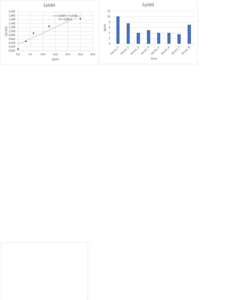
Application: ELISASample Tested: Serum and PlasmaSpecies: HumanVerified Customer | Posted 07/05/2022works well for ELISA in combination with mAb

There are no reviews that match your criteria.
Protocols
Find general support by application which include: protocols, troubleshooting, illustrated assays, videos and webinars.
- 7-Amino Actinomycin D (7-AAD) Cell Viability Flow Cytometry Protocol
- Antigen Retrieval Protocol (PIER)
- Antigen Retrieval for Frozen Sections Protocol
- Appropriate Fixation of IHC/ICC Samples
- Cellular Response to Hypoxia Protocols
- Chromogenic IHC Staining of Formalin-Fixed Paraffin-Embedded (FFPE) Tissue Protocol
- Chromogenic Immunohistochemistry Staining of Frozen Tissue
- ClariTSA™ Fluorophore Kits
- Detection & Visualization of Antibody Binding
- Extracellular Membrane Flow Cytometry Protocol
- Flow Cytometry Protocol for Cell Surface Markers
- Flow Cytometry Protocol for Staining Membrane Associated Proteins
- Flow Cytometry Staining Protocols
- Flow Cytometry Troubleshooting Guide
- Fluorescent IHC Staining of Frozen Tissue Protocol
- Graphic Protocol for Heat-induced Epitope Retrieval
- Graphic Protocol for the Preparation and Fluorescent IHC Staining of Frozen Tissue Sections
- Graphic Protocol for the Preparation and Fluorescent IHC Staining of Paraffin-embedded Tissue Sections
- Graphic Protocol for the Preparation of Gelatin-coated Slides for Histological Tissue Sections
- IHC Sample Preparation (Frozen sections vs Paraffin)
- Immunofluorescent IHC Staining of Formalin-Fixed Paraffin-Embedded (FFPE) Tissue Protocol
- Immunohistochemistry (IHC) and Immunocytochemistry (ICC) Protocols
- Immunohistochemistry Frozen Troubleshooting
- Immunohistochemistry Paraffin Troubleshooting
- Intracellular Flow Cytometry Protocol Using Alcohol (Methanol)
- Intracellular Flow Cytometry Protocol Using Detergents
- Intracellular Nuclear Staining Flow Cytometry Protocol Using Detergents
- Intracellular Staining Flow Cytometry Protocol Using Alcohol Permeabilization
- Intracellular Staining Flow Cytometry Protocol Using Detergents to Permeabilize Cells
- Preparing Samples for IHC/ICC Experiments
- Preventing Non-Specific Staining (Non-Specific Binding)
- Primary Antibody Selection & Optimization
- Propidium Iodide Cell Viability Flow Cytometry Protocol
- Protocol for Heat-Induced Epitope Retrieval (HIER)
- Protocol for Liperfluo
- Protocol for Making a 4% Formaldehyde Solution in PBS
- Protocol for VisUCyte™ HRP Polymer Detection Reagent
- Protocol for the Characterization of Human Th22 Cells
- Protocol for the Characterization of Human Th9 Cells
- Protocol for the Preparation & Fixation of Cells on Coverslips
- Protocol for the Preparation and Chromogenic IHC Staining of Frozen Tissue Sections
- Protocol for the Preparation and Chromogenic IHC Staining of Frozen Tissue Sections - Graphic
- Protocol for the Preparation and Chromogenic IHC Staining of Paraffin-embedded Tissue Sections
- Protocol for the Preparation and Chromogenic IHC Staining of Paraffin-embedded Tissue Sections - Graphic
- Protocol for the Preparation and Fluorescent IHC Staining of Frozen Tissue Sections
- Protocol for the Preparation and Fluorescent IHC Staining of Paraffin-embedded Tissue Sections
- Protocol for the Preparation of Gelatin-coated Slides for Histological Tissue Sections
- Protocol: Annexin V and PI Staining by Flow Cytometry
- Protocol: Annexin V and PI Staining for Apoptosis by Flow Cytometry
- R&D Systems Quality Control Western Blot Protocol
- TUNEL and Active Caspase-3 Detection by IHC/ICC Protocol
- The Importance of IHC/ICC Controls
- Troubleshooting Guide: Fluorokine Flow Cytometry Kits
- Troubleshooting Guide: Immunohistochemistry
- Troubleshooting Guide: Western Blot Figures
- Western Blot Conditions
- Western Blot Protocol
- Western Blot Protocol for Cell Lysates
- Western Blot Troubleshooting
- Western Blot Troubleshooting Guide
- View all Protocols, Troubleshooting, Illustrated assays and Webinars
Loading...