
Gas1 (Growth Arrest Specific 1) is one of six structurally unrelated proteins that were identified by their increased expression in growth-arrested cells relative to actively proliferating cells (1, 2). Following mitogenic stimulation, Gas1 expression is transcriptionally suppressed by c-Myc as cells transit from G0 to G1 phases of the cell cycle (3, 4). Overexpression of Gas1 prevents S phase entry and DNA synthesis (5). Gas1-mediated blockade of the cell cycle is p53-dependent but does not require the transactivating domain of p53 (6). The human Gas1 cDNA encodes a 345 amino acid (aa) precursor that includes a 39 aa signal sequence, a 279 aa mature protein, and a 27 aa C-terminal propeptide. Gas1 contains Ala-rich and Asp-rich regions as well as an RGD sequence (5). Mature human and mouse Gas1 share 85% aa sequence identity. Human Gas1 is a 40 kDa GPI-linked glycoprotein that is uniformly distributed on the cell surface (7). In contact-inhibited vascular endothelial cells, Gas1 is induced by VE-Cadherin and VEGF expression and mediates the anti-apoptotic effect of VEGF (8). In contrast, Gas1 is induced in hippocampal neurons after NMDA exposure but functions as a pro-apoptotic effector of NMDA-mediated excitotoxicity (9). Gas1 exhibits a range of developmental actions including either promoting or inhibiting growth and differentiation of somite, limb, cerebellar, and eye tissues (10‑14). Gas1 contributes to the antagonistic effect of Wnt proteins toward Shh function by binding the N-terminal region of Shh (11). The dependence of Gas1 function on the cellular context has been addressed by suggesting that Gas1 could function as a co-receptor for GDNF family ligands (15). This speculation is supported by R&D Systems data which demonstrate direct binding of Gas1 to Artemin and Neurturin.


Scientific images may be resized, cropped, or adjusted for brightness or contrast for presentation purposes. These updates are limited to presentation and do not affect the underlying scientific interpretation of the data. Where available, additional source data may be provided upon request.
Key Product Details
Species Reactivity
Validated:
Cited:
Applications
Validated:
Cited:
Label
Antibody Source
Product Specifications
Immunogen
Leu40-Ser318
Accession # P54826
Specificity
Clonality
Host
Isotype
Scientific Data Images for Human Gas1 Antibody
Human Gas1 ELISA Standard Curve
Recombinant Human Gas1 (Catalog # 2636-GS) was serially diluted and captured by Rat Anti-Human Gas1 Monoclonal Antibody (Catalog # MAB2636) coated on a Clear Polystyrene Microplate (Catalog # DY990). Goat Anti-Human Gas1 Antigen Affinity-purified Polyclonal Antibody (Catalog # AF2636) was biotinylated and incubated with the protein captured on the plate. Detection of the standard curve was achieved by incubating Streptavidin-HRP (Catalog # DY998)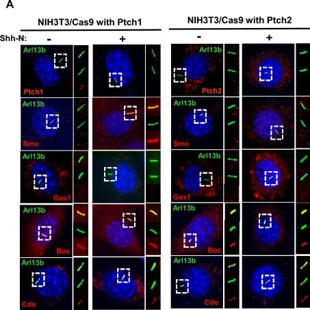
Detection of Gas1 by Immunocytochemistry/ Immunofluorescence
PTCH1 and PTCH2 dictate the differential localisation of SMO. (A) NIH3T3/Cas9 cells expressing PTCH1 or PTCH2 individually were treated with or without Shh-N to determine the intracellular location of the respective proteins (red) in relevance to primary cilia (labelled with Arl13b, green). (B) Cells with ciliary co-localisation (yellow in the merged images shown in the top panel of (A)) in each protein/treatment group are scored by counting 50 ~ 100 cells from 5 random fields. Data presented as mean ± SD with unpaired t test (***P < 0.001; ns, not significant). Image collected and cropped by CiteAb from the following open publication (https://pubmed.ncbi.nlm.nih.gov/37516749), licensed under a CC-BY license. Not internally tested by R&D Systems.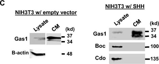
Detection of Gas1 by Western Blot
PTCH2/SHH signalling induces GAS1 expression. (A) NIH3T3/Cas9 cells expressing PTCH1 or PTCH2 individually were treated with ( +) or without (–) Shh-N and analysed by Western blotting for the endogenous proteins as indicated. B-actin as a loading control. (B) The mRNA levels of Boc, Cdo and Gas1 in WT NIH3T3 cells cultured in either 10% newborn calf serum-containing medium (NCS) or 0.5% serum-containing medium (SFM) for 24 h were analysed by RT-PCR and agarose gel electrophoresis. H2O was used as the template in the negative control (Left panel). Gas1 mRNA levels in WT NIH3T3 cultured in NCS or SFM were quantitatively assessed by real-time qRT-PCR (n = 3; unpaired student’s t test, P = 0.000066) (Right panel). (C) Western blot analyses of the cell lysate and the conditioned medium (CM) collected from WT NIH3T3 cells which were transfected with either an empty vector or SHH expression construct and cultured in SFM for 48 h. The full-length (37 kd) and the cleaved form (34 kd) of GAS1 protein are detected in CM. (D) Western blot analyses of the CM or cell lysates of NIH3T3 cells treated with varying concentrations of TMI005 (ADAM17 specific inhibitor) as indicated, for 48 h in SFM. The pro-form (135kd) and the mature form (100kd) of ADAM17 are detected in the lysate. Image collected and cropped by CiteAb from the following open publication (https://pubmed.ncbi.nlm.nih.gov/37516749), licensed under a CC-BY license. Not internally tested by R&D Systems.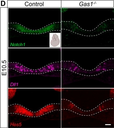
Detection of Gas1 by Immunohistochemistry
GAS1 deficiency disrupts NOTCH signaling in the murine embryonic forebrain. Detection of Notch1, Dll1 and Hes5 transcripts on coronal sections of control and Gas1−/− embryos at the indicated embryonic stages (plane of section indicated in the insets produced using BioRender.com). (A,B) While Notch1 (green; upper panels) and Dll1 (purple; middle panels) transcript levels in the rostral neuroepithelium are comparable between genotypes, transcript levels for Hes5 (red; bottom panels) are normal at 8-9 somites (A) but decreased at 10-11 somites (B) in Gas1−/− embryos compared with controls. n=4-6 embryos per genotype and somite stage (C,D) Transcript levels for Notch1 (green; upper panels), Dll1 (purple; middle panels) and Hes5 (red; bottom panels) are reduced in the rostral ventral diencephalon of E9.5 (C) and E10.5 (D) Gas1−/− embryos when compared with controls. n=8 embryos per genotype and embryonic stage. Scale bars: 50 µm. Dotted lines indicate rostral ventral neuroepithelium. Image collected and cropped by CiteAb from the following open publication (https://pubmed.ncbi.nlm.nih.gov/34698766), licensed under a CC-BY license. Not internally tested by R&D Systems.Applications for Human Gas1 Antibody
Western Blot
Sample: Recombinant Human Gas1 (Catalog # 2636-GS)
Formulation, Preparation, and Storage
Purification
Reconstitution
Reconstitute at 0.2 mg/mL in sterile PBS. For liquid material, refer to CoA for concentration.
Formulation
Shipping
Stability & Storage
- 12 months from date of receipt, -20 to -70 °C as supplied.
- 1 month, 2 to 8 °C under sterile conditions after reconstitution.
- 6 months, -20 to -70 °C under sterile conditions after reconstitution.
Calculators
Background: Gas1
References
- Schneider, C. et al. (1988) Cell 54:787.
- Mullor, J.L. and A.R. Altaba (2002) BioEssays 24:22.
- Del Sal, G. et al. (1994) Proc. Natl. Acad. Sci. USA 91:1848.
- Lee, T.C. et al. (1997) Proc. Natl. Acad. Sci. USA 94:12886.
- Del Sal, G. et al. (1992) Cell 70:595.
- Del Sal, G. et al. (1995) Mol. Cell. Biol. 15:7152.
- Stebel, M. et al. (2000) FEBS Lett. 481:152.
- Spagnuolo, R. et al. (2004) Blood 103:3005.
- Mellstrom, B. et al. (2002) Mol. Cell Neurosci. 19:417.
- Lee, K.K.H. et al. (2001) Dev. Biol. 234:188.
- Lee, C.S. et al. (2001) Proc. Natl. Acad. Sci. USA 98:11347.
- Liu, Y. et al. (2002) Development 129:5289.
- Liu, Y. et al. (2001) Dev. Biol. 236:30.
- Lee, C.S. et al. (2001) Dev. Biol. 236:17.
- Schueler-Furman, O. et al. (2006) Trends Pharmacol. Sci. 27:72.
Long Name
Alternate Names
Gene Symbol
UniProt
Additional Gas1 Products
Product Documents for Human Gas1 Antibody
Certificate of Analysis
To download a Certificate of Analysis, please enter a lot or batch number in the search box below.
Note: Certificate of Analysis not available for kit components.
Product Specific Notices for Human Gas1 Antibody
For research use only
Related Research Areas
Citations for Human Gas1 Antibody
 Powered by Bioz
Customer Reviews for Human Gas1 Antibody
There are currently no reviews for this product. Be the first to review Human Gas1 Antibody and earn rewards!
Have you used Human Gas1 Antibody?
Submit a review and receive an Amazon gift card!
$25/€18/£15/$25CAN/¥2500 for a review with an image
$10/€7/£6/$10CAN/¥1110 for a review without an image
Submit a review
Protocols
Find general support by application which include: protocols, troubleshooting, illustrated assays, videos and webinars.
- Cellular Response to Hypoxia Protocols
- R&D Systems Quality Control Western Blot Protocol
- Troubleshooting Guide: Western Blot Figures
- Western Blot Conditions
- Western Blot Protocol
- Western Blot Protocol for Cell Lysates
- Western Blot Troubleshooting
- Western Blot Troubleshooting Guide
- View all Protocols, Troubleshooting, Illustrated assays and Webinars