UGT1A1 (UDP-glucuronosyltransferase 1-A1; also UDPGT 1-1 and HUG-BR1) is a 52-57 kDa member of the UGT1A subfamily, UGT family of enzymes. UGT1A1 is expressed by the liver, and catalyzes the conjugation of glucuronic acid (GA) from UDPGA to lipophilic acceptors such as (anti-cancer) drugs and bilirubin. Addition of glucuronic acid increases target solubility and facilitates elimination. Mature human UGT1A1 is a 508 amino acid (aa) type I transmembrane ER glycoprotein. It contains a 465 aa luminal domain (aa 26-490) plus a 26 aa cytoplasmic region. The luminal domain is unusual in that aa 157-176 are embedded in the ER membrane. Amino acids 29-444 contain the enzyme active site. The cytoplasmic tail appears to mediate noncovalent homodimerization, and heterodimerization with UGT2B. The signal sequence (aa 1-25) is normally cleaved, but a Lys15Arg mutation blocks insertion into the ER membrane. There are multiple point mutations that impact enzyme activity. One potential splice form is reported that shows a six aa substitution for aa 289-533. Over aa 60-186, human UGT1A1 shares 64% aa identity with mouse UGT1A1.
Human Glucuronosyltransferase 1A1/UGT1A1 Antibody (856754)
R&D Systems | Catalog # MAB6490

Key Product Details
Species Reactivity
Human
Applications
Western Blot, Immunocytochemistry, Simple Western
Label
Unconjugated
Antibody Source
Monoclonal Mouse IgG1 Clone # 856754
Loading...
Product Specifications
Immunogen
E. coli-derived recombinant human Glucuronosyltransferase 1A1/UGT1A1
Leu60-Thr168
Accession # P22309
Leu60-Thr168
Accession # P22309
Specificity
Detects human Glucuronosyltransferase 1A1/UGT1A1 in ELISAs.
Clonality
Monoclonal
Host
Mouse
Isotype
IgG1
Scientific Data Images for Human Glucuronosyltransferase 1A1/UGT1A1 Antibody (856754)
Detection of Human Glucuronosyltransferase 1A1/UGT1A1 by Western Blot.
Western blot shows lysates of COLO 205 human colorectal adenocarcinoma cell line. PVDF membrane was probed with 2 µg/mL of Mouse Anti-Human Glucuronosyltransferase 1A1/UGT1A1 Monoclonal Antibody (Catalog # MAB6490) followed by HRP-conjugated Anti-Mouse IgG Secondary Antibody (Catalog # HAF018). A specific band was detected for Glucuronosyltransferase 1A1/UGT1A1 at approximately 50 kDa (as indicated). This experiment was conducted under reducing conditions and using Immunoblot Buffer Group 1.
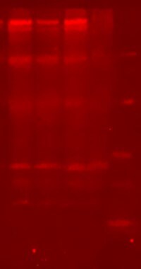
Glucuronosyltransferase 1A1/UGT1A1 in HepG2 Human Cell Line.
Glucuronosyltransferase 1A1/UGT1A1 was detected in immersion fixed HepG2 human hepatocellular carcinoma cell line using Mouse Anti-Human Glucuronosyltransferase 1A1/UGT1A1 Monoclonal Antibody (Catalog # MAB6490) at 10 µg/mL for 3 hours at room temperature. Cells were stained using the NorthernLights™ 557-conjugated Anti-Mouse IgG Secondary Antibody (red; Catalog # NL007) and counterstained with DAPI (blue). Specific staining was localized to cytoplasm. View our protocol for Fluorescent ICC Staining of Cells on Coverslips.
Detection of Human Glucuronosyltransferase 1A1/UGT1A1 by Simple WesternTM.
Simple Western lane view shows lysates of COLO 205 human colorectal adenocarcinoma cell line, loaded at 0.5 mg/mL. A specific band was detected for Glucuronosyltransferase 1A1/UGT1A1 at approximately 59 kDa (as indicated) using 20 µg/mL of Mouse Anti-Human Glucuronosyltransferase 1A1/UGT1A1 Monoclonal Antibody (Catalog # MAB6490). This experiment was conducted under reducing conditions and using the 12-230 kDa separation system.Applications for Human Glucuronosyltransferase 1A1/UGT1A1 Antibody (856754)
Application
Recommended Usage
Immunocytochemistry
8-25 µg/mL
Sample: Immersion fixed HepG2 human hepatocellular carcinoma cell line
Sample: Immersion fixed HepG2 human hepatocellular carcinoma cell line
Simple Western
20 µg/mL
Sample: COLO 205 human colorectal adenocarcinoma cell line
Sample: COLO 205 human colorectal adenocarcinoma cell line
Western Blot
2 µg/mL
Sample: COLO 205 human colorectal adenocarcinoma cell line
Sample: COLO 205 human colorectal adenocarcinoma cell line
Reviewed Applications
Read 1 review rated 3 using MAB6490 in the following applications:
Formulation, Preparation, and Storage
Purification
Protein A or G purified from hybridoma culture supernatant
Reconstitution
Sterile PBS to a final concentration of 0.5 mg/mL. For liquid material, refer to CoA for concentration.
Loading...
Formulation
Lyophilized from a 0.2 μm filtered solution in PBS with Trehalose. *Small pack size (SP) is supplied either lyophilized or as a 0.2 µm filtered solution in PBS.
Shipping
Lyophilized product is shipped at ambient temperature. Liquid small pack size (-SP) is shipped with polar packs. Upon receipt, store immediately at the temperature recommended below.
Stability & Storage
Use a manual defrost freezer and avoid repeated freeze-thaw cycles.
- 12 months from date of receipt, -20 to -70 °C as supplied.
- 1 month, 2 to 8 °C under sterile conditions after reconstitution.
- 6 months, -20 to -70 °C under sterile conditions after reconstitution.
Calculators
Background: Glucuronosyltransferase 1A1/UGT1A1
Long Name
UDP Glucuronosyltransferase 1A1
Alternate Names
GNT1, UDPGT 1-1, UG-BR1, UGT1, UGT1A
Gene Symbol
UGT1A1
UniProt
Additional Glucuronosyltransferase 1A1/UGT1A1 Products
Product Documents for Human Glucuronosyltransferase 1A1/UGT1A1 Antibody (856754)
Certificate of Analysis
To download a Certificate of Analysis, please enter a lot or batch number in the search box below.
Note: Certificate of Analysis not available for kit components.
Product Specific Notices for Human Glucuronosyltransferase 1A1/UGT1A1 Antibody (856754)
For research use only
Related Research Areas
Customer Reviews for Human Glucuronosyltransferase 1A1/UGT1A1 Antibody (856754) (1)
3 out of 5
1 Customer Rating
Have you used Human Glucuronosyltransferase 1A1/UGT1A1 Antibody (856754)?
Submit a review and receive an Amazon gift card!
$25/€18/£15/$25CAN/¥2500 Yen for a review with an image
$10/€7/£6/$10CAN/¥1110 Yen for a review without an image
Submit a review
Customer Images
Showing
1
-
1 of
1 review
Showing All
Filter By:
-
Application: Western BlotSample Tested: Liver tissueSpecies: humanised mouse and MouseVerified Customer | Posted 10/20/2017

There are no reviews that match your criteria.
Protocols
Find general support by application which include: protocols, troubleshooting, illustrated assays, videos and webinars.
- Appropriate Fixation of IHC/ICC Samples
- Cellular Response to Hypoxia Protocols
- ClariTSA™ Fluorophore Kits
- Detection & Visualization of Antibody Binding
- ICC Cell Smear Protocol for Suspension Cells
- ICC Immunocytochemistry Protocol Videos
- ICC for Adherent Cells
- Immunocytochemistry (ICC) Protocol
- Immunocytochemistry Troubleshooting
- Immunofluorescence of Organoids Embedded in Cultrex Basement Membrane Extract
- Immunohistochemistry (IHC) and Immunocytochemistry (ICC) Protocols
- Preparing Samples for IHC/ICC Experiments
- Preventing Non-Specific Staining (Non-Specific Binding)
- Primary Antibody Selection & Optimization
- Protocol for VisUCyte™ HRP Polymer Detection Reagent
- Protocol for the Fluorescent ICC Staining of Cell Smears - Graphic
- Protocol for the Fluorescent ICC Staining of Cultured Cells on Coverslips - Graphic
- Protocol for the Preparation and Fluorescent ICC Staining of Cells on Coverslips
- Protocol for the Preparation and Fluorescent ICC Staining of Non-adherent Cells
- Protocol for the Preparation and Fluorescent ICC Staining of Stem Cells on Coverslips
- Protocol for the Preparation of a Cell Smear for Non-adherent Cell ICC - Graphic
- R&D Systems Quality Control Western Blot Protocol
- TUNEL and Active Caspase-3 Detection by IHC/ICC Protocol
- The Importance of IHC/ICC Controls
- Troubleshooting Guide: Western Blot Figures
- Western Blot Conditions
- Western Blot Protocol
- Western Blot Protocol for Cell Lysates
- Western Blot Troubleshooting
- Western Blot Troubleshooting Guide
- View all Protocols, Troubleshooting, Illustrated assays and Webinars
Loading...