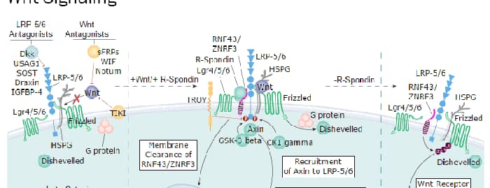
The Glypicans (glypiated proteoglycans) are a small multigene family of GPI-linked heparan sulfate (HS) proteoglycans that likely play a key role in embryonic morphogenesis (1-4). There are currently six known mammalian Glypicans. They all share a common-sized protein core of 60‑70 kDa, an N-terminus which likely forms a compact globular domain, 14 conserved cysteines that form multiple intrachain disulfide bonds, and a number of C‑terminal N- and O-linked carbohydrate attachment sites. Based on exon organization and the location of O-linked glycosylation sites, at least two subfamilies of Glypicans are known, with one subfamily containing Glypicans 1, 2, 4 and 6, and another subfamily containing Glypicans 3 and 5 (3, 5). Human Glypican 6 (GPC-6) is synthesized as a 554 amino acid (aa) preproprecursor that contains a 23 aa signal sequence, a 505 aa mature region and a 26 aa C-terminal prosegment (5, 6). There are four consecutive Ser-Gly repeats that serve as a heparin sulfate attachment site. GPC-6 is reported to be as large as 110 kDa in size. This translates into approximately 50 kDa of proteoglycan (5). Human to mouse, there is 97% aa identity over the entire GPC-6 molecule. Cells known to express GPC-6 are adult ovary and embryonic vascular and visceral smooth muscle, plus mesenchyme (embryonic connective tissue) in multiple organs (1, 5, 6). The function of GPC-6 is essentially unknown. As a Glypican family member, it may facilitate heparin-binding growth factor signaling and polyamine uptake into expressing cells (7, 8). In this regard, it would appear that GPC-6 with its attendant HS is down‑regulated by triiodothyronine during cartilage maturation, thus limiting the availability of sites for FGF sequestration and activity (9).
Human Glypican 6 Antibody (348701)
R&D Systems | Catalog # MAB2845


Scientific images may be resized, cropped, or adjusted for brightness or contrast for presentation purposes. These updates are limited to presentation and do not affect the underlying scientific interpretation of the data. Where available, additional source data may be provided upon request.
Key Product Details
Species Reactivity
Validated:
Cited:
Applications
Validated:
Cited:
Label
Antibody Source
Product Specifications
Immunogen
Asp24-Val527
Accession # Q9Y625
Specificity
Clonality
Host
Isotype
Scientific Data Images for Human Glypican 6 Antibody (348701)
Detection of Glypican 6 in HepG2 Human Cell Line by Flow Cytometry.
HepG2 human hepatocellular carcinoma cell line was stained with Mouse Anti-Human Glypican 6 Monoclonal Antibody (Catalog # MAB2845, filled histogram) or isotype control antibody (Catalog # MAB002, open histogram), followed by Allophycocyanin-conjugated Anti-Mouse IgG Secondary Antibody (Catalog # F0101B). To facilitate intracellular staining, cells were fixed with Flow Cytometry Fixation Buffer (Catalog # FC004) and permeabilized with Flow Cytometry Permeabilization/Wash Buffer I (Catalog # FC005). View our protocol for Staining Intracellular Molecules.Applications for Human Glypican 6 Antibody (348701)
CyTOF-ready
Intracellular Staining by Flow Cytometry
Sample: HepG2 human hepatocellular carcinoma cell line fixed with Flow Cytometry Fixation Buffer (Catalog # FC004) and permeabilized with Flow Cytometry Permeabilization/Wash Buffer I (Catalog # FC005)
Western Blot
Sample: Recombinant Human Glypican 6 (Catalog # 2845-GP)
Flow Cytometry Panel Builder
Bio-Techne Knows Flow Cytometry
Save time and reduce costly mistakes by quickly finding compatible reagents using the Panel Builder Tool.
Advanced Features
- Spectra Viewer - Custom analysis of spectra from multiple fluorochromes
- Spillover Popups - Visualize the spectra of individual fluorochromes
- Antigen Density Selector - Match fluorochrome brightness with antigen density

Formulation, Preparation, and Storage
Purification
Reconstitution
Reconstitute at 0.5 mg/mL in sterile PBS. For liquid material, refer to CoA for concentration.
Formulation
Shipping
Stability & Storage
- 12 months from date of receipt, -20 to -70 °C as supplied.
- 1 month, 2 to 8 °C under sterile conditions after reconstitution.
- 6 months, -20 to -70 °C under sterile conditions after reconstitution.
Calculators
Background: Glypican 6
References
- Song, H.H. and J. Filmus (2002) Biochim. Biophys. Acta 1573:241.
- Filmus, J. (2001) Glycobiology 11:19R.
- De Cat, B. and G. David (2001) Semin. Cell Dev. Biol. 12:117.
- Filmus, J. (2003) Glycoconj. J. 19:319.
- Veugelers, M. et al. (1999) J. Biol. Chem. 274:26968.
- Paine-Saunders, S. et al. (1999) Genomics 57:455.
- Fransson, L-A. et al. (2004) Cell Mol. Life Sci. 61:1016.
- Fransson, L-A. (2003) Int. J. Biochem. Cell Biol. 35:125.
- Bassett, J.H.D. et al. (2006) Endocrinology 147:295.
Alternate Names
Gene Symbol
UniProt
Additional Glypican 6 Products
Product Documents for Human Glypican 6 Antibody (348701)
Certificate of Analysis
To download a Certificate of Analysis, please enter a lot or batch number in the search box below.
Note: Certificate of Analysis not available for kit components.
Product Specific Notices for Human Glypican 6 Antibody (348701)
For research use only
Related Research Areas
Citations for Human Glypican 6 Antibody (348701)
 Powered by Bioz
Customer Reviews for Human Glypican 6 Antibody (348701)
There are currently no reviews for this product. Be the first to review Human Glypican 6 Antibody (348701) and earn rewards!
Have you used Human Glypican 6 Antibody (348701)?
Submit a review and receive an Amazon gift card!
$25/€18/£15/$25CAN/¥2500 for a review with an image
$10/€7/£6/$10CAN/¥1110 for a review without an image
Submit a review
Protocols
Find general support by application which include: protocols, troubleshooting, illustrated assays, videos and webinars.
- 7-Amino Actinomycin D (7-AAD) Cell Viability Flow Cytometry Protocol
- Cellular Response to Hypoxia Protocols
- Extracellular Membrane Flow Cytometry Protocol
- Flow Cytometry Protocol for Cell Surface Markers
- Flow Cytometry Protocol for Staining Membrane Associated Proteins
- Flow Cytometry Staining Protocols
- Flow Cytometry Troubleshooting Guide
- Intracellular Flow Cytometry Protocol Using Alcohol (Methanol)
- Intracellular Flow Cytometry Protocol Using Detergents
- Intracellular Nuclear Staining Flow Cytometry Protocol Using Detergents
- Intracellular Staining Flow Cytometry Protocol Using Alcohol Permeabilization
- Intracellular Staining Flow Cytometry Protocol Using Detergents to Permeabilize Cells
- Propidium Iodide Cell Viability Flow Cytometry Protocol
- Protocol for Liperfluo
- Protocol for the Characterization of Human Th22 Cells
- Protocol for the Characterization of Human Th9 Cells
- Protocol: Annexin V and PI Staining by Flow Cytometry
- Protocol: Annexin V and PI Staining for Apoptosis by Flow Cytometry
- R&D Systems Quality Control Western Blot Protocol
- Troubleshooting Guide: Fluorokine Flow Cytometry Kits
- Troubleshooting Guide: Western Blot Figures
- Western Blot Conditions
- Western Blot Protocol
- Western Blot Protocol for Cell Lysates
- Western Blot Troubleshooting
- Western Blot Troubleshooting Guide
- View all Protocols, Troubleshooting, Illustrated assays and Webinars
Associated Pathways